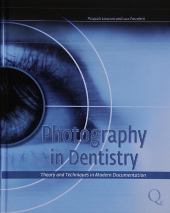
It is generally accepted that well-composed images are necessary for proper diagnosis and treatment planning. In their book, Photography in Dentistry: Theory and Techniques in Modern Documentation , Drs Pasquale Loiacono and Luca Pascoletti address almost every aspect of clinical dental photography, from optics to patient positioning.
Attempting to cover all aspects of clinical photography is a daunting task, and Drs Loiacono and Pascoletti are to be praised for their efforts. The images and illustrations are well placed and of excellent quality, and their discussions of concepts such as f-stops and magnification make complex topics easy to understand. However, in more than a few instances, the book covers general photographic principles in far greater depth than necessary for dental photography. As an avid photographer, I enjoyed such discussions, but I suspect that many readers will not find them quite as engaging.
The real strength of this book is found in the final 100 pages, which are dedicated to equipment and intraoral photographic techniques. The authors did an admirable job of outlining the equipment and positioning necessary to capture 30 basic images with a 2-operator approach. Although it is far more challenging than a single-operator technique and not as effective, the combination of images and text creates a systematic way of moving through any photographic protocol if 2 operators are to work together.
Nonetheless, a significant disappointment was that representative images of many techniques were not clinically ideal. For instance, the guidelines for “Quadrants in occlusion for orthodontic documentation” (full lateral arch images) do not extend anteriorly to include the contralateral incisor, preventing a proper view of the inclination of the incisors. This is as disappointing as the improperly captured interarch relationship in the same image or the lips against the teeth in many occlusal images.


