Fig. 4.1
Clinical radiographs demonstrating (a) immediate post-operative view of non-surgical root canal treatment of tooth 47 treated over the course of two visits using an intra-canal medicament. Preparation was carried out using ProTaper Universal files 25.06 and obturation using vertical compaction (System B and Obtura using AH Plus cement). Note adequate cleaning and shaping have allowed the lateral canal to be sealed in the mesial root. The acute apical distal curvature was maintained and apical patency confirmed with a seal puff. (b) Six-month post-operative view demonstrating well-placed coronal permanent cast restoration to ensure biological seal is maintained. Note the distal peri-radicular radiolucency has resolved. Nonsurgical root canal treatment has a 100 % capability of healing provided biological chemomechanical preparation and obturation have been carried out with strict adherence to common protocols. Nickel-titanium technology can produce predictable radicular preparations that can be easily obturated
However, in the last 30 years, despite these developments, there have not been any real improvements in terms of the outcome of treatment (success) and prognosis. Various cross-sectional surveys assessing root canal treatment outcome have repeatedly demonstrated a significant correlation between the presence of an apical radiolucency and a radiographically inadequate root filling. A recent systematic review confirmed a high prevalence of peri-radicular radiolucencies (PR) that vary from 0.5 to 13.9 % overall [4–7].
It is therefore obvious that certain treatment factors must potentially have an impact on treatment outcome. A recent study attempted to identify these factors and their influence on outcome measured by the periapical status of the teeth. This prospective study involved annual clinical and radiographic follow-up of non-surgical root canal treatment of 702 teeth in 534 patients carried out by endodontic postgraduate students for 2–4 years. Pre-, intra- and postoperative data was collected prospectively using customised data collection forms and investigated using multiple logistic regression models. Ten prognostic factors were identified. Four preoperative factors (the preoperative absence of a periapical lesion, in the presence of a periapical lesion the smaller the size the better the prognosis, the absence of a preoperative sinus tract and the absence of tooth/root perforation) and six intraoperative factors (achievement of patency at the canal terminus, extension of canal cleaning (and therefore filling) as close as possible to the apical terminus, abstaining from the use of 2 % chlorhexidine as an adjunct to sodium hypochlorite solution, absence of inter-appointment flare-up, the absence of root filling extrusion and the presence of a satisfactory coronal restoration) were all found to influence success based on periapical health associated with roots following non-surgical root canal treatment [8]. Interestingly the specific type of instrument utilised in mechanical preparation (stainless steel or nickel-titanium) had no direct effect on outcome. More importantly the biological factors indirectly related to preoperative and perioperative factors are what determine whether a case is successful or fails.
In order to improve the success rate of root canal treatment in general dental practice, the referral of difficult cases to dentists with advanced knowledge and training in endodontics may be appropriate. The decision to treat a case lies with the practitioner and should be based on whether the case is perceived to be treatable or beyond their abilities requiring a referral to a specialist endodontic practitioner. One can assume that, if a practitioner treats a case beyond his or her level of expertise, then there may be a greater likelihood of iatrogenic incidents that can result in an unsuccessful treatment outcome. The General Dental Council [9], the European Society of Endodontology [10] and the American Association of Endodontists [11] provide guidance for clinicians when making a referral. The American Association of Endodontists has published a form, which describes 17 areas that should be assessed when evaluating the potential difficulty of an endodontic case. Difficult cases that deserve particular attention and possibly the need to refer include patient factors (complex medical history, difficulty with achieving anaesthesia, prominent gag reflex and significant pain and swelling), radiographic factors (difficulty obtaining radiographs, appearance of calcified canals, pulp stones and obstructions) and tooth factors (tooth type, inclination, canal curvature, canal morphology) (see Tables 4.1, 4.2 and 4.3).
Endodontic instrumentation plays an important role in the process of microbial elimination from root canal systems. Stainless steel instruments have been the principal material for fabrication of endodontic files until nickel-titanium alloy was proposed in 1989 [12]. Since its inception, nickel-titanium rotary files have become a routine part of the endodontic armamentarium enabling clinicians to treat straightforward cases in a timely and efficient manner. If one were to compare effectiveness with regard to elimination of biofilm from within the root canal system, no study to date has proved that nickel-titanium files are superior when compared to stainless steel [13]. Traditional stainless steel instruments are standardised to a taper of 0.02 mm/1.00 mm in length (2 % taper). Rotary instruments on the other hand have developed tapers ranging from 2 to 10 %. Due to the inherent inflexibility of larger-sized stainless steel instruments and to avoid canal transportation in curved canals, standardised preparation techniques proposed a final apical instrument size of #20 or #25. The clinician must be aware that nickel-titanium although inherently flexible due to the super elasticity is prone to canal transportation due to its other property of shape memory. Use of a 6 % or 8 % tapered instrument in a curved canal can lead to either alteration in original canal anatomy or risk of file separation. Recently a proprietary method of thermo-mechanical treatment of nitinol wire was developed known as “M-wire”. Vortex Blue rotary files (Dentsply, Tulsa Dental Specialities) alter the machined nitinol wire by a series of complex heating and cooling treatments resulting in a shape memory alloy that makes the instruments extremely flexible at room temperature [14]. Studies have demonstrated that rotary instruments manufactured from M-wire had significantly higher fatigue resistance, torsional strength, flexibility and higher deformation to failure when compared to conventional NiTi instruments [15, 16].
Nickel-titanium instrument development continues to strife forward in an attempt to improve flexibility and resistance using novel manufacturing processes [17, 18]. Novel instrumentation techniques such as the self-adjusting file (SAF) system (ReDent, Raanana, Israel) represent new concepts in root canal cleaning based on a hollow file that adapts itself to the shape of the canal rather than the conventional concept of shaping every canal to a round cross section and fixed taper according to the file system used [19]. Commercially available systems that have embraced the concept of reciprocation as opposed to the tried and tested rotation of files include Reciproc (VDW, Munich, Germany) and WaveOne (Dentsply Maillefer, Ballaigues, Switzerland). These systems utilise a “single file concept” through a counterclockwise cutting motion and clockwise rotation that have gained widespread appeal due to their simplicity [20]. At the same time, interest in irrigation research has not waned given the professions focus on the ease and predictability of rotary instruments to plane the canal walls efficiently. In spite of the ability to create tapered shapes of various sizes, studies have demonstrated the great extent to which canal walls remain un-instrumented. The delivery of intra-canal irrigants and its importance as well as an awareness of the biological impediments, in the form of biofilms, will no doubt lead to the introduction of new standards of irrigation in this field in the future [21].
Root canal preparation can be challenging in narrow calcified canals requiring a systematic approach for successfully negotiating canals to length. Coronally the presence of pulp stones can hinder access to the root canals and their subsequent shaping. Pulp stones have been described as compact degenerative masses of calcified tissue, which can be classified according to structure (true and false) and location (embedded, adherent or free). Embedded or adherent pulp stones within the root canal can result in significant occlusion of the canals or difficulty with negotiation if present at the curvature. Free pulp stones found in the pulp chamber may block access to canal orifices and alter the internal anatomy. Correct access cavity design, ultrasonic file use, hypochlorite irrigation and magnification should aid in the removal of pulp stones and present little difficulty during root canal treatment [22].
Pulp canal obliteration (PCO) or calcific metamorphosis occurs commonly in 4–25 % of traumatised teeth resulting in apparent loss of the pulp space radiographically and yellow discolouration of the crown. These teeth provide particular endodontic challenges with increased difficulties with location of the canal, negotiation of the canal, irretrievable instrument fracture and potential iatrogenic perforation [23, 24]. According to the American Association of Endodontist case assessment criteria [11], these cases fall into the high difficulty category.
Reactive and degenerative changes in the pulp as a direct consequence of pulpal irritants, insults and wear and tear can result in the phenomena of “calcified” or “sclerosed” canals. Pulp space diminishes throughout life by the regular deposition of secondary dentine. Pulp space can be further reduced by reactionary or reparative dentine (previously classified as tertiary or irritation dentine) as a means of reducing the porosity of dentinal tubules exposed to carious, traumatic or chemical insults. Reactionary dentine represents material laid down by existing surviving odontoblast cells. Reparative dentine, on the other hand, is a new material laid down by new odontoblast-like cells, which have differentiated and migrated to the site of injury following the death of primary odontoblast cells. [25] In addition fibrotic changes within the root canal system can pose further challenges to canal negotiation with the increased potential of fibrotic pulp tissue compaction leading to obstructions that can be very difficult to overcome [26]. According to the American Association of Endodontist case assessment criteria [11], following radiographic assessment if the canals are obviously reduced in size or their path is indistinct or not visible, then these cases fall into the category of moderate to high difficulty.
Understanding root canal curvature is also of prime importance when carrying out instrumentation techniques in order to assess which taper will effectively shape the canal without the risk of either structural deformation of the instrument (and ultimately fracture) or iatrogenic errors including apical transportation, ledges, elbows, zips, perforations and loss of working length. Several methods have been described in the endodontic literature to determine canal curvature and more specifically the angle and radius of curvature [27]. Schneider proposed a radiographic method to determine canal curvature based on the angle that is obtained by two straight lines (Fig. 4.2). The first line is parallel to the long axis of the root canal. The second line passes through the apical foramen (point B) until it intersects with the first line at which the curvature starts (point A). The angle formed (X) allows the degree of curvature of root canals to be categorised into straight (≤5°), moderate (10–20°) or severe (25–70°) [28].
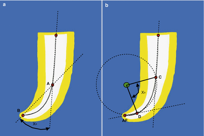

Fig. 4.2
Diagrammatic representation of the (a) Schneider measuring method and (b) Pruett et al. measuring method for determination of canal curvature. Note (a) a line is scribed on the radiograph parallel to the long axis of the canal. A second line was drawn from the apical foramen (Point B) to intersect with the first at the point where the canal begins to level the long axis of the tooth (Point A). These two lines intersect at an angle (X) as a measure of the change in direction of the canal in relation to the long axis of the tooth. Note (b) a straight line is drawn along the long axis of the portion of the canal. There is a point on each of these lines at which the canal deviates to the brain (Point C) or end (Point D) the canal curvature. The curved portion of the canal is represented by a circle with tangents at points C and D. The centre of the circle is thus the point at which the vertical lines intersect the tangents through C and D. This method allows calculation of the radius of curvature and angle of curvature of two independent parameters
However, this method of canal curvature assessment did not consider the radius of curvature as a second independent parameter. Pruett and colleagues reported that the two root canals measured at the same angle in degrees by Schneider’s method could have two very different radii or abruptness of curvatures. This radius of curvature had a significant factor on the number of cycles to failure of a NiTi engine-driven rotary instrument. As the radius of curvature decreased, instrument strain and stress increased and the fatigue life decreased. In other words, the smaller the radius of curvature, the more likely canal transportation or instrument fracture to occur. The authors graphically determined radius of curvature by drawing a straight line along the long axis of the coronal portion of the canal (see Fig. 4.2).
Along this line there lies a point at which the canal deviates to begin (point C) or end (point D) the canal curvature. A circle represents the curved portion of the canal, with tangents at points C and D. The centre of the circle is thus at the point where the vertical lines intersect the tangents through C and D. The change in direction of the long axis of the canal is described by the radius of the circle and by an angle or the arc length of this circle. The radius of curvature represents how abruptly or severely a specific angle of curvature occurs as the canal deviates from a straight line. The smaller the radius of curvature, the more abrupt is the canal deviation, which can result in file deformation and fracture [29].
Schäfer and colleagues proposed a modification of the Pruett method for measuring the angle of curvature, radius of curvature and also the length of curvature (Fig. 4.3) [30].
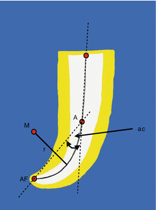

Fig. 4.3
Diagrammatic representation of the modified method proposed by Shafer et al. (2002) for measuring angle, radius and length of curvatures. A straight line is drawn along the outer side of the tooth canal in the coronal portion; this line is parallel to the long axis of the canal. The point where the canal deviates from this line to begin the canal curvature is marked (Point A). A second line is drawn to intersect the apical foramen (Point AF) with the point where the canal began to leave the long axis (Point A). The angle of curvature is formed between these two lines. The line between the point where the canal began to leave the long axis and the apical foramen is an arc of the hypothetical circle that defines the curved part of the canal. The curved part of the root canal between these two points A and AF is the circular arc of the hypothetical circle, which is specified by its radius (r). The radius of curvature can be calculated on the basis of the measured length of the arc based on geometrical principles of an isosceles triangle
From a clinical standpoint, conventional radiography limits calculation on curvatures since unseen curvatures (in the bucco-lingual direction) can also play a significant role in the cleaning and shaping of canals. The use of cone-beam CT scanning has been advocated for determining canal curvatures, particularly in the labio-palatal direction, which cannot be seen using standardised radiographic techniques [31].
Despite the introduction of flexible nickel-titanium instruments and torque-controlled motors, root canal preparation of severely curved canals remains an endodontic challenge with risks of canal transportation, procedural errors or instrument fracture [32].
ProTaper NEXT instruments (Dentsply, Maillefer) manufactured from M-wire alloy and characterised by an innovative off-centre rectangular cross section, which gives the files a snake-like swagger movement during preparation, have been introduced specifically suited for the preparation of curved root canals [33]. ProTaper NEXT instruments have been shown to prepare severely curved canals (25–39° curved canals) up to a size 40 without an increased risk of canal straightening or canal transportation [34].
Recently, several different single file systems have been marketed with the ability to shape a canal with the use of only one instrument. WaveOne (Dentsply, Maillefer, Ballaigues, Switzerland) is manufactured from M-wire alloy, which is characterised by an increased flexibility and cyclic fatigue resistance in comparison with martensitic NiTi. These instruments are used in an alternating reciprocal motion of clockwise (CW) and counterclockwise (CCW) rotations [35]. These instruments have been shown to be safe to use even in severely curved root canals in extracted teeth [36].
4.2 Endodontic Case Assessment
Patient factors that may require endodontic referral include medical history issues, anaesthesia difficulties, uncooperative anxious patients, patients with limited mouth opening, patients with a prominent gag reflex or initial presentation of patients requiring further surgical management to alleviate acute symptoms (see Tables 4.1 and 4.2).
Table 4.1
Patient risk factors that need to be considered when assessing case difficulty that may warrant further endodontic referral
|
Moderate risk
|
High risk
|
|
|---|---|---|
|
Medical history
|
One or more medical problems
|
Complex medical history or serious illness/disability
|
|
Anaesthesia
|
Vasoconstrictor intolerance
|
Difficulty achieving anaesthesia
|
|
Patient disposition
|
Anxious
|
Uncooperative
|
|
Mouth opening ability
|
Slight limitation in opening
|
Significant limitation
|
|
Gag reflex
|
Gags occasionally
|
Severe gag reflex
|
|
Presence of swelling/pain
|
Moderate pain or swelling
|
Severe pain or swelling
|
Table 4.2
Treatment considerations that need to be considered when assessing case difficulty that may warrant further endodontic referral
|
Moderate risk
|
High risk
|
|
|---|---|---|
|
Radiographic assessment
|
Difficulty obtaining
radiographs due to high floor of mouth, narrow or low palatal vault, presence of tori
|
Extreme difficulty in obtaining adequate radiographs
|
|
Tooth
position in the arch
|
1st molar
Moderate inclination (10–30°)
Moderate rotation
(10–30°)
|
2nd or 3rd molar
Extreme inclination (>30°)
Extreme rotation (>30°)
|
|
Tooth
isolation
|
Pretreatment modification required for rubber dam isolation
|
Extensive pretreatment modification required
|
|
Crown
morphology
|
Full coverage
Bridge abutment
Moderate toot/root deviation
Teeth with extensive coronal destruction
|
Restoration does not reflect normal anatomy/alignment
Significant deviation from normal tooth/root
|
|
Canal and root
morphology
|
Moderate curvatures (10–30°)
Crown axis differs from root axis
Wide open apex (1–1.5 mm)
|
Extreme curvature (>30°)
Premolars with more than two canals
Lower incisors with two canals
Canal division in middle/apical 1/3rd
Very long teeth (>25 mm)
Open apex (>1.5 mm)
|
|
Radiographic appearance of canals
|
Canal and chamber visible but reduced in size
Pulp stones present
Partial canal calcification
|
Indistinct canal paths
Canal (s) not visible
PCO present
|
|
Resorption
|
Apical resorption present
|
Extensive apical resorption
Internal resorption
External resorption
|
In routine cases such as obvious peri-radicular pathosis associated with a tooth, the diagnosis is usually straightforward. However, in certain situations such as irreversible pulpitis, where the patient cannot easily identify the offending tooth or cases where signs and symptoms are confusing as is synonymous with orofacial pain, then prompt referral may be necessary to ensure correct management and appropriate treatment is selected.
Treatment considerations warranting particular attention prior to commencing treatment include radiographic assessment of potential difficulties, tooth position in the arch, isolation problems, crown morphology, canal and root morphology, radiographic appearance of canals and extent of resorption present (see Table 4.2).
Additional considerations that should be assessed include history of trauma, previous endodontic history and periodontal-endodontic conditions that may all affect outcome if not correctly managed from the outset (see Table 4.3).
Table 4.3
Additional considerations when assessing case difficulty that may warrant further endodontic referral
|
Moderate risk
|
High risk
|
|
|---|---|---|
|
Trauma history
|
Complicated crown fracture
Subluxation
|
Complicated crown fracture immature tooth
Horizontal root fracture
Alveolar fracture
Intrusion
Luxation
Extrusion
Avulsion
|
|
Endodontic treatment history
|
Previous access without complications
|
Previous access with complications
Perforations
Unlocated canal
Ledging
Canal transportation
Instrument fracture
|
|
Periodontal-endodontic condition
|
Concurrent moderate periodontal disease
Draining sinus
|
Severe periodontal disease
Cracked teeth
Combined perio-endo lesions
Root amputation
|
4.3 Pulp Stones and Coronal Calcifications
In order to locate calcified canal orifices, the clinician must first visualise and assess the pulp space dimensions on a two-dimensional radiographic image. Access preparation should be initiated, with this view in mind, and access burs directed towards the presumed location of the pulp chamber (see Chapter 1 for a more detailed discussion).
Accurate preoperative radiographs should be taken with a long-cone paralleling beam aiming device so that an initial measurement can be taken to ascertain depth of bur penetration (usually a distance of approximately 6.5 mm from the occlusal surface to the projected pulp chamber floor). Teeth that have undergone significant calcifications and diminished pulp volumes require particular care during access preparations. Depth of penetration to reach the underlying pulp will be less and overzealous preparation could lead to iatrogenic perforation. Teeth that have been crowned also require appropriate assessment of the crown-root orientation to ensure correct access preparation results in location of the canals (see Figs. 4.4, 4.5 and 4.6).
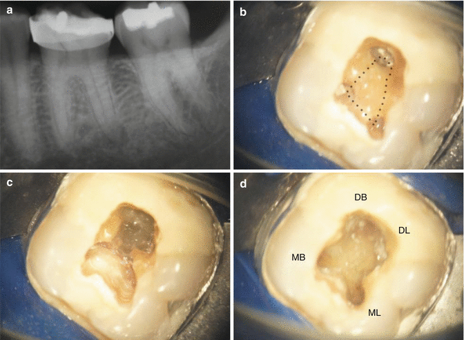
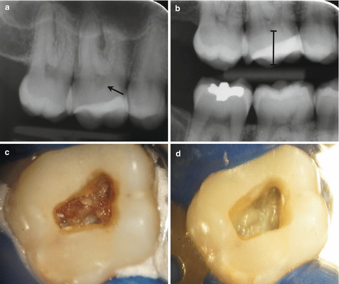
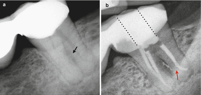

Fig. 4.4
Clinical radiograph and photographs demonstrating (a) pre-operative radiograph showing peri-radicular radiolucency associated with tooth 36. Referring clinician has carried out emergency pulp extirpation but had difficulties locating the ‘calcified canals’. Intra-operative dental operating microscopic views show (b) intact root of pulp chamber and (c, d) unroofing and ultrasonic troughing carried out to reveal internal anatomy with straight line access of all four canals. Canals could be easily negotiated to length

Fig. 4.5
Clinical radiographs and photographs demonstrating (a) pre-operative long-cone parallel radiograph demonstrating calcified coronal pulp chamber (black arrow). Tooth 16 presented with irreversible pulpitis and history of cracked tooth syndrome. (b) Parallel bitewing radiograph confirms radiopaque mass in coronal pulp chamber suggestive of pulp stones. Depth of bur penetration to roof of pulp chamber can be measured prior to access preparation. (c) Following access preparation pulp stones present in pulp chamber and (d) following use of ultrasonic refinement and troughing root canal anatomy can be seen

Fig. 4.6
Clinical demonstrating (a) pre-operative long-cone parallel radiograph demonstrating peri-radicular infection associated with distal bridge abutment tooth 47. Note inter-radicular lucency (black arrow). (b) Post operative view demonstrating final obturation following two visit non surgical endodontic therapy. Access preparation was completed through the bridge abutment. Orientation for access was made using the long axis of the tooth (dotted lines). Note lateral canal obturated in distal root resulting in sealer extrusion (red arrow). Distal apical curvature maintained to ensure patency and obturation to length
- 1.
Once the pulp chamber has been located, the entire roof of the pulp chamber should be removed to facilitate straight-line access and unimpeded access to all internal anatomy. Use of a non-end cutting bur is essential, and the combination of ultrasonic, magnification and illumination is invaluable (see Fig. 4.4 and Chap. 1 also for more detailed discussion).
- 2.
Any pulp stones encountered in the coronal pulp chamber should be removed prior to initial instrumentation of any canals to avoid the risk of any stones becoming dislodged into the root canals and becoming compacted apically resulting in blockage and further difficulties with negotiation (see Fig. 4.5). If a pulp stone accidently falls into one of the root canals, then early identification is essential. Never introduce a large-sized file (#20 or more), which will further increase the chance of blockage by pushing the stone further apically. Instead small hand files (#08 and #10 Hedstroem files) should be introduced, initially bypassing the blockage, and then used in a pull motion in an attempt to engage the stone more coronally.
- 3.
Intermittent flushing with sodium hypochlorite solution is advised to facilitate this process. If all coronal calcifications and pulp stone remnants are removed effectively, then this minimises any risks during canal cleaning and shaping procedures.
4.4 Location of Canal Orifices
The pulpal floor should be carefully examined with prior knowledge from preoperative radiographs and likely anatomy based on tooth type to indicate probable number of canals expected. A DG-16 probe should be used with firm probing for signs of any “sticking” associated with the presence of a canal orifice. Once a suspected orifice is found, the canal should be checked for patency using small fine instruments such as a #08 or #10 K file. Use of a #6 file can be reserved for very fine calcified canals, but due their inherent lack of stiffness, any calcifications encountered during file penetration will result in the file bending and deflecting.
Stay updated, free dental videos. Join our Telegram channel

VIDEdental - Online dental courses


