L
Laboratory analog Copy of a prosthetic or implant element used in laboratory fabrication procedures.1 See also: Analog/analogue.
Laboratory screw Element used in dental laboratory procedures in the fabrication of the prosthesis. Laboratory screws can be modified, eg, elongated or made from a different alloy, from the definitive design.2
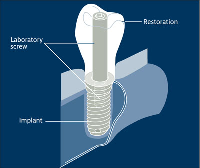
Laboratory screw.
(Redrawn from Yanase and Preston2 with permission.)
Lamellar bone Adult, mature bone consisting of 3- to 5-μm-wide layers of mineralized collagen fibrils. The orientation of fibrils changes from layer to layer; this construction is often compared with that of plywood. It appears birefringent in polarized light and is found in mature cortical as well as trabecular bone.
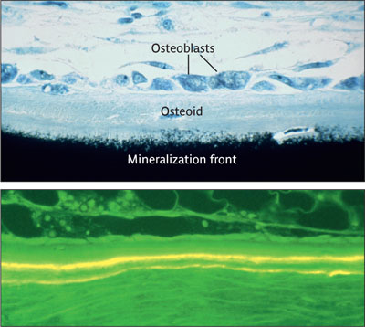
Lamellar bone. (Top: Von Kossatolnidine blue stain; magnification x800. Bottom: Fluorescence after double tetracycline labeling with 1-week-interval-mineralization rate 1-2 micron per day.)
(Reprinted from Buser et al3 with permission.)
Laminate Layered material. In dentistry, a thin layer or veneer of restorative material applied to the surface of a tooth, usually for cosmetic purposes. See also: Facing; Veneer.
Lapping tool Instrument used with or without abrasives to improve the adaptation of two opposing surfaces. In the laboratory, an instrument, often rotating, used to remove casting irregularities by means of grinding or polishing.4
Laser Acronym for light amplification by stimulated emission of radiation; a source that emits photons in a coherent beam that can propagate over long distances without significant divergence and can be focused on very small areas. Can have a very narrow bandwidth, compared with the broad spectrum emitted by most lamps. Light may be emitted continuously or in the form of short or ultrashort pulses, with durations from microseconds to a few femtoseconds. These properties may be the consequence of the very high coherence of laser radiation. Usually includes an optical cavity (resonator), in which light can circulate (eg, between two end mirrors) and which holds a gain medium that serves to amplify the light. See also: Er:YAG laser; Low-level laser therapy; Nd:YAG laser; Pulsed-mode laser.
Laser etching Creation of an altered surface by the application of laser energy; frequently used to apply permanent markings to metal objects, such as surgical instruments.
Late implant loss Outcome related to the loss of an implant that occurred after implant osseointegration. Compare: Early implant loss.
Late implant placement Implant placement at least 6 months following tooth extraction. The chosen time period should allow for sufficient bone regeneration of the extraction socket, and consequently, implant placement without bone augmentation procedure. Late implant placement bears the risk of bone atrophy in the orofacial direction, particularly in the anterior maxilla.
Latency period See: Consolidation period.
Lateral Bennett shift See: Bennett movement.
Lateral cephalograph Extraoral radiograph showing the region of the skull that comprises the bones of the face (ie, the viscerocranium). It requires a 18 x 24-cm or 20 x 25-cm image receptor and is intended for diagnosis in orthodontics. It also has been used in the preoperative implant examination to determine size and length of implants to be placed in the interforaminal region.
Lateral window technique
Stay updated, free dental videos. Join our Telegram channel
VIDEdental - Online dental courses


