This article reports the successful treatment of a patient with a malocclusion and missing maxillary lateral incisors with an unusual implant-prosthetic rehabilitation in place of the canines. A man, 25 years 5 months of age, was referred by his general dentist with the chief complaint of retained maxillary deciduous canines. He had a skeletal Class I and an Angle Class I malocclusion with an open-bite tendency and prolonged retention of both deciduous canines. The malocclusion was treated with extraction of the maxillary deciduous canines, a fixed edgewise appliance, and implant-prosthesis rehabilitation in place of the canines. A functional and an esthetic occlusion was achieved.
The primary problem associated with the treatment of malocclusion involving agenesis of the maxillary lateral incisors is identifying the procedure that will result in the best esthetic and functional results; this involves more than mere decisions regarding when to open or close the edentulous spaces.
In cases involving congenitally missing maxillary lateral incisors, an interdisciplinary treatment approach is beneficial for obtaining the most predictable outcomes. The management of a patient with such a malocclusion involves multiple treatment options, including acceptance of the space, orthodontic closure of the space (eg, with the maxillary canine substituting and camouflaging this appearance), single-tooth implants, bridges, and tooth-supported restorations.
The most satisfactory results are achieved when the spaces are closed with mesial movement of the canines. However, canine substitution is often associated with difficulty in achieving acceptable esthetic results because of the inherent size and shape of the teeth and differences between the maxillary canines and the lateral incisors. Therefore, for patients who do not meet the specific qualifications necessary to be considered optimal candidates for canine substitution, an alternative form of treatment must be considered. Restorative treatment alternatives can be divided into 2 categories: a single-tooth implant and a tooth-supported restoration. Today, such alternative treatments are a great choice because of osseointegrated implants and the difficulty associated with obtaining a satisfactory cosmetic result via the closure of edentulous spaces, especially with regard to unilateral agenesis.
This case report illustrates a successful treatment involving canine substitution and implant-prosthetic rehabilitation in a patient who was missing both maxillary lateral incisors.
Diagnosis and etiology
A man, 25 years 5 months of age, was referred by his dental implantologist to an orthodontist with the chief complaint of the presence of the maxillary deciduous canines. He requested that the treatment time not last for more than 1 year.
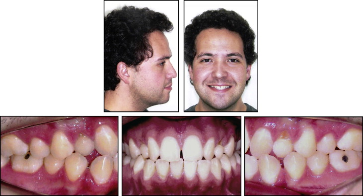
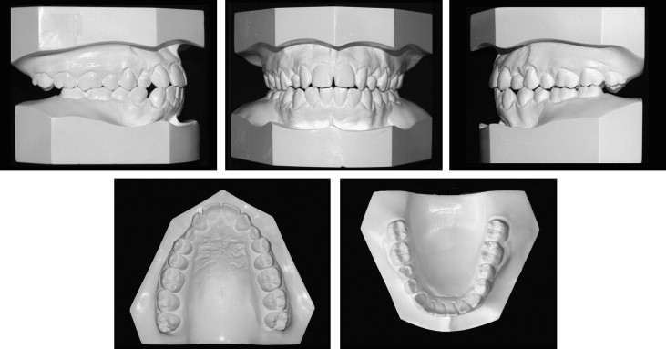
The patient was diagnosed with an Angle Class I malocclusion with an open-bite tendency, missing maxillary lateral incisors, and prolonged retention of the deciduous canines. The permanent canines had erupted in place of the lateral incisors. Both maxillary deciduous canines were in crossbite with the mandibular first premolars. The patient also had a slight midline diastema between the maxillary central incisors, which also fell between the mandibular first premolars and canines. The maxillary midline was deviated 1 mm to the right, and the mandibular midline was coincident with the facial midline. The mandibular third molars were absent, but the maxillary molars were overerupted. There were no signs or symptoms of temporomandibular joint dysfunction. The patient had a straight profile; although the lower lip was more prominent than the upper lip, a good facial profile was observed ( Figs 1 and 2 ).


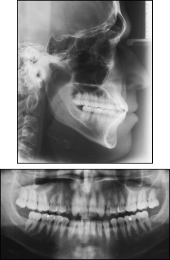
The panoramic radiograph showed that all permanent teeth except the maxillary lateral incisors and the mandibular third molars were present, as well as the prolonged retention of his deciduous canines with some evidence of external root resorption. The maxillary third molars displayed mild extrusion. Root parallelism was evident, including the maxillary canines and first premolars ( Fig 3 ). The cephalometric values showed a skeletal Class I relationship, a slight proclination of the maxillary incisors, and proclined mandibular incisors ( Table ).

| Measurement | Pretreatment | Postreatment | 1-year retention |
|---|---|---|---|
| SNA angle (°) | 89 | 90 | 91 |
| SNB angle (°) | 87 | 87 | 88 |
| ANB angle (°) | 2 | 3 | 3 |
| Convex angle (°) | 1 | 1 | 2 |
| Y-axis (°) | 53 | 52 | 54 |
| Facial angle (°) | 92 | 90 | 91 |
| GoGn-SN (°) | 32 | 30 | 29 |
| FMA (°) | 26 | 25 | 25 |
| IMPA (°) | 95 | 88 | 89 |
| 1-NA (°) | 27 | 31 | 28 |
| 1-NA (mm) | 7 | 9 | 8 |
| 1-NB (°) | 32 | 25 | 24 |
| 1-NB (mm) | 8 | 7 | 8 |
| Interincisal angle (°) | 127 | 123 | 125 |
| LS-S (mm) | −3 | 0 | −1 |
| LI-S (mm) | 0 | 0 | 0 |
| Z-angle (°) | 80 | 80 | 80 |
Treatment objectives
The treatment objectives for this patient were to achieve dental esthetics, which was his chief complaint, using one of the following alternative techniques: (1) opening a space between the maxillary central incisors and the canines via implants or a fixed prosthesis; (2) closing the space between the maxillary canines and the first premolars and moving all teeth forward; (3) opening a space between both premolars; (4) keeping the space between the canines and the first premolars and replacing the canines with implants; (5) achieving canine guidance; (6) obtaining anterior guidance; (7) extracting the maxillary third molars; and (8) ultimately establishing a healthy occlusion.
The short-term treatment goals were the maintenance of good anteroposterior relationships and improvement of the overbite as well as alignment and leveling of all teeth with a slight posterior dentoalveolar expansion. Other goals involved retraction of the mandibular anterior teeth because of a space between the canines and first premolars; this would improve the position of the lower lip and increase the space for future implants in the maxillary canine area. The final esthetic goals involved closure of the midline diastema, replacing the permanent canines with implants, and reshaping both canines into lateral incisors.
Stay updated, free dental videos. Join our Telegram channel

VIDEdental - Online dental courses


