Introduction
Root contact by mini-implants should be avoided. Prolonged contact can damage the root and might cause external root resorption. To reduce the proximity of a mini-implant to the root, information about positioning of the implants and the variability of inclination is useful. The purpose of this study was to investigate root proximity and variability of the placement inclination of a mini-implant according to placement position.
Methods
Fifty patients with 147 implants (diameter, 1.6 mm; length, 8 mm) were included. Cone-beam computed tomography images were taken of the area around the implant-placement site. The distances between the root and the mini-implant, and the vertical and horizontal inclinations of the placed implants, were measured.
Results
Of 147 implants, approximately 20% were in contact with a root. The vertical inclinations of the mini-implants were 48.3° to 50.4° in the maxilla and 57.5° to 63.3° in the mandible. In the right maxilla, the incidence of root contact with the distal adjacent tooth was significantly greater than that with the mesial tooth.
Conclusions
One fifth of the mini-implants in this study contacted adjacent roots. During placement of mini-implants in the buccal maxillary right alveolar bone, contact with the root of the distal adjacent tooth should be avoided.
Orthodontic mini-implants contribute to a positive treatment outcome without reciprocal movement. Screw-type mini-implants are popular because of their ease of use and minimal invasiveness. However, screw implants are usually placed in small gaps between adjacent teeth, possibly inducing root contact that might contribute to the failure of orthodontic mini-implants. Min et al used cone-beam computed tomography (CBCT) to examine the correlation between root proximity and the success rate of mini-implants and concluded that root proximity was significantly related to the success rate. Kim et al used CBCT to evaluate mini-implants and reported that root proximity was not a major risk factor for mini-implant failure. Nevertheless, root contact by mini-implants should be avoided because prolonged contact can damage the root and cause external root resorption. To reduce the proximity to the root, information about positioning of the implants and the variability of inclination of placed implants is potentially useful.
Inaba investigated the initial stability of mini-implants when placed skewed or perpendicular to the bone surface and demonstrated that implants inclined to the bone surface have increased primary stability, associated with the extended bone-implant contact. This inclined implant is also thought to be less likely to contact the adjacent root because the horizontal embedded depth of the implant is shorter, although this theory has not been verified.
Evaluations before and after mini-implant placement are generally performed using 2-dimensional images, such as panoramic or dental radiographs, to evaluate the position of the implant in relation to adjacent tooth roots. In recent years, CBCT has become widely accepted in dentistry. It provides accurate and undistorted images of the dentition that allow visualization of the area surrounding mini-implants placed into small gaps between adjacent roots.
In this study, we investigated root proximity and the variability of placement inclination of mini-implants to evaluate technical bias during placement and attempt to decrease contact with adjacent roots. For this purpose, we used CBCT images taken postplacement.
Material and methods
Fifty patients (15 male, 35 female; average age, 21.8 ± 5.7 years; range, 13-34 years) who received mini-implants participated in this study. This study was approved by the ethics committee of Nihon University School of Dentistry, Tokyo, Japan, and all patients consented to participate.
One hundred forty-seven implants, excluding cases of second premolar extraction or in which the mini-implant penetrated the maxillary sinus, were evaluated using CBCT. All mini-implants were placed in the buccal alveolar bone between the second premolar and the first molar and were used as anchors for orthodontic treatment.
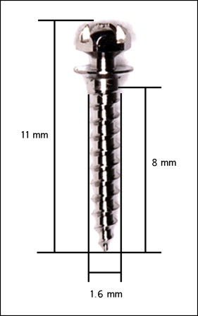
Commercial mini-implants (diameter, 1.6 mm; length, 8 mm; ISA orthodontic mini-implants; Biodent, Tokyo, Japan) were used ( Fig 1 ). After local anesthesia had been administered, a pilot hole was drilled in the buccal alveolar bone between the second premolar and the first molar of the maxilla or the mandible without creation of a flap. The pilot hole was formed so that it inclined 45° to 60° vertically to the adjacent tooth axis. To improve the success rate, bone drills with diameters of 1.0 mm in the maxilla and 1.3 mm in the mandible were used to control the placement torque within the recommended range (5-10 Ncm), based on published results. The mini-implants were placed by 3 skilled clinicians (M.M., Y.U., N.S.) who each had over 7 years of clinical experience. Immediately after placement, we applied an orthodontic force of approximately 2 N to the mini-implant and then took CBCT images for postplacement evaluations. Each patient was prescribed 3 days of antibiotic therapy after the procedure.
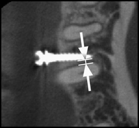
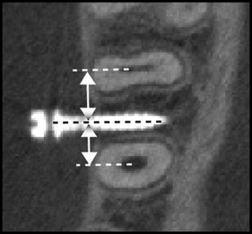
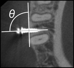
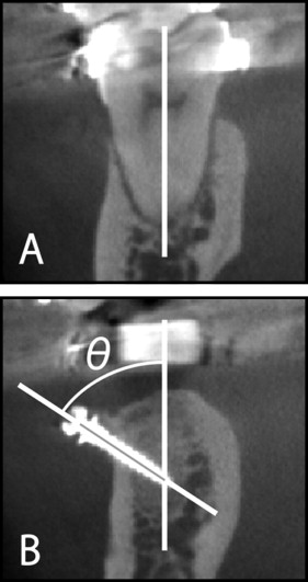
CBCT (3D Accuitomo; J. Morita, Kyoto, Japan), using 1.000-mm slices with a voxel size of 0.125 mm 3 , an x-ray tube voltage of 80 kV, and current of 5.5 mA, was used for diagnostic imaging of the area around the site. An examiner (Y.U.) measured the distance between the root and the mini-implant, and the inclinations of the mini-implants using a 3-dimensional viewer program (One Volume Viewer, version 1.6.1.13; J. Morita). The nearest root of the adjacent teeth and the shortest distance from the mini-implant were recorded ( Fig 2 ). A distance between the root and the implant of 0 mm denoted root contact. Mesiodistal placement location was examined by measuring the mesial and distal distances between the mini-implant and the root ( Fig 3 ). Next, the horizontal and vertical inclinations of the implant were measured. A tomographic cross-section was fixed parallel to the occlusal plane, corresponding to the implant placement site horizontally. On the horizontal cross-section, the angle between the bone surface and the long axis of the mini-implant was recorded as the horizontal inclination ( Fig 4 ). In the vertical measurement, a tomographic cross-section was fixed perpendicular to the occlusal plane corresponding to the mini-implant. On the vertical cross-section, the angle between the long axis of the mini-implant and the tooth axis of the first molar was recorded as the vertical inclination ( Fig 5 ). To evaluate intraexaminer error, we randomly selected the CBCT images of 10 subjects and remeasured the root proximity and the vertical and horizontal inclinations of the mini-implant 1 month after the initial measurements. Measurement error was calculated using the Pearson correlation coefficient. The Scheffé test was used to evaluate the root proximity and the variability of the placement inclination of the mini-implant according to placement position. Chi-square or Fisher exact tests were used to compare the incidence of root contact according to placement site. The Pearson correlation coefficient was calculated to evaluate the relationship of root proximity with the vertical and horizontal placement inclinations. These analyses were performed with the SPSS statistical program (version 16.0; SPSS Japan, Tokyo, Japan); P <0.05 was considered to indicate statistical significance. Mini-implants that endured an orthodontic force for 6 months or more without mobility were considered successful.
Results
Ten subjects were randomly selected and remeasured 1 month after the initial measurements. This reliability test showed a significant correlation (r = 0.85-0.92; P <0.01), verifying the accuracy of the measurements. The success rates of the mini-implants in this study were 95.6% in the maxilla and 93.7% in the mandible; this difference was not significant ( Table I ). Failure rates of mini-implants with and without root contact were 20.7% and 1.7%, respectively, with a significant difference ( Table II ; P <0.001). The rate of root contact varied according to the mesiodistal position of each placement site, which ranged from 0.0 to 24.2% ( Table III ). Significant differences were observed between the distal and mesial sites in the right maxilla, and between the distal sites in the right maxilla and the mesial sites in the left mandible. The rates of root contact according to placement site ranged from 10.3% to 27.3%; however, there was no significant difference. The rate of root contact for the 147 implants was about 20%. The average root proximity values ranged from 0.6 to 1 mm, and there was no significant difference in relation to placement site ( Table IV ). The average mesial and distal distances ranged from 3.5 to 4.3 mm, and the distal distances in the right and left maxilla were significantly smaller than the mesial distance ( Table IV ). The vertical inclinations of the placed mini-implants were 48.3° to 50.4° in the maxilla and 57.5° to 63.3° in the mandible. Comparison of the maxilla and mandible showed significant differences, but there was no significant difference between the right and left sides ( Table IV ). The horizontal inclinations ranged from 83° to 89°, and the angle in the left mandible was significantly larger than the angles in the left maxilla and the right mandible. The correlation coefficients between root proximity and vertical inclination were −0.505, −0.439, −0.299, 0.259, and 0.303 for the mesial aspects of the right and left maxilla, the distal aspect of the right mandible, and the mesial aspects of the right and left mandible, respectively ( P >0.05). The correlation coefficients between root proximity and horizontal inclination were −0.310 and 0.267 distally in the right mandible and mesially in the left mandible, respectively ( P >0.05).


