Fig. 3.1
Root caries and external apical root resorption in a mandibular anterior tooth
The most susceptible roots to fracture are those in which the mesiodistal diameter is narrow compared to the buccolingual dimension (oval, triangular, kidney-shaped, ribbon-shaped) [2, 7]. These include the maxillary and mandibular premolars, mesial roots of mandibular molars, mandibular incisors, and the mesiobuccal root of the maxillary molars. In a retrospective prevalence study of fractured roots among this group, Tamse et al. [8] found that the most frequently fractured roots and teeth were those with this specific anatomy (79 %). To minimize the risk of VRF, familiarity with the root anatomy and morphology is essential for appropriate instrumentation, obturation, and restoration of these teeth [7].
Root depression in the interproximal aspect of the mesial root of the mandibular molars and in the buccal root of the bifurcated maxillary premolar is an anatomical entity that can predispose the likelihood for fractures and root perforations when excessive removal of dentin occurs. Thus, these areas should be considered as “danger zones” (Figs. 3.2 and 3.3) [9–11]. The prevalence of the furcation groove in the buccal root of the bifurcated maxillary premolar is high (62–100 %) (Figs. 3.4, 3.5 and 3.6) [9–11]. The initial dentin thickness can be as small as 1 mm [9, 11] in these areas. In an in vitro study that evaluated the amount of remaining dentin thickness after root canal preparation and post space preparation in both roots of the bifurcated maxillary premolar, the remaining dental thickness was less than 1 mm in 77 % in the buccal roots in the bifurcation aspect [12]. Therefore, the clinician should express extra caution with the endodontic and prosthetic procedures in this area.
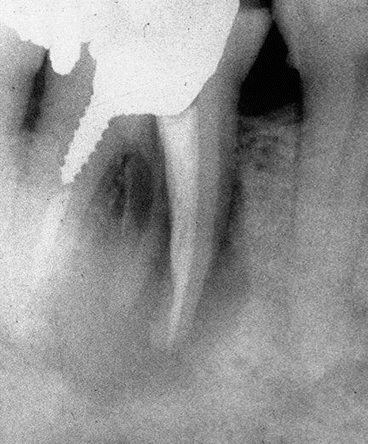
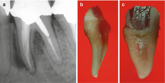
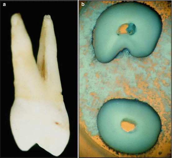
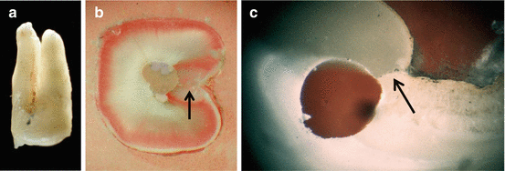
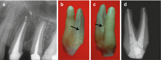

Fig. 3.2
Excessive removal of root dentin during root canal preparation in the mesial root of a mandibular molar caused a vertical root fracture. Separation of the root segments and large bone resorption can be seen in the radiograph

Fig. 3.3
A periapical radiograph of endodontically treated mandibular molar. (a) Due to excessive removal of the root dentin, both a VRF (b) and strip perforation (c) had occurred

Fig. 3.4
The typical depression on the bifurcation aspect of the buccal root of the bifurcated maxillary premolar can be seen in an extracted tooth. (a) Cross section of the tooth (b) is showing the typical kidney-shaped buccal root of the maxillary bifurcated premolar and the depression facing the bifurcation aspect

Fig. 3.5
Another extracted, bifurcated, endodontically treated maxillary premolar is showing the depression in the buccal root. (a) A cross section in the middle of the buccal root (b) is showing the irregular preparation of the root canal sealed with laterally condensed gutta-percha. The black arrow is pointing to the VRF. A different buccal root of the maxillary premolar is showing a round preparation for a dowel, leaving minimal residual dentin between the canal wall and the depression. The black arrow is pointing at the VRF (c)

Fig. 3.6
A periapical radiograph of an endodontically treated maxillary premolar. (a) A large lateral radiolucency can be seen in the mesial aspect. Few exacerbations occured in recent years, and a deep buccal probing was noted in midbuccal area. The extracted tooth is showing the complete VRF in the buccal root. (b, c) The black arrows are pointing at the VRF. The bench periapical radiograph of the extracted tooth (d) is showing the large amount of dentin removed in the buccal root
The canal and root shape combined with dentin thickness affect tensile stress distribution during root canal procedures [13]. The root canal shape is the most important factor of the two since the area of reduced curvature radius (the buccal and lingual root aspects) is strongly influenced by stress concentrations. More than likely, this is the reason why these roots fracture in a buccolingual direction and not mesiodistally. This is the case although more dentin is usually removed from the mesial and distal aspects at the stage of canal instrumentation and post space preparation.
In many cross sections of single-rooted teeth, the mesial and distal aspects are more calcified and harder than the buccolingual one showing a “butterfly effect.” This may also explain the high prevalence of vertical root fractures that are directed buccolingually [14].
Another important predisposing etiology is the amount of sound tooth structure pretreatment in the crown and root as a result of caries or trauma. Combined with the reduced amount of radicular dentin as a result of the various intracanal procedures (initial root canal therapy, retreatment, postspace preparation), sound tooth structure is directly correlated to the ability of the endodontically treated tooth to resist fractures [6, 7, 15–18].
A common clinical speculation is that an endodontically treated tooth is more brittle compared to one with a vital pulp [19] and that the dentin undergoes changes in collagen cross-linking after root canal treatment [20]. However, these studies have not been validated [21]. In endodontically treated teeth, moisture loss compared with teeth with vital pulps is not a major etiological factor but rather a predisposing one for root fracture [22].
Small cracks have been reported to be present in the dentin parallel or perpendicular to the root canal space in intact teeth [23–25]. During intracanal procedures when dentin is removed, especially in the mesiodistal areas, such cracks may turn into incomplete fractures and then later during the life of the tooth may progress in buccal and/or lingual directions to form a complete fracture [25].
The specific biochemical properties of dentin are also predisposing factors in VRFs. In a study on the stress–strain response in human dentin, Kishen et al. [5] found that the dentin adaptation to functional strain–stress distribution results in greater mineralization in the buccolingual areas. This may increase the likelihood for a fracture to propagate in this direction compared with less mineralization and more collagen in the mesiodistal areas.
By evaluating radiographs, it was found that a correlation exists between the height of the alveolar crest and the stress in the apical part of a post [26]. It may be assumed that loss of bone support due to periodontal disease and pre-endodontic and prosthetic treatment can result in reduced tooth ability to withstand functional stresses.
Contributing Etiological Factors
The iatrogenic factors are etiological factors that contribute to the susceptibility of a root to fracture. These include removal of large amounts of sound dentin during the endodontic and restorative procedures in the root canal (Figs. 3.7, 3.8 and 3.9), reduction of tooth stiffness as a result of the endodontic and restorative procedures and the stress they generate [27], and lateral condensation of gutta-percha followed by dowel selection and placement. These are all examples for such stress-generating procedures in the root canal [28–31].
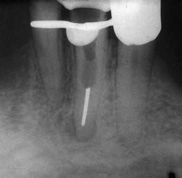

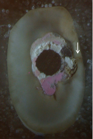

Fig. 3.7
An attempt to retract a previously endodontically treated mandibular premolar sealed with a silver cone, resulting in minimal remaining dentin and poor endodontic and restorative prognosis for the tooth

Fig. 3.8
Excessive removal of root dentin is shown in the two periapical radiographs (a, b). A bench periapical radiograph (c) is showing the large amount of dentin removed which was a major contributing etiology for the VRF in this tooth. (d) A black arrow is pointing at the fracture

Fig. 3.9
An extremely large amount of dentin removed from the root canal of a single-rooted maxillary premolar during the endodontic post preparation. Gutta-percha, sealer, and a metal post can be seen in this cross section. The preparation’s left irregular canal walls and the VRF to the mesial aspect is shown with the white arrow
The design of the NiTi instruments is an important factor when evaluating the ability of roots to resist fractures [18, 32]. These studies address the correlation of canal preparation to fracture susceptibility. Roots may be significantly weakened with larger instrument tapers which tend to remove more dentin, but by the same token, the greater taper may cause less stress during obturation with gutta-percha [18]. Recently, an additional flaw of the NiTi instruments, which is gaining more attention, is the ability of various root canal instruments to induce dentinal microcracks due to their design (Fig. 3.10) [33–35]. The small cracks in the canal wall or at the root surface seen in in vitro studies following instrumentation with various NiTi instruments together with previous studies regarding the existence of cracks in the dentin even in the untreated tooth [23–25] are considered precursors to the development of incomplete and complete fractures at a later stage [36]. Dentinal cracks also occur after root end preparation with ultrasonic retrotips [37]. These cracks can be an etiological factor in the future success of endodontic surgical treatment.
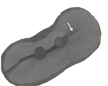

Fig. 3.10
A polished cross section of endodontically treated mesial root of a mandibular molar. Incomplete VRF is shown from the root canal to the lingual aspect, while a crack from the root canal is shown in the buccal part. Given enough time, this crack, shown by an arrow, will progress to the buccal aspect forming in this root a complete VRF from one aspect to the other
Changes in the quality of the root canal wall dentin when using various medicaments and irrigation solutions in vitro suggest that these may have an effect on the future resistance of the root to fracture [38]. Erosion of the dentinal wall has been shown with different irrigation solutions and long-term exposure to the root canal dentin to calcium hydroxide and various other chemical agents [39].
Tooth restorative procedures following root canal therapy such as postspace preparation, selection of dowels, and traumatic sitting of the dowels, especially in susceptible teeth and roots, are additional iatrogenic contributing etiologies for VRFs in endodontically treated teeth [40, 41]. In the AAE Colleagues for Excellence [42], it was emphasized that “The use of a post carries with it a certain attendant risk of root fracture, particularly if sound dentin is removed during preparation. Premolars require clinical judgment because of their transitional morphology.”
Recently, the use of fiber posts in the endodontically treated tooth is increasing because their modulus of elasticity is similar to dentin, which allows them to flex with the root when under stress [42]. Cagidiaco et al. [43] have shown that the placement of the fiber post did improve the survival rate of endodontically treated premolars. Other studies show a preference of fiber posts with different coronal coverage [44, 45]. Ferrari et al. [46] have pointed out that the preservation of at least one coronal wall during the restoration of the endodontically treated tooth is also a significant factor in the reduction of fractures in maxillary premolars.
Factors that affect fracture resistance of post-restored teeth are post length, diameter, design, material and fitting, the core material, the ferruling effect, the luting cement, coronal coverage, remaining coronal tooth structure, the loading conditions, and the alveolar bone support [47].
Conclusions
The potential vertical root fracture in the endodontically treated restored tooth can be reduced by the clinician by being aware of the various etiological factors, especially in VRF-susceptible teeth. These are the minimal removal of sound crown and root dentin, controlling the force when sealing the root canals with lateral condensation of gutta-percha, using dowels only when necessary to retain core buildup, using the more recommended fiber-reinforced resin dowels if needed and incorporating them, and adequate ferrule when restoring the tooth with a crown.
Etiology of VRFs: A Fracture Mechanics Perspective
Vertical Root Fractures (VRF) is a leading failure mode in teeth which has been studied extensively. VRF may be caused by wedging forces or pressure transmitted to the root canal surface during root canal obturation or from cyclic occlusal forces. In this section, we explore VRF from a mechanistic viewpoint as a fracture mechanics problem incorporating unique geometry, material, and loading. Examination of horizontal sections of roots extracted due to VRF reveals interesting morphological features which are helpful in any analytic modeling. A previously developed two-dimensional fracture mechanics model of VRF for single-canal roots subjected to apical condensation of gutta-percha is reviewed. The model is used to determine the full crack path due to the application of uniform pressure on the canal surface. A simple relationship is then used to connect this pressure to the apical condensation force, yielding an analytic expression for the critical load needed to cause VRF in terms of the system’s geometric and material variables, e.g., canal shape and taper, root wall thickness, and dentin’s failure stress σ F and toughness K C.
VRF is a term associated with a fracture extending over part or all of the root length, see Fig. 3.11. Such failure, often necessitating tooth extraction, is conclusively associated with endodontic treatment involving root canal cleaning and shaping followed by condensation of gutta-percha (gp), a rubbery material used for canal filling. VRF may occur during the treatment itself from excessive apical forces or more commonly later in time from cyclic occlusal forces [4, 48]. The growth history of a crack in the dentin leading to VRF involves complex geometry, material, and loading aspects. Analytic studies of VRF commonly employ 3D finite element analysis (FEA) and are generally limited to elucidating the stress distribution in the root due to some loading applied to the canal surface, e.g., localized or distributed (pressure), or occlusal surface [18, 31, 49–56]. Lertchirakarn et al. [31, 53] and Sathorn et al. [54, 55] presented tensile stress contour plots for circular and elliptical canal sections subjected to wedging forces or uniform pressure on the canal surface. The results show that the tensile stress responsible for crack initiation is maximized at the inner canal surface where the radius of curvature is the smallest, which is consistent with clinical observations. However, there appears to be no full-fledged FEA addressing the issue of crack growth in the dentin or relating canal pressure to apical or occlusal force.
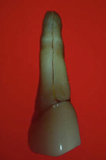

Fig. 3.11
Buccal view of endodontically treated mandibular premolar tooth extracted from a patient due to VRF in our laboratory
Recently, a simple 2D fracture mechanics analysis for determining crack growth behavior due to uniform canal pressure was presented [57]. The analysis shows that dentinal cracks tend to grow continuously with pressure and that the apical condensation force needed to cause VRF derived from the canal pressure using a simple formula correlates quite well with values obtained from in vitro tests on extracted teeth. In this chapter, we will discuss such tests as well as analytic concepts aimed at understanding and preventing VRF.
In Vitro VRF Testing
Several studies have conducted in vitro tests on extracted single-canal human roots [30, 31, 54, 58–60]. Such tests provide a useful database for analytic studies. After supporting the roots by a soft medium they were instrumented, filled by laterally condensed gp to a certain height L above the root apex, and continuously loaded to fracture by a spreader. Fracture or VRF was generally taken to occur when a noticeable load drop in the load vs. displacement output was recorded. Figure 3.2 in Chai and Tamse [57] depicts the mean (F max) and standard deviation load values vs. L. No distinction between the different instrumentation procedures used was made because such detail did not seem to be significant. The effect of loading distance L was moderate. The mean VRF load ranged from 80 to 170 N (1 KG = 9.8 N) with F max for oval canals somewhat smaller than for round ones. Pitts et al. [61], Holcomb et al. [29], and Soros et al. [62] conducted similar tests on maxillary and mandibular human incisors and mandibular goat teeth in that order except that gp condensation took place in repeating ramp cycles at increasing load levels. The fact that the resulting mean VRF loads (i.e., 149, 70, and 133 N in that order) are similar to the values obtained under continuous loading indicates the clinical relevance of the single-cycle loading tests.
Root Sectioning
During our experimental studies, we accumulated numerous teeth extracted due to VRF. Figure 3.12 shows three sequences of horizontal cross sections of premolar teeth, all previously subjected to root canal treatment with gp, which embed some essential characteristics of the fracture morphology. The sequences in (a) and (b) are for single-canal roots while that in (c) is for a double-canal root. Note that each of the two canals in (c) contains a post which extends to the middle part of the root. As shown, the canal sections are generally irregular, tending to change size and orientation along the root axis whether naturally or due to endodontic treatment. The sequence in (a) shows an incomplete VRF and that in (b) a complete one except in the apical section. In the case of (c), a complete VRF occurs only in the apical part of the root. In addition to major fractures leading to VRF, Fig. 3.12 shows some dentinal cracks of a limited extent, e.g., the first image in (b) and first two images in (c) from left to right. This indicates a competition for crack growth on the canal surface. A dominant feature in all micrographs in Fig. 3.12 is that cracks tend to initiate on the canal surface where the radius of curvature is the smallest irrespective of the outer root surface curvature.
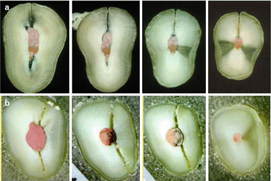
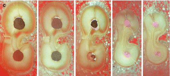


Fig. 3.12
Three micrograph sequences of horizontal cross sections for maxillary premolars extracted due to VRF in our laboratory: (a) Single-canal root showing incomplete VRF, (b) single-canal root showing complete VRF, (c) double-canal root with posts extending to the middle part of the root; a complete VRF occurs only in the apical part of the root. The fracture conclusively initiates at the root canal surface where the radius of curvature is the smallest (Courtesy Dr A. Raizman)
Several less apparent morphological features can also be seen from Fig. 3.12. The relatively large gap between the fractured parts in the two middle images in Fig. 3.12b suggests that fracture may have initiated from this region. Observing the lingual canal in Fig. 3.12c, one notes the fracture in the lingual part of the root where the canal is filled with gp while no fracture can be seen in the region of the post. It may be speculated that VRF in this case has initiated from the apical part and propagated coronally and that the presence of post did not add additional damage to the root.
Stay updated, free dental videos. Join our Telegram channel

VIDEdental - Online dental courses


