Introduction
For most orthodontic patients, esthetic concerns are as important as functional demands. The purpose of this study was to assess the effect of self-etching primer and conventional acid etching on tooth color after orthodontic treatment.
Methods
A total of 34 patients were enrolled in a clinical trial and divided into 2 groups based on age: adolescents (≤17 years) and adults (>17 years). Tooth color of all maxillary and mandibular anterior teeth was measured before bonding and after debonding using a spectrophotometer (Vita Easyshade Compact; Vita Zahnfabrik, Bad Säckingen, Germany). Two types of etching techniques were used for orthodontic bonding, self-etching primer and conventional acid etching, in a randomized split-mouth design. Tooth color measurements were done according to the system of the Commission Internationale de l’Eclairage (lightness, red/green, and yellow/blue). The corresponding tooth color differences between pretreatment and posttreatment, etching groups, sexes, and age groups were calculated.
Results
Tooth color was significantly changed in all (L, a, b) color parameters ( P <0.05). The lightness value decreased by 2.16 units, and the red/green and yellow/blue values increased by 0.32 and 1.78, respectively. The average tooth color difference after orthodontic treatment was 2.85 units. No significant difference was found between self-etching primer and conventional acid etching in their effects on tooth color ( P >0.05). Men and adolescents had more color change than did girls and adults ( P <0.05).
Conclusions
Fixed orthodontic appliances caused tooth color changes; self-etching primer and conventional acid etching had similar effects on tooth color; men and adolescents had greater color changes than did girls and adults.
Orthodontic treatment in the past had focused mainly on improving occlusal functions, but now esthetic concerns are as important as functional demands. One factor that contributes to optimal tooth esthetics is color. Tooth color results from the interaction between light and the enamel surface that is perceived by the human eye.
In-vivo tooth color can be assessed by 2 means: visual and instrumental assessments. The visual color assessment was until recently considered the key for dental color determination and matching. This approach is considered quick and cost-effective. However, a main drawback for this approach is that it is highly subjective. Also, the range of shades available for tooth matching does not cover all natural colors.
The transformation of the system of the Commission Internationale de l’Eclairage into numeric data and the advances in computer and optical technologies made the instrumental tooth color measurement an applicable method.
Several studies were conducted to evaluate and compare the visual and instrumental color determination methods. Authors reported that instrumental methods were more accurate and more reproducible compared with human visual shade assessments and can provide precise measurements of tooth color in vivo.
The effect of fixed orthodontic treatment on tooth enamel had been widely investigated. One reported adverse effect of fixed orthodontic treatment on tooth enamel is retention and discoloration of resin tags after cleaning.
The relationship between fixed orthodontic treatment and tooth color has not been studied intensively; only 1 in-vivo study has been conducted to assess the effect of fixed orthodontic treatment on enamel color, in which the authors concluded that fixed orthodontic treatment resulted in tooth color changes after bracket debonding. In-vitro studies also reported tooth color changes after fixed orthodontic treatment.
Acid etching of enamel aims to dissolve enamel rods and to create surface porosity that allows for mechanical retention of adhesive resin. Several studies have been conducted to compare self-etching primer and conventional etching techniques in terms of bond strength, clinical chairside time, and enamel loss at debonding. However, there are still no data in the literature on the effect of different etching techniques on tooth color.
Therefore, the aims of this prospective study were to (1) assess the effect of fixed orthodontic treatment on tooth enamel color, (2) evaluate the effect of different acid etching techniques on tooth enamel color by comparing self-etching primer with conventional etching, and (3) determine other factors that might affect tooth enamel color changes during fixed orthodontic treatment such as tooth type, age, and sex.
Material and methods
This prospective clinical study was conducted on patients who required fixed orthodontic treatment. These patients received their orthodontic treatment at the dental teaching clinics of Jordan University of Science and Technology in Irbid, Jordan. Ethical approval for this study was obtained from the institutional review board.
Thirty-eight consecutive patients (24 female, 14 male; age range, 12-26 years; average age, 18 years 5 months) were included in this study. Consequently, the patients were divided into 2 groups: adolescent subjects (≤17 years) and adult subjects (>17) years.
The patients were selected according to the following inclusion criteria: (1) no history of orthodontic treatment; (2) need for maxillary and mandibular fixed orthodontic treatment; (3) permanent dentition; (4) no missing, impacted, or extracted teeth; (5) mild or no dental crowding (<4 mm); (6) adequate oral hygiene when first seen, with no plaque accumulation or gingival inflammation; (7) no dental caries, decalcifications, or restorations in the teeth under examination; (8) no smoking habit; (9) medically and mentally fit with no disabilities; and (10) signed the consent form to participate in this study.
An organized protocol for patients’ data recording before and after orthodontic treatment was used. All patients were examined at the same examination and treatment clinic with good lighting conditions for the visual examinations. All patients were examined, and the data were recorded in the morning under the same fluorescent lamp of the dental unit. Cheek retractors were used, and all teeth were polished with nonfluoridated paste and then rinsed thoroughly with water. The teeth were kept wet to prevent color changes from dryness.
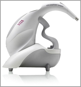
The spectrophotometer Vita Easyshade Compact (Vita Zahnfabrik, Bad Säckingen, Germany) ( Fig 1 ) was used to assess the color of the teeth before and after treatment. This device provided precise color measurement. Color assessment was based on the system of the Commission Internationale de l’Eclairage involving 3 color parameters: lightness (L), red/green chromaticity (a), and yellow/blue chromaticity (b). This system allows for numeric information that relates well to actual visual response and makes it the most popular one for color measurement.
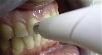
Teeth included in the study were the maxillary and mandibular central incisors, lateral incisors, and canines. For all study teeth, 3 consecutive measures for each color parameter (L, a, b) were recorded. The pretreatment and posttreatment colors of teeth were determined as the average value of the 3 consecutive measures for each tooth. During color measurement, the sterile intraoral device tip (mouthpiece) was held at a right angle to the labial surface of each tooth, and the spectrophotometric color assessment involved a standardized circular area in the center of the middle third of the labial surface of each tooth ( Fig 2 ).
The device was calibrated before each session on the white table supplied with it. During measurements, each tooth was illuminated by the same internal light at the probe tip of the device. Calibration was required to prevent any deviation in the quantity of light from internal light sources.
All measurements were taken by the same operator (A.A.A.O.), who was calibrated for using the device for 3 weeks before the study.
A randomized split-mouth design was used for bonding the maxillary and mandibular anterior teeth with 2 etching techniques: self-etching primer and conventional etching. This design was selected so that the patients would act as their own controls to prevent any effect from differences in their daily diets.
The split-mouth design was selected for each patient by randomly using closed envelopes and asking the patient to choose an envelope that contained the design to be used.
After polishing the teeth with nonfluoridated paste, moisture was controlled using a continuous saliva ejector and cotton rolls.
The assigned quadrants of teeth for conventional acid etching were etched with 37% phosphoric acid (3M ESPE, Seefeld, Germany) for 15 seconds, followed by rinsing with water for 15 seconds, and then dried with an oil-free and moisture-free compressed air flow until the etched surfaces exhibited a frosty white appearance with no traces of moisture.
A thin coat of Transbond XT primer (3M Unitek, Monrovia, Calif) was then applied on the whole labial surface of each tooth followed by a gentle air burst for primer thinning according to the manufacturer’s instruction. Transbond Plus Self Etching Primer (3M Unitek) was applied on the whole labial surface of the teeth in the quadrants assigned for the self-etching primer with a rubbing motion for 5 seconds followed by a gentle air burst for primer thinning according to the manufacturer’s instruction. Since bracket positioning is variable from tooth to tooth and from patient to patient, it was thought that application of the primer to the whole labial surface would make the process more standardized. Metal orthodontic brackets (0.022 × 0.028-in, Roth system; GAC International, Bohemia, NY) were then bonded with Transbond XT adhesive (3M Unitek).
All excess resin flash was removed, and the brackets were light-cured with a curing light (Biolux, CFON 1163; BIO-ART Dental Equipment, São Carlos, Brazil) for 10 seconds per tooth according to the manufacturer’s instructions.
Posterior teeth were bonded using the conventional etching technique. All patients were treated by the same orthodontist (A.A.A.O.) from bonding to debonding the appliances.
During orthodontic treatment, the patients were instructed to brush and floss their teeth regularly and maintain good oral hygiene. To prevent potential tooth discoloration and staining, chlorhexidine mouth rinse was not used during treatment.
After completion of orthodontic treatment, the fixed orthodontic appliances were debonded and the adhesive remnants were cleaned using a spiral 12-fluted tungsten carbide bur (1172RA; Ortho-Care, Bradford, United Kingdom) on a slow-speed hand piece. The extent of resin removal was assessed visually by the same operator (A.A.A.O.). The teeth were then polished with nonfluoridated paste and rinsed thoroughly with water.
The color difference (ΔE) for each etching group was calculated using the difference in the L, a, and b values before and after orthodontic treatment according to the following equation: ΔE = [(L 1 − L 2 ) 2 + (a 1 − a 2 ) 2 + (b 1 − b 2 ) 2 ]1/2. Subscript letters 1 and 2 indicate before and after treatment, respectively.
Because of poor oral hygiene and associated gingival enlargement and formation of white spot lesions, 4 patients were excluded from the study after debonding. This left only 34 patients (21 female, 13 male) as the study sample. The pretreatment average age of these patients was 18 years 7 months (range, 12-26 years). They were divided into 2 groups: adolescents (≤17 years) and adults (>17 years).
Only maxillary and mandibular anterior teeth (canine to canine) were studied. Accordingly, 408 stainless steel brackets were bonded with 1 of 2 etching techniques in a randomized split-mouth design; 204 brackets were bonded with self-etching primer, and 204 brackets were bonded with conventional etching.
The examiner (A.A.A.O.) was calibrated for 3 weeks before starting the study to ensure intraexaminer reliability for color measurement. At the end of the calibration period, 7 patients were randomly selected and reexamined (before starting orthodontic treatment) by the same operator 1 week later. The differences between the first and second measurements of tooth color were assessed by the Pearson correlation coefficient and paired t test.
Statistical analysis
Statistical analysis was performed using the Statistical Package for the Social Sciences for Windows program (version 17.0; SPSS, Chicago, Ill). Descriptive statistics including means and standard deviations were calculated for all variables in the study. A paired t test was used to detect changes in all tooth color parameters before and after treatment. A 2-way analysis of variance (ANOVA) test was used to assess the effects of etching technique, tooth type, and their interactions on tooth color changes. Independent t tests were used to detect changes in tooth color between the sexes and between the age groups. Results were considered significant at P <0.05.
Results
In the error testing, a significant correlation was found between the first and second readings (0.942), and there was no significant difference between the first and second readings using the paired t test, with a significance level of P = 0.893.
Treatment duration for all patients was 12 to 15 months. Since there was no significant variation in treatment duration between patients, it was not considered as a variable for statistical analysis.
The mean pretreatment values for the L, a, and b color parameters were 80.86 ± 5.99, 0.10 ± 1.51, and 22.36 ± 5.87, respectively.
The mean posttreatment values for the L, a, and b color parameters were 78.7 ± 6.06, 0.42 ± 1.50, and 24.14 ± 5.86, respectively.
By comparing the pretreatment and posttreatment means, the color parameters showed statistically significant changes after orthodontic treatment. The mean L value decreased by 2.16 units ( P <0.001), and the means of both a and b increased by 0.32 and 1.78 units, respectively ( P <0.001), as shown in Table I .
All measured teeth showed significant color changes after fixed orthodontic treatment; the color differences of all measured teeth ranged from 1.75 to 3.50 units. The mean total tooth color difference for all teeth was 2.85 units (SD, ± 0.3).
Color difference was assessed with respect to etching techniques, tooth types, and their interactions using the 2-way mixed ANOVA test.
The results showed that etching technique, tooth type, and their interactions had no statistical significant effect on tooth color differences ( Table II ).


