Chapter 11
Direct pulp therapy for young permanent molars
Patrice B. Wunsch
Introduction
As previously discussed in Chapter 10, young immature permanent teeth respond more favorably to insult and treatment therapies. The same principle holds true when considering direct pulp therapy in young immature permanent teeth. Young permanent teeth have immature pulps and incomplete root formation. Not only are the root ends not fully closed, but the root canal walls are thin and fragile. In the event of pulp exposure, if the tooth exhibits signs of reversible pulpitis, every effort should be made to maintain pulp vitality and allow the tooth to continue to develop naturally (apexogenesis). If direct pulp therapy is not possible and nonsurgical root canal treatment is indicated before the tooth has fully developed, an apexification procedure would be performed in an effort to obtain root end closure and an adequate apical stop. However, the apexification procedure does not allow for continued development of the root. A tooth that is not able to fully develop will result in having thin root canal walls and is at risk for fracture (Ward, 2002).
Background
Direct vital pulp therapy in cariously involved teeth is diagnostically and technique dependent. The following are critical for success (Ward, 2002):
- An accurate diagnosis of reversible pulpitis
- Caries removal to include the infected pulp tissue
- Use of aseptic technique (rubber dam isolation, irrigation)
- Prevention of bacterial leakage with a permanent restoration
As early as 1993, Mejare and Cvek wrote a paper reviewing the partial pulpotomy in carious teeth. They surmised that bacteria are able to gain access to the pulp lumen only after some portion of the coronal pulp has become necrotic (Mejare & Cvek, 1993). In order to preserve the remaining pulp, the infected dentin and inflamed pulp must be removed and a biocompatible material which promotes the reparative process is placed over the pulp. In the past calcium hydroxide (Ca(OH)2) has been the material of choice for direct pulp therapy. Long-term follow-up of cariously involved permanent molars that received Ca(OH)2 pulpotomy treatments resulted in a 44.5% failure rate at 5 years and 79.7% failure rate at 10 years (Witherspoon et al., 2006). Recently, mineral trioxide aggregate (MTA) has proven to be successful in vital pulp therapy. Analogous to Ca(OH)2 (pH of 12), MTA is highly basic (pH of 12.5) and is biocompatible. The goals of a successful pulp capping material are as follows:
- the ability to kill bacteria
- the ability to promote mineralization
- the ability to withstand bacterial invasion (bacterial tight seal).
MTA to a great extent fulfills the desired goals of a pulp capping/pulpotomy material. It has the ability promote mineralization and a dentinal bridge faster and more effectively than Ca(OH)2. It does however have a weak antibacterial effect; in fact it is less antibacterial than Ca(OH)2. Regardless, MTA does have the advantage of providing an enhanced nonresorbable seal over the vital pulp. Its ability to seal out bacteria is to a great extent why the material has been so successful. According to El Meligy and Avery (2006), Ca(OH)2 does not provide any protection from microleakage.
MTA has many advantages but it does have the disadvantage of being rather expensive; it costs $425 for one system (approximately $300 for five packets of the MTA powder alone).
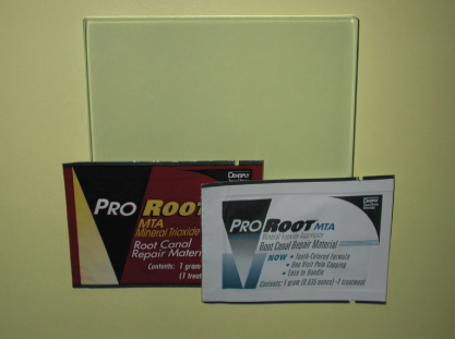
Another disadvantage, one of great concern if treating an anterior tooth, is that MTA (even white MTA) can result in tooth discoloration (Hutcheson et al., 2012). This discoloration is amenable to internal bleaching (Table 11.1 and Figure 11.1).
Table 11.1 MTA system contents and ingredients (Pro Root MTA)
| One MTA system contains | MTA ingredients |
| Five 1-g treatments of MTA material | Tricalcium silicate |
| Six water ampules | Tricalcium aluminate |
| Four carriers | Dicalcium silicate |
| Six mixing sticks | Calcium sulfate dihydrate |
| Ten dispensing sleeves | Bismuth oxide |

Figure 11.1 Gray and white MTA by Proroot (always mixed on a glass slab) (contributed by Dr. Patrice Wunsch, VCU Department of Pediatric Dentistry).
Permanent tooth pulp capping
In the event of a small mechanical or traumatic exposure, the direct application of Ca(OH)2 on the nonbleeding pulp has proven to be very successful in young immature teeth (direct pulp cap or Cvek pulpotomy). Some believe that while using Ca(OH)2 for direct pulp capping is an option, over time, it washes out or disintegrates where MTA does not (Farsi et al., 2006). Some now advocate the use of MTA for direct pulp capping over the use of Ca(OH)2 (Figure 11.2).

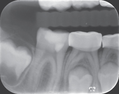
Figure 11.2 MTA direct pulp cap on tooth #30 (contributed by Dr. Claudia Colorado, VCU Department of Endodontics).
Permanent tooth pulpotomy
When the carious exposure is large, as discussed earlier, it is important to remove the infected, necrotic pulp by performing a pulpotomy. Ca(OH)2 has been used to preserve the apical vitality of a carious exposed pulp. But it has been shown that once root end closure has been obtained, the root canals would require obturation in order to prevent dystrophic calcification in the canals, a condition that would later prevent successful root canal therapy (Ranly & Garcia-Godoy, 2000).
It is important to recall the patient every 6 months and obtain a periapical radiograph on a yearly basis. Once root end closure has been obtained and the tooth remains clinically and radiographically within normal limits, it is then determined that nonsurgical root canal treatment is not indicated.
Some studies have shown that the control of pulpal bleeding plays a significant role in the success of the pulpotomy treatment. If a clot is allowed to form, this can result in treatment failure and thus irrigation is an important part of the treatment therapy. Use of either sterile saline or sodium hypochlorite is recommended. However, sodium hypochlorite has the added advantage of being able to provide hemostasis, disinfection and removal of dentinal chips that remain after excavation (Farsi et al., 2006)
Stay updated, free dental videos. Join our Telegram channel

VIDEdental - Online dental courses


