12
Digital Applications in Endodontics
Ashraf F. Fouad
12.1 Introduction
Endodontics is the dental specialty involved in the diagnosis and treatment of diseases of the dental pulp and periapical tissues in a manner that aims to preserve the health and integrity of the natural tooth. The pulp and periapical tissues are unique among oral tissues in many respects. Under normal and most disease conditions, they are invisible to the clinician even with magnification. The pulp space is very small with complex anatomy and neurophysiology. Moreover, the pulp is frequently rendered devoid of vital tissues and host responses by irreversible pathosis. The periapical tissues respond to pulpal disease and its treatment but are not directly manipulated clinically, except during surgical procedures.
Endodontic disease results from infection of the pulp and periapical tissues that is secondary to caries, trauma, advanced periodontal disease, congenital anomalies of teeth, root resorption, and severe abrasion or attrition. Therefore, the treatment of endodontic pathosis focuses on prevention or elimination of infection in the pulp and apical tissues. The instrumentation of the canal space is intended to provide adequate cleaning of this space and makes it feasible to obturate it. The obturation is frequently done in a way that allows retreatment or revision of initial therapy, as the conditions of the coronal restoration, tooth function, and future disease risk change.
This chapter will describe digital technologies that support the basic goals of endodontic diagnosis and treatment and provide some information on their efficacy and effectiveness where appropriate. In describing these technologies, it is essential to remember that the most important objective of these technologies is to improve diagnostic accuracy and treatment outcomes. Naturally, other goals include efficiency and convenience for the practitioner; however, these additional goals would only be justified if they do not interfere with the first objective and provide safety and cost‐effectiveness for the patient.
12.2 Digital Diagnostic Technologies
12.2.1 Pulp Vitality Versus Sensibility Testing
One of the most important steps in endodontic diagnosis is pulp testing. This basic procedure, which has been available for many decades, allows the clinician to determine whether the pulp is vital or necrotic and occasionally allows the clinician to reproduce the patient’s chief complaint. The most common pulp‐testing methods allow the determination of pulp sensitivity, also known as sensibility, rather than vitality. The assumption is that the vital pulp contains actively conducting neurons that can be easily stimulated by thermal or electrical stimuli. However, this assumption is not always true, such as in the case of traumatic injuries. Following trauma, pulp vitality is frequently maintained despite the loss of sensibility. In addition, common pulp‐testing technologies do not differentiate reversible from irreversible pulpitis with accuracy. For example, lingering pain following cold application is commonly diagnosed as irreversible pulpitis, when it is not known how long and how intensive the pain needs to be maintained for this diagnosis to be accurate. Frequently, clinicians misinterpret brief hyperalgesia that is common with reversible pulpitis as irreversible pulpitis. This is important because this distinction is essential in determining whether conservative operative procedures or vital pulp therapy versus root canal treatment is necessary. This is true whether or not the patient has pulpal symptoms, as it is commonly known that symptoms limited to the pulp and not in the apical tissues are effectively managed with pulpotomy (Hasselgren and Reit 1989; Eren et al. 2018). Finally, restorations, caries, difficulty in moisture control, and mineralization within the pulp–dentin complex frequently modify the response of the patient yielding limited test reliability.
Other digital technologies have been used to measure pulp vitality, by attempting to detect an intact vascular supply in the pulp. Laser Doppler flowmetry is the oldest of these technologies, but continues to be a viable option, when available, and with adequate tooth isolation (Setzer et al. 2013). Laser Doppler has been to be able to detect resumption of vitality of a traumatized tooth long before the EPT can register a response (Gazelius et al. 1988; Mesaros and Trope 1997). However, this technology has not gained widespread adoption because the probe needs to be stabilized with respect to the tooth to avoid erroneous measurement, may detect gingival blood flow and requires that the pulp chamber extend into or close to the crown of the tooth to allow direct reflection of the Doppler signal (Polat et al. 2004). Moreover, the size of the pulp chamber and the presence of restorations have been shown to significantly affect the pulpal blood flow, as measured by laser Doppler flowmetry (Chandler et al. 2010).
Pulse oximetry was introduced for the measurement of pulp vitality over 20 years ago (Schnettler and Wallace 1991; Kahan et al. 1996; Noblett et al. 1996). This method relies on the measurement of oxygen saturation with pulpal blood as a method of assessing pulp vitality. One study showed that a custom‐made pulse oximeter had a sensitivity of 1.00, compared with 0.81 for cold test and 0.71 for the electrical test (Gopikrishna et al. 2007). More recently, it was shown that the mean oxygen saturation levels were as follows: normal pulp: 92.2%, reversible pulpitis: 87.4%, irreversible pulpitis: 83.1%, pulp necrosis: 74.6%, and endodontically treated tooth: 0% (Setzer et al. 2012a). A recent systematic review and meta‐analysis of all current pulp‐testing methods showed that laser Doppler flowmetry and pulse oximetry were the most accurate pulp‐testing methods (Mainkar and Kim 2018).
12.2.2 Allodynia Measuring Device
Dentists commonly percuss teeth as part of endodontic diagnosis. However, they rely on the patient’s subjective response in assessing the results of this test. Patients are frequently not sure if the percussion caused sensitivity or was just different in sensation from a neighboring normal tooth. Moreover, there are factors related to percussion that may lead to differences in response such as the magnitude of the force, the location on the tooth of percussion, and the direction of percussion (occlusal or buccal). Percussion sensitivity is referred to as allodynia (or mechanical allodynia), since under normal circumstances, teeth do not hurt with percussion. It is useful in identifying periapical inflammation, periodontal abscess, cracked teeth (bite test), and occlusal trauma. One device was proposed to measure the degree of occlusal force that is exerted by the patient on a tooth, compared with a normal contralateral tooth, as an objective measure of mechanical allodynia (Khan et al. 2007a, 2007b). A follow‐up study using this device showed that mechanical allodynia is associated with 57% of cases with irreversible pulpitis (Owatz et al. 2007; Kayaoglu et al. 2020). The diagnosis of mechanical allodynia in conjunction with symptomatic irreversible pulpitis is of clinical importance because it necessitates that the emergency visit to relieve the symptoms include total pulpal debridement, rather than just a pulpotomy, if allodynia is absent. Quantitative assessment of mechanical allodynia was recently shown to identify central sensitization in endodontically involved teeth (Alelyani et al. 2020), and to predict postoperative pain (Jang et al. 2021).
12.2.3 Optical Coherence Tomography
Optical coherence tomography (OCT) is an imaging technology that analyzes the scattered reflections of light close to the infra‐red region, to determine the structure of a biological tissue. An advantage of OCT is that it is not biologically hazardous and is not invasive. However, technologies that employ OCT in dentistry are still in their infancy. In endodontics, OCT has been used to detect cracks in enamel or dentin (Imai et al. 2012; Nakajima et al. 2012; Segarra et al. 2017) and vertical root fractures (Shemesh et al. 2008; Yoshioka et al. 2013; de Oliveira et al. 2017). It has also been used for the detection on periapical lesions not perforating the cortical bone (Ding et al. 2019). However, all these efforts have been used in ex vivo models and no clinical devices are currently available for this purpose. This is an important clinical area as these clinical conditions are frequently difficult to diagnose without removal of restorations or surgical exploration.
12.2.4 Cone Beam Computed Tomography
In the past two decades, cone beam computed tomography (CBCT) has gained wide acceptance and implementation in endodontics. It allows observation of the tooth and its surrounding structures in the coronal, sagittal, and axial planes, which have many advantages in clinical endodontics (Cotton et al. 2007) (Figure 12.1). Not only does CBCT allow the detection of small lesions not visible on other forms of radiographic imaging, it allows enhanced imaging and visualization of the number of root canals present, aberrant or unusual canal anatomy, congenital anomalies, presence of root resorption, procedural mishaps, maxillary sinus pathosis, and traumatic injuries, among other conditions and anatomical structures (Kruse et al. 2015).

Figure 12.1 (a) Traditional radiograph; (b) cone beam computed tomography (CBCT) radiograph of maxillary left first molar (in a) with previous endodontic treatment and a separated instrument in the mesio‐buccal (MB) canal; note that the CBCT image showed a large periapical lesion that the periapical radiograph did not show; root end surgery confirmed that there was a thick cortical plate of bone and a lesion present in the medullary bone; (c) and (d) show two angles using periapical radiography in a different case that suggest a missed MB2 canal and a periapical lesion; (e) CBCT confirms that a missed canal and a lesion existed and shows their location and extent (arrows); (f) completed retreatment of MB1 and MB2.
Cone beam computed tomography was shown to have higher accuracy compared with traditional radiography in detecting periapical lesions (Estrela et al. 2008; de Paula‐Silva et al. 2009; Kruse et al. 2017; Kruse et al. 2019), assessment of healing following treatment (Christiansen et al. 2009; Patel et al. 2012; Kruse et al. 2016), and detection of root resorption (Durack et al. 2011). Studies have shown that for cases with previous root canal treatment and persistent disease, CBCT imaging would change the providers’ decisions on the treatment plan by 40–60% (Ee et al. 2014; Kruse et al. 2018; Bhatt et al. 2021). The resolution of images varies widely among different CBCT machines. Studies have shown that the higher the resolution, the more accurate the ability to detect aspects of endodontic interest, such as root resorption (Liedke et al. 2009) or the presence of mesio‐buccal (MB)2 canal (Bauman et al. 2011). Cone beam computed tomography cannot detect most vertical root fractures, but generally allow accurate determination of bone loss patterns that are consistent with vertical root fractures (Byakova et al. 2019; Bhatt et al. 2021). Cone beam computed tomography scans that yield image stack with voxel sizes of 76–120 μm and reduced edge streak artifacts provide the best use for endodontic applications. Newer machines have algorithms to correct motion artifacts (Spin‐Neto et al. 2020) or reduce beam hardening (de Rezende Barbosa et al. 2016), and may improve diagnostic accuracy in future. Although several studies have now evaluated outcomes of surgical and non‐surgical endodontic treatment using CBCT (von Arx et al. 2016; Al‐Nuaimi et al. 2018; Parmar et al. 2019), the benefits and risks of using CBCT in endodontics must be considered carefully and must follow available guidelines (Bhatt et al. 2021).
12.2.5 Magnetic Resonance Imaging
Magnetic resonance imaging (MRI) represents a non‐invasive imaging technology that does not use ionizing radiation and therefore does not have health hazards. It relies on the use of a strong magnetic field that excites hydrogen atoms within tissues. The resonant frequencies of the atoms are detected as they return to equilibrium state. Attempts have been made to image teeth with MRI for a long time (Lockhart et al. 1992). However, it was not until the advent of newer modifications of the technology, such as SWIFT‐MRI, that image details potentially useful in endodontic applications could be shown (Idiyatullin et al. 2011) (Figure 12.2).
Dental or medical MRI systems have been used to differentiate periapical granulomas and cysts (Lizio et al. 2018), to detect cracks and root fractures (Schuurmans et al. 2019), and to detect regenerated dental pulp tissues (Iohara et al. 2016; El‐Kateb et al. 2020). MRI is also being explored as for guided endodontic treatment, given its safety (Leontiev et al. 2021).
12.2.6 Ultrasound Real‐Time Imaging of Periapical Lesions
Another non‐invasive imaging technology that does not utilize ionizing radiation is ultrasound imaging combined with color power Doppler. This technology was introduced at the turn of the twenty‐first century, with evidence of being able to not only detect the presence of periapical lesions, but also to differentiate periapical granulomas from cysts (Cotti et al. 2002; Cotti et al. 2003). The ability of ultrasound to differentiate cysts and granulomas was confirmed in a more recent trial (Aggarwal et al. 2008). More recently, it was shown to aid in tracing the path of sinus tracts of endodontic origin (Cotti et al. 2019). Some have even used it to monitor healing of periapical lesions following non‐surgical treatment (Rajendran and Sundaresan 2007; Maity et al. 2011) or endodontic surgery (Curvers et al. 2018), showing that ultrasound may detect healing earlier than radiography. However, it is still generally recognized that radiographic techniques are more accurate than ultrasound, and the differentiation of granuloma and cyst does not yield any difference in treatment planning at this time.
There has been some interest in the use of ultrasound to aid in the healing of dental lesions because of its anti‐inflammatory and disease‐suppressing capabilities; however, this area has not been sufficiently studied in dentistry (Scheven et al. 2009a, 2009b).
12.3 Electronic Technologies in Local Anesthesia
The Wand (Henry Schein Dental) (also known as Computer‐Assisted Anesthesia System or Single Tooth Anesthesia) was introduced to minimize pain during anesthetic process (Fukayama et al. 2003). The Wand consists of a foot pedal‐operated pump delivery system, which delivers a constant slow rate of anesthesia, and does not utilize a traditional syringe. In addition to reducing pain from the pressure of anesthesia, particularly in painful injections such as palatal infiltration and incisive blocks, the lack of a traditional syringe is purported to reduce apprehension of patients, particularly children, who may be scared by the sight of a dental syringe, and elevation of pulse with some injections such as periodontal ligament or intraosseous injections. Studies have shown that The Wand may be beneficial in reducing the pain of palatal anesthesia in children (Gibson et al. 2000). The Wand was comparable to slow intraosseous injection in maintaining normal patient’s pulse, compared with a fast intraosseous injection that elevated the pulse (Susi et al. 2008). As far as pain with injection in adults, one study showed no benefit (Kandiah and Tahmassebi 2012), but another study, in which patients were used as their own controls, showed reduced painful anesthesia and physiologic stress during injection (Campanella et al. 2018).

Figure 12.2 (a) Typical magnetic resonance image of posterior teeth and surrounding tissues, showing contrast for different tissues but low resolution to discern dentin and pulp detail; (b) SWIFT‐MRI shown in comparison with traditional radiography and cone beam computed tomography (CBCT): the photograph depicts the maxillary teeth that are also imaged with a traditional two‐dimensional radiograph used to detect interproximal caries; (1)–(4) the dotted lines correlate with the cross‐sectional CBCT and SWIFT images at those levels, from being more superior closer to the root tip, moving inferiorly to the crown of the teeth; note the higher resolution for SWIFT‐MRI (field of view diameter of 110 mm and an isotropic voxel size of 430) and the lack of streaking artifacts associated with metallic restoration that are present in CBCT.
Source: Image reproduced with permission from Idiyatullin et al. (2011).
Another electronic device that is claimed to reduce the pain of anesthetic injection is the DentalVibe (DentalVibe Inc.). This is a two‐pronged fork that is applied to soft tissues around the area of needle injection and activated to deliver soft vibrations that minimize the sensation of the needle penetration of the tissues. One study has shown that this device was significantly effective in reducing pain with mandibular nerve block in children (Hassanein et al. 2020).
12.4 Digital Technologies in Root Canal Treatment
12.4.1 Magnification Technologies: Microscopes, Videoscopes, and Endoscopes
The use of magnification technologies in endodontics has gained considerable popularity in the past three decades. While magnifying loupes, especially those with adjunctive lighting sources, have become standard among numerous dentists, endodontic applications call for a higher level of magnification and illumination, which can only be accomplished by surgical operating microscopes and endoscopes (AAE 2012). One study showed that there were no differences among endodontists between those who used microscopes and those who used dental loupes in the incidence of locating MB2 canals in maxillary molars, but that both technologies allowed the detection rate to be almost threefold that of no magnification (Buhrley et al. 2002). More recently, it was shown that not using microscopes in treating maxillary first molars was, in fact, associated with a significant increase in missed MB2 canals and the prevalence of an apical radiolucency of the MB root (Khalighinejad et al. 2017).
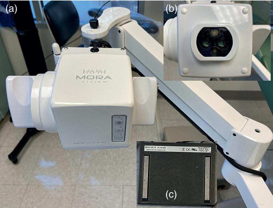
Figure 12.3 The Mora Vision video imaging system is an example of digital illumination, magnification, and recording during dental (including endodontic) procedures: (a) the main mount and arm system of the camera; (b) the lenses and illumination interface of the multi‐lens video camera system; (c) the foot pedal.
Studies have shown that the use of the microsurgical technique in endodontic surgery has been associated with a dramatic improvement of the prognosis of endodontic surgery in recent years (Azarpazhooh 2010, Setzer et al. 2012b). It should be noted, however, the microsurgical technique involves reduced resection angle of the root, ultrasonic root end preparation (which reduces the size of the osteotomy) and filling with mineral trioxide aggregate, which is much more biocompatible than older materials. Randomized analysis of the effect of endoscopes vs. dental loupes on outcomes of surgery did not show significant differences (Taschieri et al. 2006). However, it was shown that endoscopy at 64× magnification was more accurate in diagnosing root end cracks than microscopy at 16× and 24× magnification (von Arx et al. 2010). The use of microscopes in treatment, in addition to the obvious magnification advantage, allows the incorporation of a number of digital technologies for better documentation of cases and communication with patients and referring dentists.
One additional digital technology that is gaining some popularity is videoscopy or video microscopy. This technology allows illumination magnification at a comparable power to microscopy. In addition, it allows full recording of procedures, which are projected on one or two high resolution monitors for the provider, assistants, and/or trainees to be able to observe the procedure. The magnification and localization are controlled by foot pedals to allow unencumbered practice (Figure 12.3).
In addition to objective demonstrations for the importance of magnification in endodontics, endodontic practitioners have found magnification to be useful in identifying calcified and bifurcating canals, removal of obstructions such as pulp stones, posts, and separated instruments, visualizing the root apex or complex anatomical features during surgical endodontics and a multitude of other tasks performed in endodontics.
12.4.2 Sonic, Ultrasonic, and Multisonic Technologies
Sonic and ultrasonic digital devices are utilized extensively in endodontic practice. Traditionally, active root canal instrumentation was undertaken with files that were activated by analog sonic or ultrasonic devices. However, these devices were shown to not be more effective than regular instrumentation, and in some cases were more aggressive than traditional hand instrumentation. Currently, ultrasonic devices are commonly used in endodontic access preparation to locate canals by removing pulp stones and tertiary dentin, remove posts and root canal obstructions, and prepare root end cavities during endodontic surgery. Passive sonic or ultrasonic irrigation and needle activation during sonic or ultrasonic irrigation have also been used in devices designed to enhance root canal debridement and cleaning.
The advantages of ultrasonic tips include that they are narrow allowing precise cutting and permitting visualization under the microscope without interference by the handpiece head. Ultrasonic drivers are usually set to an intensity that ranges from 1 to 10. The higher the intensity, the more energy is given. For example, high energy is necessary for activating the tip on the left in Figure 12.4a vertically, over a post to loosen it. Other tips may be used at reduced intensity to carve out a space around the post to isolate it, remove the composite in the pulp chamber without gouging and/or negotiate intracanal obstruction at lower intensities.
Stay updated, free dental videos. Join our Telegram channel
VIDEdental - Online dental courses


