Clinical Photography
Christopher C.K. Ho
Principles
Photography is an essential diagnostic and communication tool for the aesthetic clinician. The old adage ‘A picture paints a thousand words’ is often quoted, and in aesthetic dentistry photographs help educate patients to understand the proposed treatment, are important clinical records and aid in the treatment planning process.
Historically we have witnesed the development from conventional to digital cameras. In the 1990s there was a rapid introduction of the intra-oral camera. It was the ability to show patients their dental problems ‘tooth by tooth’ that led to the rapid utilisation of this technology. However, the disadvantages were being able to show only a few teeth at a time, and the low resolution of subsequent picture reproduction.
The latest generation of digital photography with digital SLR and prosumer cameras is easy to use, provides good lighting and can take portrait and intra-oral shots from whole arch to two or three teeth with excellent resolution.
Benefits of Photography
The benefits of photography include the following:
- Improved patient communication. Being able to display what is in a patient’s mouth is a huge advantage compared to trying to describe their problem with words. If you let a patient see what is in their mouth, they can co-examine/diagnose their own situation.
- Laboratory communication. (Figure 2.2.1) Well-exposed clinical photographs can effectively communicate the optical characteristics of teeth and can show the shape, surface, morphology, value, shade, translucency and chroma. It was customary for ceramists to take a shade in person and convey it in words, but when building the crown trying to interpret what they wrote down was difficult and could be frustrating for both dentist and ceramist when colour matches were incorrect. Being able to access the images at any time makes the task of matching restorations much easier and can only improve the final result for both dentist and ceramist.
- Diagnostic tool and treatment planning aid. Being able to recall images of a patient with the ability to magnify pictures enables the clinician sometimes to see what they may have missed in their clinical examination. The ability to look at images of patients, records and diagnostic models after they have left the practice also gives the clinician the ability to plan treatment for the patient as if they had the patient sitting in the chair.
- Marketing library of before-and-after images. Photos of patients who have undergone treatment can be both an educational and a powerful marketing tool.
- Medico-legal considerations. Unfortunately, with the increase in litigation that is evident in our community, it is advantageous to have photographic records of patients pre-treatment, during treatment and post-treatment.
- Self-improvement. Documenting your cases allows you to critique your own dentistry and helps you improve.
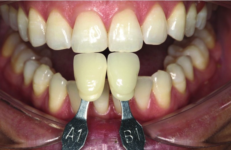
Figure 2.2.1 Laboratory communication: use of shade guides conveyed in photograph to laboratory – note that the tabs are placed in the same vertical plane and angles as the teeth, with the incisal edge facing the incisal edges, as the ginigival portion of the tab is always shaded more like dentine.
Stay updated, free dental videos. Join our Telegram channel
VIDEdental - Online dental courses


