and Silvio Mazziotti1
(1)
Department of Radiological Sciences, University of Messina, Messina, Italy
Abstract
While clinical evaluation plays a fundamental role in the diagnosis and the evaluation of the extension of the dental caries, it is clear that radiological examination, in a considerable percentage of cases, mainly plays a complementary role. Nevertheless, orthopantomography is sufficient to distinguish between the damage of the enamel and the dentine’s one and between the latter and a possible involvement of the pulp chamber.
Caries, or dental decay is a pathological destructive process which can affect all the components of the dental elements (enamel, dentine, cement), always in an irreversible way.
It is caused by a disturbance in the homeostasis existing between the floral bacteria (streptococcus, lactobacillus, etc.) and the local and systemic immune defences.
An increase in bacterial load or a reduction of the immune defences breaks this balance and determines the onset of caries. Caries is also furthered by the sugar fermentation activity performed by bacteria, with a consequent production of local hyperacidity. The latter has the effect of destroying the hydroxyapatite crystals and therefore the enamel integrity.
In the relevant literature, numerous studies show that radiological evaluation significantly (about 30–50 %) increases detection of caries lesions, with respect to the clinical examination.
While clinical evaluation plays a fundamental role in the diagnosis and in the evaluation of the extension of the caries lesion, it is clear that radiological examination, in a considerable percentage of cases, mainly plays a complementary role.
All of the literature concerning this topic shows different classification systems of caries, both on clinical and radiological basis.
Radiological categorisation of caries evaluates the effects on the enamel (whether in partial or full thickness), the dentine and eventually the pulp chamber.
With regard to this, it must be noted that such classifications derive from the evaluations of the endoral radiological examination which, as is known, is characterised by a resolution power which is superior to the OPT one.
As a consequence, the evaluation of caries through OPT suffers from some limitations, especially related to the quantification of damage.
From a practical point of view however, in orthopantomographic examination it is sufficient to distinguish between the damage of the enamel and the dentine’s one and between the latter and a possible involvement of the pulp chamber.
This last eventuality gives rise to ‘penetrating caries’ which, because of this contamination of the chamber, along with the pulp canal and its content, will result in a series of complications.
A further progression of the damage, followed by a much wider affection of the crown, will give rise to so-called destructive caries, obviously easily detectable on the clinical point of view.
From the radiological point of view, caries, regardless of its evolutionary stage, presents itself as a radiolucent lesion which affects both the crown and the cementoenamel junction. Its morphology varies in relation to the projective conditions with which it is represented.
Indeed, it seems evident that carious lesions captured in profile will be characterised by sharply defined outlines and by a semilunar morphology. Conversely, frontally hit lesions, though maintaining the sharpness of their outlines, present a roundish morphology.
Finally, caries which, due to their position, are captured in three-quarter view present irregular morphology and blurred, badly definable outlines (Fig. 4.1a–c).
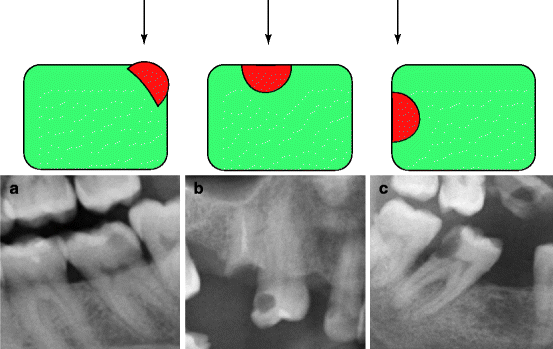
Fig. 4.1
Variations of the radiological morphology of the carious lesion in relation to the different projective conditions. (a) Caries captured in three-quarter view. (b) Caries captured frontally. (c) Caries captured in profile
Depending on its localisation, caries must be distinguished into occlusal, interproximal (or interstitial caries), palatal and vestibular caries.
Occlusal caries originate at the level of the tooth’s masticatory surface.
The caries are easily detectable with inspection, and radiological evaluation is useful to estimate the extent of their depth (Figs. 4.2a–c and 4.3a–c).
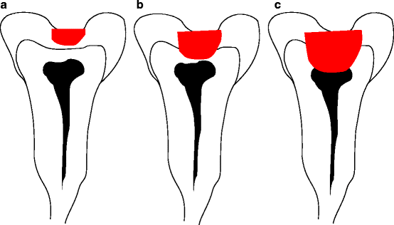
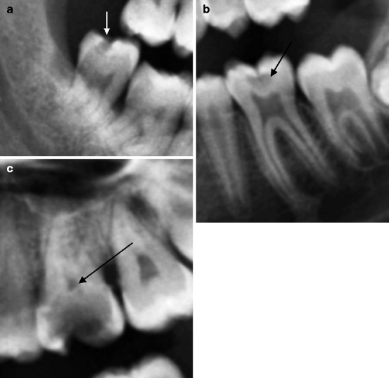
Fig. 4.2
Schematic diagram which illustrates the progression in depth of the occlusal caries. (a) Affection of the enamel. (b) Affection of the dentine. (c) Affection of the pulp chamber
Fig. 4.3
Different evolutionary stages of the occlusal caries (indicated by short arrows). (a) The lesion is limited to the enamel. (b) The lesion extends itself towards the dentin. (c) The lesion affects the pulp chamber (directed by long arrow)
Occlusal caries develops in an area where the enamel presents maximum thickness. Consequently it is characterised by a later involvement of the pulp chamber when compared with other localisations.
Indeed, slow evolution allows the formation of the so-called reactive dentine which in turn contributes to further delay of this destructive process (Fig. 4.4).
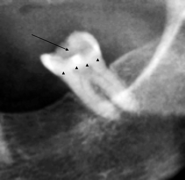
Fig. 4.4
Wide occlusal caries (indicated by arrow) separated from the pulp chamber by a band of major opacity (arrowheads), expression of the presence of reactive dentine
Interproximal caries originate at the level of the contact surfaces between the elements. Therefore, they can appear both in the mesial side and in the distal side (Figs. 4.5a–c and 4.6a–d).
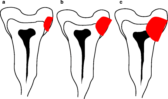
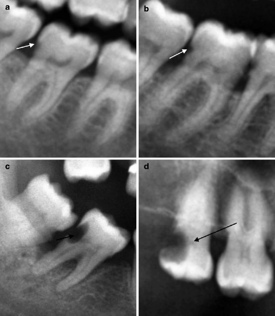
Fig. 4.5
Different evolutionary stages of the interproximal caries. (a) Affection only of the enamel. (b) Affection of the dentine. (c) Affection of the pulp chamber
Fig. 4.6
Different evolutionary stages of the interproximal caries (indicated by short arrows). (a) Distal caries limited to the enamel. (b) Distal caries affecting only the dentine. (c) Affection in depth of the dentine. (d) Affection of the pulp chamber (indicated by long arrow)
Interproximal caries are less detectable in radiographical examination as the interdental surfaces are less explorable than the other portions of the tooth.
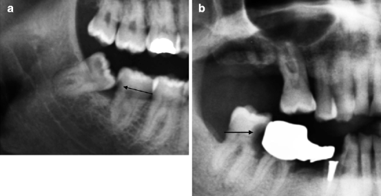
The aforementioned carious localisations can simultaneously affect the interproximal surfaces of two adjacent elements, giving rise to opposed interproximal caries (distal interproximal caries and mesial interproximal caries). Such caries are often characterised by a different evolutionary stage (Fig. 4.7a–c).
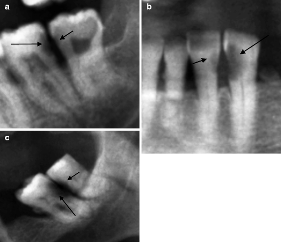
Fig. 4.7
Interproximal caries both in the medial side and distal side. (a) Distal caries (indicated by short arrow) affecting only the enamel and distal caries (indicated by long arrow) also affecting the dentine. (b) Mesial caries (indicated by long arrow) also affecting the pulp chamber and distal caries (indicated by short arrow) affecting only the dentine. (c) Distal caries (indicated by long arrow) affecting the pulp chamber and mesial caries (indicated by short arrow) limited to the dentine
In their pathogenesis, interproximal caries can show anatomical conditions favourable to development and progression of the pathological event.
In this scenario, caries which arise after the impact of an adjacent element with abnormal angulation appear to be paradigmatic (an event which occurs typically in situations of disodontiasis of the third molars).
Stay updated, free dental videos. Join our Telegram channel
VIDEdental - Online dental courses


