Chapter 9  The Upper Limb
1 Skeleton
SHOULDER (PECTORAL) GIRDLE
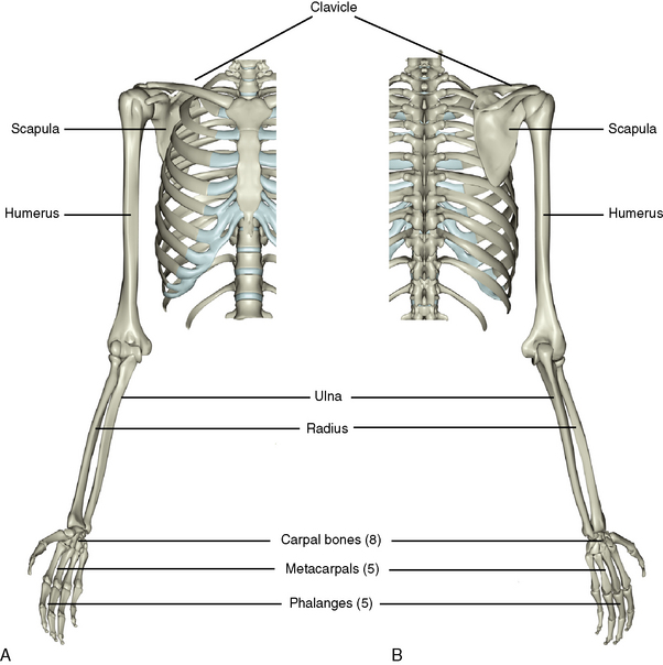
The shoulder girdle, or pectoral girdle, consists of two bones: the scapula (shoulder blade) and the clavicle (collar bone) (Figure 9-1). The scapula is anchored to the posterosuperior surface of the thoracic cage by muscles. The clavicle is attached firmly to the manubrium of the sternum by the strong but movable sternoclavicular joint and to the scapula at the weaker acromioclavicular joint. The clavicle acts as a strut to keep the shoulders pointed laterally.
Scapula
Description
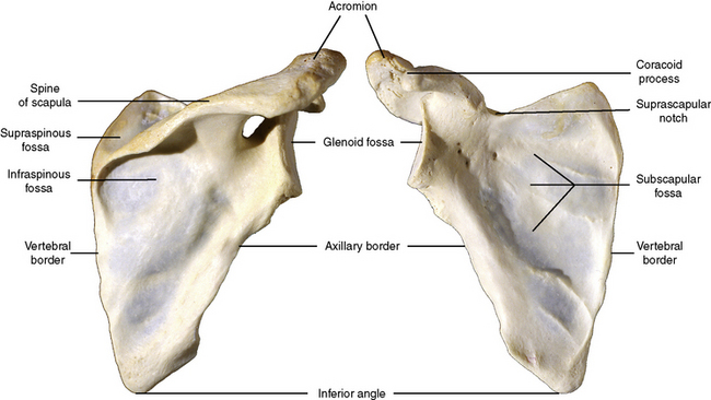
The scapula is a thin, triangular bone (Figure 9-2). Its concave anterior surface, is anchored by muscles to the posterior aspects of ribs 2 to 7. As a triangle, it possesses three sides: (1) a vertebral (medial) border that parallels the vertebral column, (2) an axillary (lateral) border that faces the axilla, and (3) a suprascapular (superior) border. The inferior angle is the apex of the triangle.
Features
Clavicle
Description
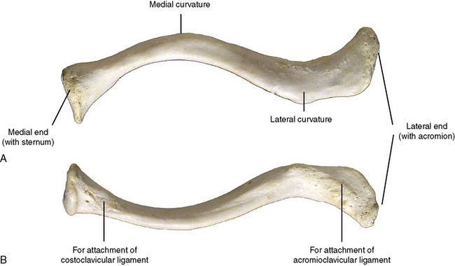
The clavicle is an elongated S-shaped bone that is subcutaneous and may be palpated over its entire length (Figure 9-3). The clavicle functions as a strut to prop the upper limb away from the body, affording the hand a greater range of positions, and transmits a portion of the weight of the upper limb through its sole articulation with the axial skeleton at the sternoclavicular joint.
Features
ARM
Humerus
Description
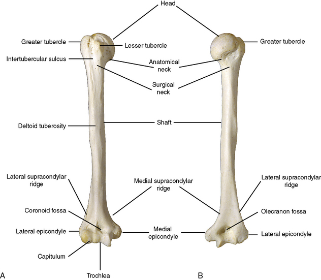
The humerus is the only bone of the arm. It articulates superiorly with the scapula of the shoulder girdle and inferiorly with the radius and ulna of the forearm (Figure 9-4).
Features
FOREARM
Features of the Radius
Features of the Ulna
THE WRIST AND HAND
Carpal Bones
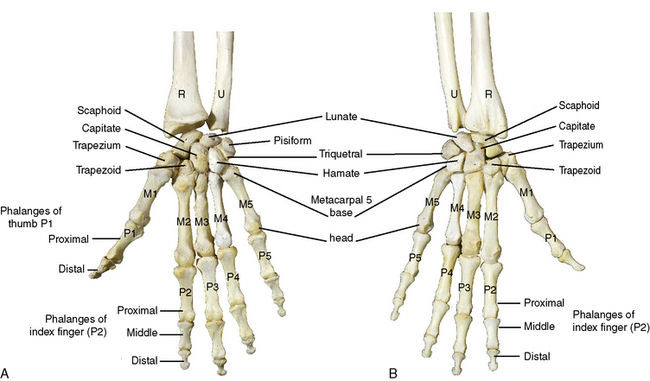
Eight carpal bones make up the bones of the wrist (Figure 9-6). They are short, cuboidal bones and are arranged in two rows of four bones each to give a degree of flexibility of movement to the wrist. The bones are named rather fancifully by their shape.
2 Joints, Movements, and Muscles
JOINTS OF THE PECTORAL GIRDLE
Features
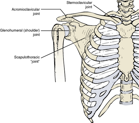
Three joints contribute to the movements of the shoulder girdle: (1) the sternoclavicular joint, (2) the acromioclavicular joint, and (3) the scapulothoracic joint (Figure 9-7).
GLENOHUMERAL JOINT
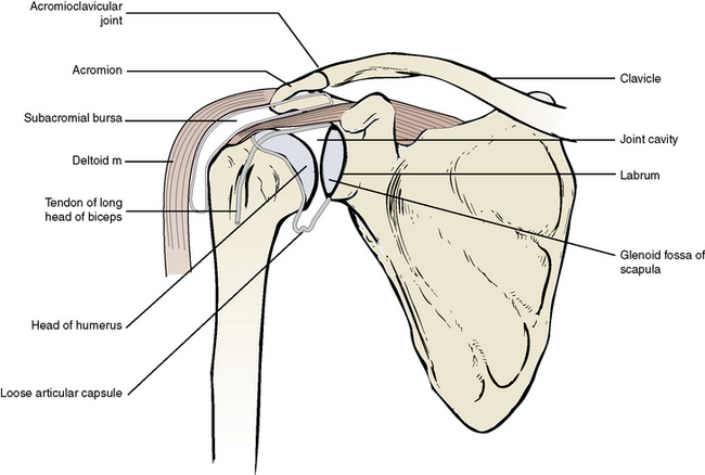
The glenohumeral, or shoulder, joint is an articulation between the spherical head of the humerus and the shallow depression of the glenoid fossa of the scapula (Figure 9-8). The shallow fossa is deepened only slightly by the glenoid labrum, a short fibrocartilage ring that encircles the rim of the fossa. The tendon of the long head of biceps passes through the cavity of the joint en route to the intertubercular sulcus of the humerus.
Movements
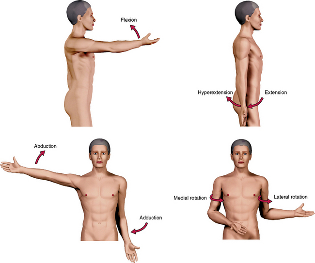
The glenohumeral joint is capable of six movements (Figure 9-9):
THE ELBOW (HUMEROULNAR) JOINT
Features
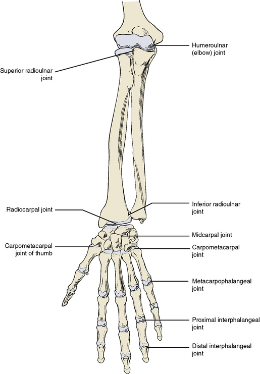
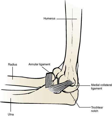
The elbow is a compound articulation between the distal end of the humerus and the proximal ends of the ulna and radius (Figures 9-10 and 9-11). The two sites of articulation take place between (1) the trochlea of the humerus and the trochlear notch of the ulna and (2) the capitulum of the humerus and the radial head of the radius. The joint capsule is lined by synovium and reinforced externally by radial (lateral) and ulnar (medial) collateral ligaments.
THE RADIOULNAR JOINTS
Features
The radius and ulna articulate with each other at the proximal and distal radioulnar joints.
Movements
The forearm is capable of twisting around its long axis (Figures 9-13 and 9-14). The head of the radius spins within its annular ligament while the distal end of the ulna curves around the distal ulna from a lateral position to a medial position and back again. This results in two possible movements at these joints:
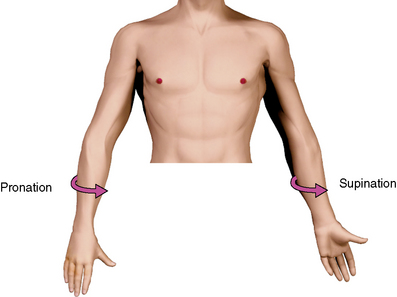
Figure 9-13 Movements at the radioulnar joints. The right forearm is pronated; the left forearm is supinated.
WRIST JOINT (RADIOCARPAL AND MIDCARPAL JOINTS)
Features
The wrist joint is the articulation between the forearm and the carpal bones of the wrist (see Figure 9-10). The ulna is separated from these bones by an articular disc so only the radius makes contact with the distal row of carpals. The main joint of the wrist is the biaxial, condyloid radiocarpal joint. The midcarpal joint
Stay updated, free dental videos. Join our Telegram channel

VIDEdental - Online dental courses


