Chapter 5 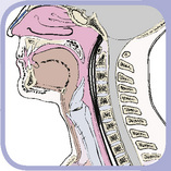 The Neck
1 Skeleton and Surface Anatomy
THE CERVICAL SKELETON
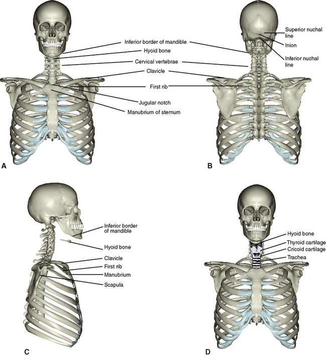
The skeleton of the neck consists of a vertebral unit and a visceral unit (Figure 5-1). In addition, bones of the upper limb girdle, inferior aspect of the skull, and the superior aspect of the thoracic skeleton help provide attachment for muscles of the neck.
Skeleton of the Vertebral Unit
Seven cervical vertebrae compose the vertebral unit of the neck. Descriptions of typical vertebrae C3 to C6 and atypical vertebrae (C1-atlas, C2-axis, and C7) are presented in Chapter 2. The cervical vertebrae should be reviewed at this point.
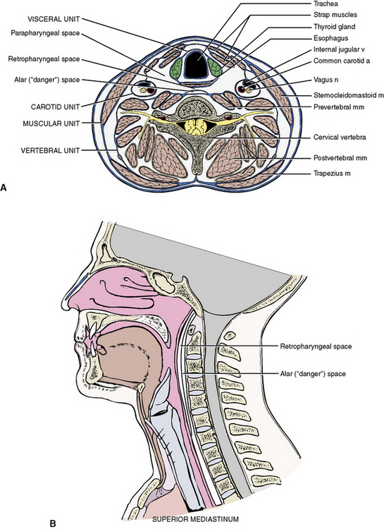
Skeleton of the Visceral Unit
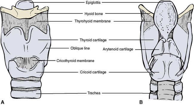
The skeleton of the visceral unit consists of the hyoid bone, the larynx, and trachea (Figure 5-2). These structures are briefly described here; more detailed descriptions are presented subsequently in Chapter 7 in the section dealing with the pharynx and larynx.
Hyoid Bone
The hyoid bone is a floating bone just below the mandible (see Figure 5-2). It is a U-shaped bone with the prongs, or horns, of the U facing posteriorly. The hyoid bone consists of three parts: (1) a rectangular body to which is appended; (2) a pair of lesser horns, which project upward and backward; and (3) a pair of greater horns, which project posteriorly.
Other Bones
Additional bones that belong to other regions are noted here because they provide attachments for various cervical structures. These are the base of the skull and the mandible (see Chapter 6), the scapula and the clavicle of the upper limb girdle (see Chapter 9), and the manubrium of the sternum and the first rib (see Chapter 3).
2 Coverings and Regions
FASCIAL COVERINGS
Immediately deep to the skin of the neck is a layer of superficial fascia. Deeper still is an intricate covering of deep cervical fascia. These fasciae compartmentalize the structures of the neck (Figure 5-4). Between the various compartments are spaces occupied by loose areolar tissue. These fascial compartments are potential routes through which infection can spread from one site to another.
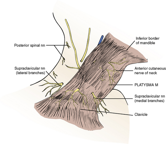
Superficial Fascia and Platysma Muscle
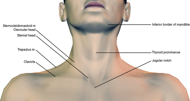
Platysma muscle is a thin, wide sheet of muscle that covers the anterior and lateral aspects of the neck (Figure 5-5). It is a superficial muscle related to the superficial muscles of facial expression, and therefore it lies within the superficial fascia of the neck. It originates from pectoral fascia below the clavicle and sweeps upward to the inferior border of the mandible. The more lateral fibers continue superiorly and medially to blend into the risorius muscle, which inserts into the angle of the mouth. The medial fibers decussate as they approach and insert into the inferior border of the mandible.
Deep Cervical Fascia
Deep Investing Fascia
Below the superficial fascial layer is a thin sheet of deep cervical fascia that wraps around the entire circumference of the neck—like a collar. This fibrous collar has superior attachments to the skull and inferior attachments to the sternum and pectoral girdle. As it encircles the neck, it splits to pass around and form the sheath of two large muscles, the sternocleidomastoid and the trapezius. The detailed attachments are rather complicated and are presented for reference purposes.*
* Superiorly the fascia attaches to the inferior border of the mandible, inferior border of the body of the hyoid bone, angle of the mandible, the inferior border of the zygomatic arch, and the mastoid and styloid processes. Because the fascia splits to enclose the sternocleidomastoid and trapezius muscles, it shares their attachment to the mastoid process, superior nuchal line, and external occipital protuberance of the skull. At the inferior aspect of the skull, the deep investing fascia also splits to pass around and help form the fibrous capsules of the parotid and submandibular glands.
Inferiorly the deep investing fascia attaches to the manubrium of the sternum, clavicles, and spines of the scapula along with the sternocleidomastoid and trapezius muscles, which it ensheathes. As it descends to the manubrium, it divides into two sheets: an anterior one attaching to the anterosuperior aspect of the manubrium and a posterior one attaching to the posterosuperior aspect of the manubrium. This creates a space, the suprasternal space (of Burns), that contains some areolar tissue, fat, and lymph nodes. It also contains some portions of the inferior thyroid veins and the anterior jugular venous arch.
Posteriorly the deep investing fascia gains attachment to the ligamentum nuchae, a membranous extension of the cervical spines.
Carotid Sheaths
Contents
Mediastinitis
Massive infections of dental origin can break through into the retropharyngeal and alar spaces and track down to the mediastinum below, resulting in infection and inflammation of the mediastinum (mediastinitis). A more detailed description of the spread of dental infection is presented in Figure 11-32.
Surgical Emphysema
Emphysema is a pathological condition in which air or gas is abnormally present within or between tissues. Inappropriate oral surgery procedures can introduce air under pressure into the fascial issue planes. If it occurs near the surface, a characteristic “crackling” is produced on palpation. If the air is forced into the retropharyngeal or alar fascial space, it can rapidly track to the mediastinum. This condition is described in Figure 11-32.
REGIONS
To facilitate the study of a seemingly complicated area, the neck is divided into two major areas, or triangles, by the sternocleidomastoid muscle (Figure 5-6). The area anterior to this muscle and below the inferior border of the mandible is the anterior triangle of the neck. The area posterior to the sternocleidomastoid muscle is the posterior triangle of the neck, which is limited posteriorly by a second large muscle, the trapezius.
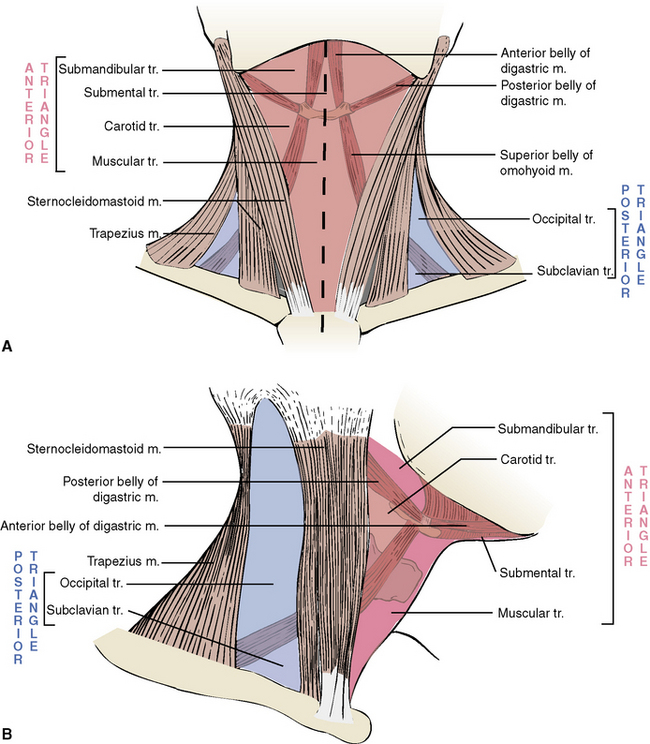
Sternocleidomastoid Muscle
Trapezius Muscle
The trapezius muscle is expansive and covers a number of regions. It is a superficial muscle of the back, a muscle of the upper limb girdle, and a cervical muscle (see Figure 2-14).
3 The Anterior Triangle
The anterior triangle occupies the anterior portion of the neck as an inverted triangle, its base consisting of the inferior border of the mandible and its apex directed downward toward the manubrium of the sternum (Figure 5-7). Like the posterior triangle the anterior triangle has depth and should be considered a region. Therefore, in addition to three boundaries, it has a roof, a floor, and several contents.

BOUNDARIES
The anterior boundary is the midline of the neck (i.e., straight line running from the base of the chin above to the jugular notch of the sternum below). The posterior boundary is formed by the anterior border of the sternocleidomastoid muscle. The superior border is the bony inferior border of the mandible. For ease of description the anterior triangle of the neck is further divided into smaller component triangles (see Figure 5-6).
ROOF
Superficial Veins
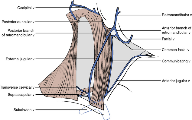
The detailed venous drainage of the face is described with the study of the face in Chapter 7. To summarize, the anterior portion of the face is drained by the facial vein, and the posterior portion of the face is drained by the retromandibular vein. Both veins leave the face and drain inferiorly to the neck. The retromandibular vein at the angle of the jaw divides into anterior and posterior divisions (Figure 5-8).
Superficial Nerves
The transverse cervical nerve of the neck originates in the posterior triangle and passes across the sternocleidomastoid muscle to supply skin overlying the anterior triangle of the neck (see Figure 5-5). The cervical branch of the facial nerve (cranial nerve VII) passes inferiorly from the parotid region above to supply the platysma muscle.
FLOOR
The floor of the anterior triangle of the neck is formed by the pharynx, the larynx, and the thyroid gland. These structures are posteroinferior extensions and relations of the oral and nasal cavities and are therefore described in Chapter 7.
CONTENTS
The anterior triangle of the neck contains a number of muscles, arteries, veins, and nerves.
Muscles
The muscles of the anterior triangle are grouped according to position and function (see Figures 5-7, 5-9, and Table 5-1). Suprahyoid muscles originate above the hyoid bone: infrahyoid muscles originate below the hyoid bone. Both sets of muscles insert directly or indirectly into the hyoid bone.
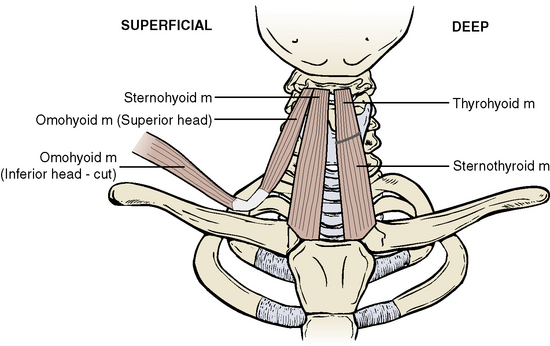
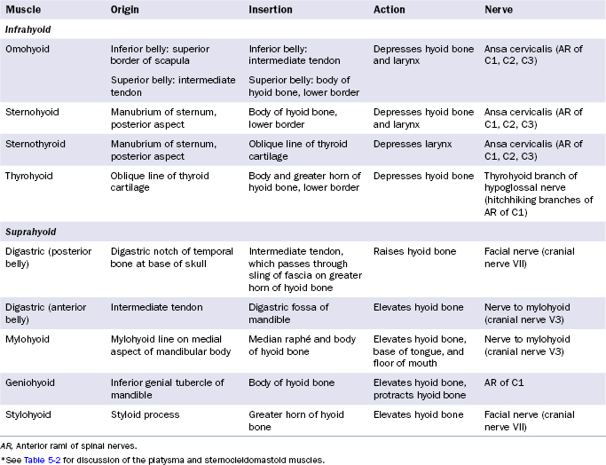
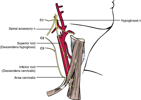
Infrahyoid Muscles
Stay updated, free dental videos. Join our Telegram channel
VIDEdental - Online dental courses


