Chapter 8  Systemic Anatomy of the Head and Neck
1 Arteries
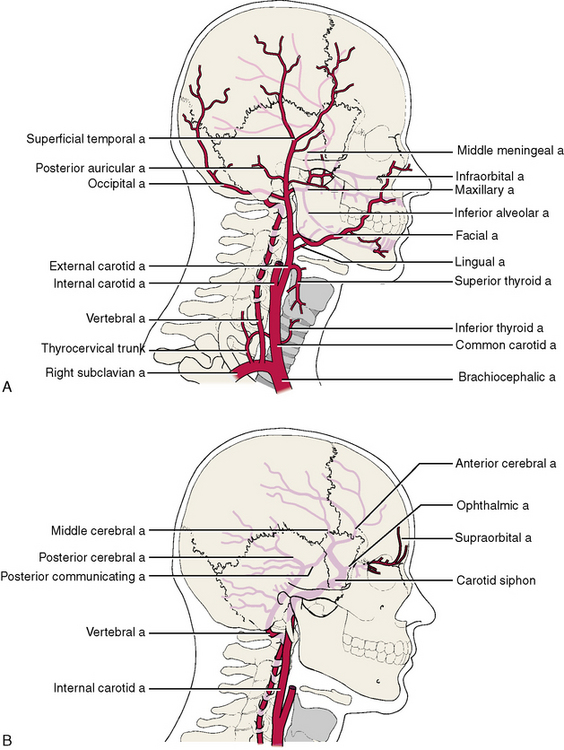
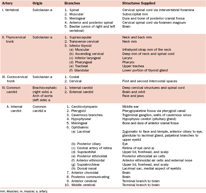
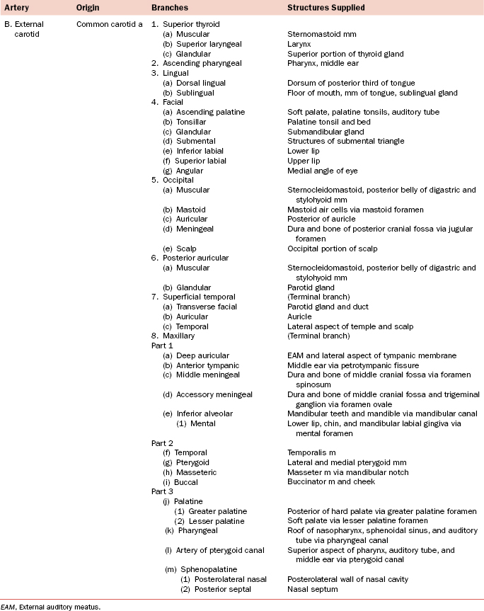
The arteries of the head and neck are illustrated in Figure 8-1 and listed in Table 8-1. Regional detailed descriptions of the arteries are found in Chapter 7.
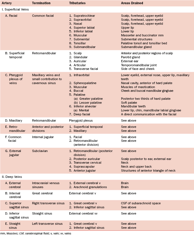
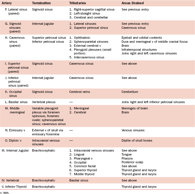
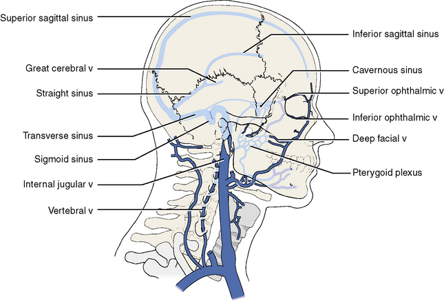
2 Veins
The veins of the head and neck are illustrated in Figures 8-2 and 8-3 and listed in Table 8-2. Regional detailed descriptions of the veins are found in Chapter 7.

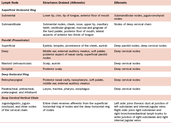
3 Lymphatics and Lymph Nodes
Knowledge of the routes by which lymph flows from the head and neck back to the venous system is essential to understand lymphatic spread of infections and cancer (Table 8-3). For a general discussion of the lymphatic system, see Chapter 1, Section 6.
| Lymph Node | Structures Drained (Afferents) | Efferents |
|---|---|---|
| Superficial Horizontal Ring | ||
| Submental | Lower lip, chin, tip of tongue, anterior floor of mouth | Submandibular nodes, jugulo-omohyoid nodes |
| Submandibular | Submental nodes, cheek, nose, upper lip, maxillary teeth, vestibular gingivae, mucosa and gingivae of the hard palate, posterior floor of mouth, lateral aspects of anterior two thirds of tongue | Nodes of deep cervical chain |
| Parotid (Preauricular) | ||
| Superficial | Eyelids, temples, prominence of the cheek, auricle | Deep parotid nodes, deep cervical nodes |
| Deep | Middle ear, external auditory meatus, soft palate, posterior aspect of nasal cavity, superficial parotid nodes | Deep cervical nodes |
| Mastoid (retroauricular) | Scalp, auricle | Deep cervical nodes |
| Occipital | Posterior scalp | Deep cervical nodes |
| Deep Horizontal Ring | ||
| Retropharyngeal | Posterior nasal cavity, nasopharynx, soft palate, middle ear, external auditory meatus | Deep cervical nodes |
| Paratracheal, pretracheal, prelaryngeal, and infrahyoid | Larynx, trachea, pharynx, esophagus | Deep cervical nodes |
| Deep Cervical Vertical Chain | ||
| Jugulodigastric, jugulo-omohyoid, and other nodes of the cervical chain | Entire chain receives afferents from the superficial horizontal ring of nodes and the deep horizontal ring of nodes | Left side: joins thoracic duct at junction of left subclavian and internal jugular veinsRight side: joins right subclavian and right bronchomediastinal lymph trunks to enter junction of right subclavian and right internal jugular veins |
The lymphatics of the head and neck, as in other areas of the body, drain toward groups of lymph nodes. The nodes act as filters and add lymphocytes to the lymph fluid. In the head and neck, the lymph nodes may be conveniently grouped into (1) a horizontal ring of superficial nodes, (2) a horizontal ring of deep nodes, and (3) two vertical chains of deep cervical nodes. Both horizontal rings drain to the two deep vertical chains (Figure 8-4).
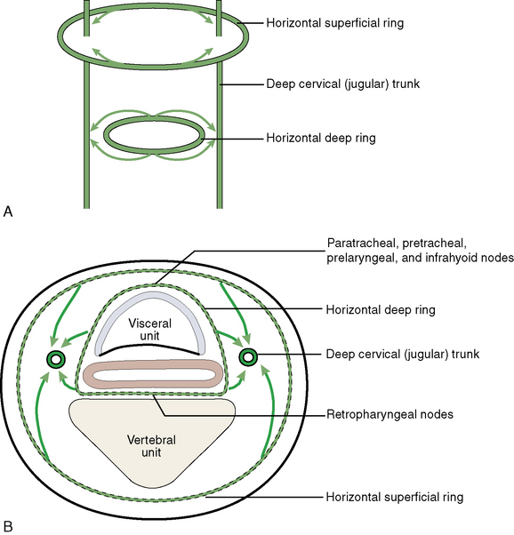
SUPERFICIAL RING OF NODES
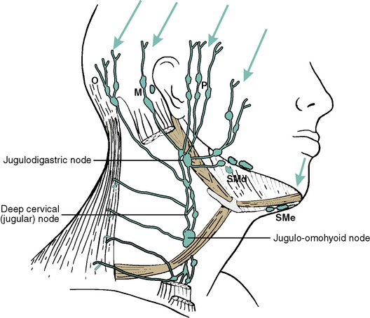
The superficial ring surrounds the transition area of neck to head and is arranged into five main groups (Figure 8-5; see Table 8-3). These nodes are palpable when infected.
DEEP RING OF LYMPH NODES
DRAINAGE OF SPECIFIC STRUCTURES AND AREAS
Gingivae
The vestibular gingivae of the mandibular and maxillary arches drain to the submandibular nodes.
Tongue
The tip of the tongue drains to the submental lymph nodes, the lateral aspects of the anterior two thirds drain to the submandibular nodes, and the medial portion of the anterior two thirds drain directly to the deep cervical nodes (Figure 8-6).
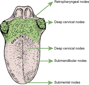
The posterior third of the tongue drains posteriorly to the retropharyngeal group of lymph nodes.
4 Cranial Nerves and Cranial Autonomics
CRANIAL NERVES
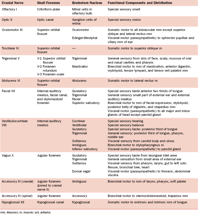
A general description of the nervous system is found in Chapter 1, Section 7, and a description of the brain and attached cranial nerves is presented in Chapter 7, Section 2. In addition, a description of each cranial nerve is given as it appears in the various regions of the head. A summary of the cranial nerves is presented in Table 8-4.
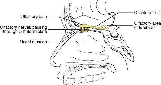
Cranial Nerve I: Olfactory Nerve
Functional Component
The special sensation of smell is the only functional component of the olfactory nerve (Figure 8-7). The olfactory nerve originates from bipolar olfactory cells within the nasal mucosa, where peripheral processes end as specialized smell receptors in the mucosa covering the superior concha and upper nasal septum. Central processes collect as 18 to 20 branches of the olfactory nerve proper. These pass upward through the cribriform plate to the anterior cranial fossa and enter the overlying olfactory bulbs. Here they synapse with mitral cells, and their central processes pass back along the olfactory tract to the olfactory area of the forebrain.
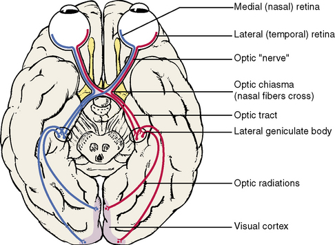
Cranial Nerve II: Optic Nerve
Functional Component. The special sensation of sight is the sole functional component of this cranial nerve (Figure 8-8). Classically (but incorrectly) the optic nerve is described as the section that passes posteriorly from the eyeball to the optic chiasma. Actually, the optic nerve proper is contained within the retina of the eye and originates from rod cells (nondiscriminating sight) and cone cells (discriminating sight and color). These receptor cells occupy the most external portion of the retina and receive incoming light. Central processes pass inward to synapse with bipolar cells, which, in turn, synapse with ganglionic cells of the innermost layer. Central processes of the ganglionic cells collect and leave the eyeball as the optic nerve. The nerve, or tract, leaves the orbit through the optic canal, and right and left nerves join at the optic chiasma. Here the fibers originating from the medial (nasal) half of the retina decussate; the fibers of the lateral (temporal) half of the retina do not decussate. The optic tract continues posteriorly from the chiasma, and this ends in the lateral geniculate body of the thalamus, where the optic fibers synapse. Postsynaptic fibers pass posteriorly through optic radiations to the visual cortex in the occipital lobe of the cerebral hemispheres.
Cranial Nerve III: Oculomotor Nerve
The oculomotor nerve arises from the interpeduncular fossa of the midbrain and passes forward to enter and traverse the cavernous sinus (Figure 8-9). The oculomotor nerve then enters the orbit through the superior orbital fissure.
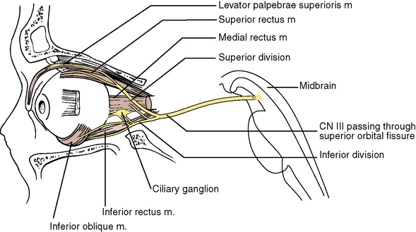
Functional Components
Cranial Nerve IV: Trochlear Nerve
The trochlear nerve arises as a slender thread from the dorsal aspect of the midbrain and sweeps anteriorly under the cover of the free edge of the tentorium cerebelli (Figure 8-10). It pierces the dura of the triangular field, enters and traverses the cavernous sinus, and enters the orbit through the superior orbital fissure.
Stay updated, free dental videos. Join our Telegram channel
VIDEdental - Online dental courses


