Chapter 2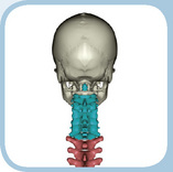 The Back
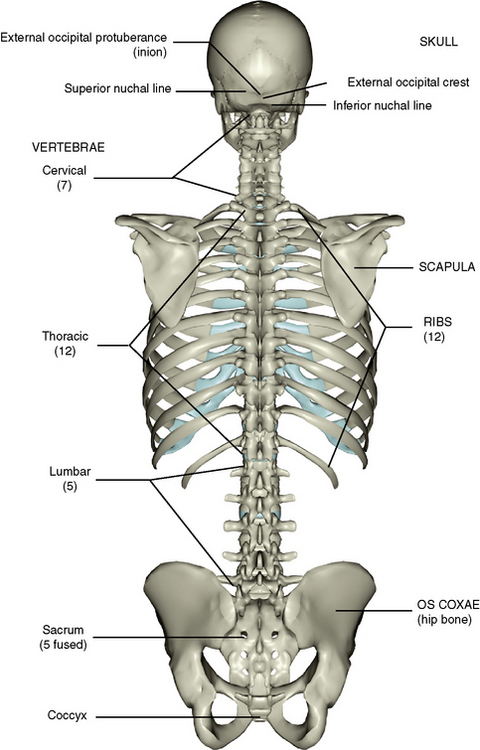
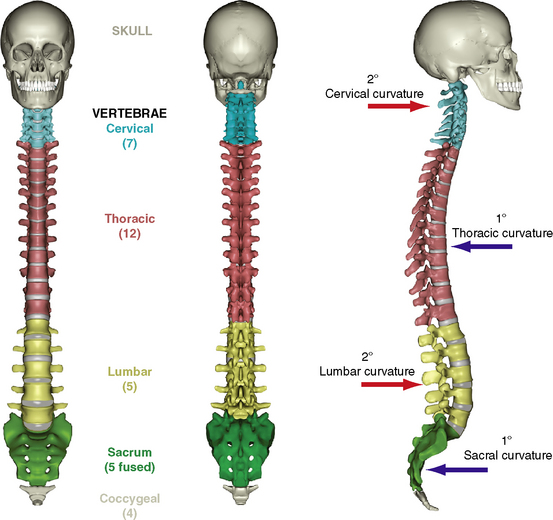
The skeleton of the back is presented in Figure 2-1. It consists of the skull, the vertebral column, and the ribs, which are appendages of the thoracic component of the vertebral column.
1 Skeletal Parts
POSTERIOR ASPECT OF THE SKULL
The various views and bones of the skull are considered in Chapter 6. Only the posterior aspect of the skull is described here. The posterior surface of the skull is convex and is commonly called the occiput. The external occipital crest is a midline ridge running superiorly from the foramen magnum. Arising from this crest are two transverse lines, the inferior nuchal line and above it the superior nuchal line. The external occipital crest ends at the midpoint of the superior nuchal line as a lump called the inion, or external occipital protuberance. Some muscles of the back extend up to this area of the skull and insert into these features.
VERTEBRAL COLUMN
Vertebrae: Typical Features
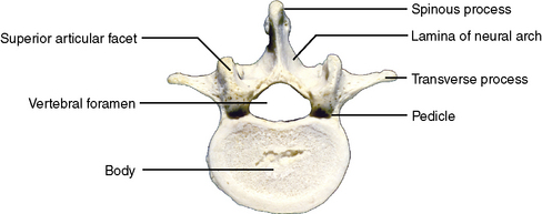
All vertebrae have certain features in common. Learning these features and then noting regional differences makes the study of vertebrae far less difficult. A midthoracic vertebra is generally chosen as exhibiting features common to most other vertebrae (Figure 2-2, A).
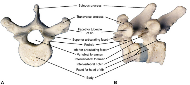
Figure 2-2 A, Superior view of a typical vertebra. B, Left lateral view of two articulated vertebrae.
Bilateral superior and inferior articulating processes are situated between the pedicle and lamina. They are bilateral articulating facets that are covered with hyaline cartilage and allow the vertebra above to articulate with the vertebra below (Figure 2-2, B).
Cervical Vertebrae
Typical Features
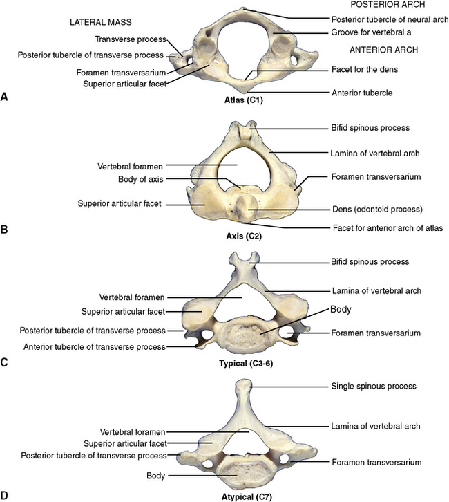
In addition to the general features, cervical vertebrae exhibit (1) a transverse foramen, a hole through the transverse processes to transmit the vertebral artery and vein; (2) a bifid spinous process; and (3) a transverse process, which ends laterally as an anterior and a posterior tubercle for attachment of cervical muscles (Figure 2-3, C).
Atypical Features
Atlas (C1)
The first cervical vertebra (Figure 2-3, A) differs from the remaining cervical vertebrae in the following ways: (1) the concave superior articulating facet is bean-shaped to accommodate the reciprocally shaped occipital condyles of the skull; (2) the body of the atlas is lost early in development and becomes fused to the body of the axis below, leaving only an anterior arch in its place; (3) there is no anterior tubercle on the transverse process; and (4) it exhibits two grooves for the vertebral arteries just posterior to the superior articulating facets.
Axis (C2)
This vertebra (Figure 2-3, B) differs from the typical vertebrae of the cervical region in two respects: (1) the original body of C1 is fused to the body of the axis as the dens, or odontoid process; and (2) the axis has no anterior tubercle on its transverse process.
C7
This vertebra (Figure 2-3, D) displays two atypical features: (1) it is the last component of the cervical segment and closely resembles the thoracic vertebrae below, with a long slender spinous process in contrast to the bifid spines of the vertebrae above; and (2) it has no anterior tubercle on its transverse process. Although it resembles a thoracic vertebra, it is distinctly cervical because it does have a transverse foramen.
Thoracic Vertebrae
The 12 thoracic vertebrae (see Figure 2-2) exhibit a number of features that distinguish them from other vertebrae: (1) the spinous process is long and slender; (2) the body has an articulating facet for the head of a rib; (3) the transverse process has an articulating facet for the tubercle of a rib; and (4) the body is heart-shaped.
Lumbar Vertebrae
Three features distinguish the five lumbar vertebrae (Figure 2-4): (1) they are relatively massive, with bean-shaped bodies; (2) they have no facets for ribs nor do the transverse processes possess transverse foramina, as in the cervical vertebrae; and (3) the spinous processes are not bifid nor slender but square-shaped.
The Sacrum
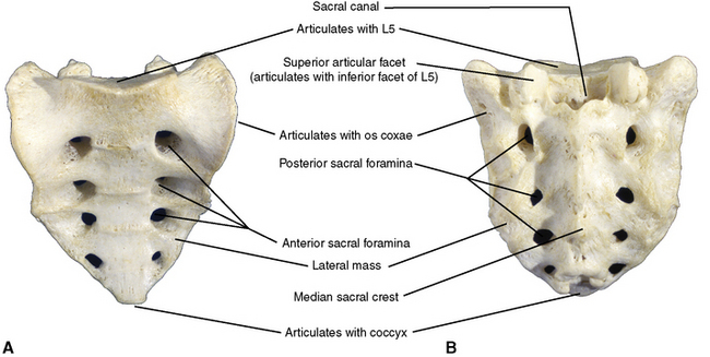
The five elements of the sacrum (Figure 2-5) are fused to form a solid triangular mass. The spinous processes are represented posteriorly by a median crest. A lateral mass representing fused transverse processes terminates laterally as two ear-shaped articular surfaces for the right and left os coxae. Superiorly, the sacral promontory projects anteriorly, and the sacral canal continues the vertebral canal inferiorly from the lumbar region. Leaving the sacral canal on either side are four anterior and posterior pelvic foramina, which transmit sacral anterior and posterior rami.
THE ARTICULATED SPINE
The stacked vertebrae and their intervening intervertebral discs make up the total length of the spinal column (Figure 2-6). In the neonate the fetal, or primary, curvature of the vertebral column is present. A convex cervical secondary curvature appears in the cervical area when the child learns to hold the head erect and to sit up. Similarly, when the child learns to walk, a secondary curvature appears in the lumbosacral region. The curvatures are formed by the intervertebral discs of the curved regions assuming a wedge shape.
JOINTS OF THE SPINE
Joints Between Bodies
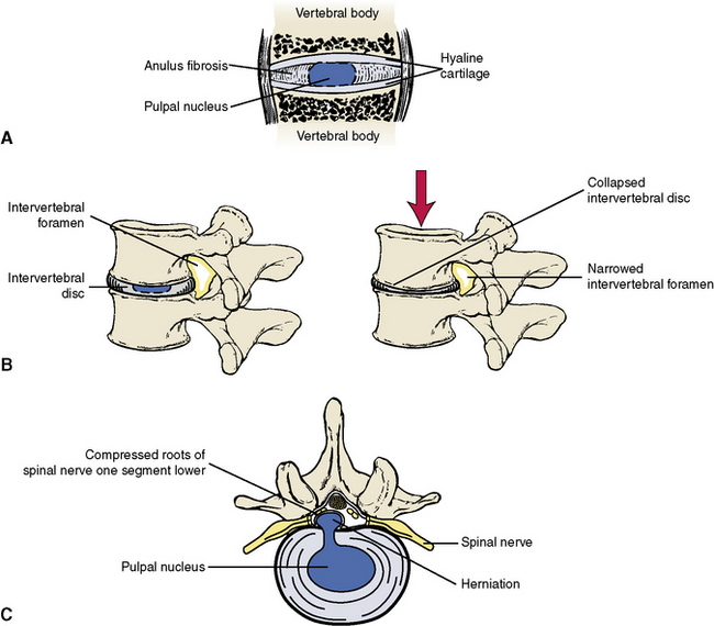
A symphysis type of joint is found between vertebral bodies (Figure 2-7). Hyaline cartilage lines the bony surfaces of the apposing bodies. Interposed is a disc consisting of concentric layers of fibrocartilaginous fibers (anulus fibrosis) surrounding a nucleus of fibrogelatinous material (pulpal nucleus), which is a remnant of the early notochord. The discs act as shock absorbers and in younger individuals (aged 20 years or younger) are quite strong. Anterior and posterior longitudinal ligaments run along the anterior and posterior surfaces of the vertebral bodies from the base of the skull to the sacrum, binding the components of the vertebral column together.
Stay updated, free dental videos. Join our Telegram channel
VIDEdental - Online dental courses


