9: Problem Solving in Working Length Determination

Chapter 9

Problem Solving in Working Length Determination

Problem-Solving List

Problem-solving issues and challenges in working length determination addressed in this chapter are:

Anatomic Basis for Challenges With Working Length Determination
Apical Root Anatomy and Its Impact on Working Length

    Working length determination: radiographic technique
    Working length determination: electronic apex locator
    Working length determination: other clinical techniques
Controversies and Challenges With the Pulpal and Periapical Status and Their Impact on Working Length Determinations and Choices
Posttreatment Implications and Outcomes of Root Canal Treatment Based on Working Length Philosophies

“The healing processes after removal of a pulp occur in the tissue immediately adjacent to the point where the pulp was severed. It is, therefore, of great importance to retain the vitality of these tissues in order to make healing possible.”< ?xml:namespace prefix = "mbp" />35

R. Kronfeld, 1933

“If the tooth is uncomfortable, however, or presents an area of rarefaction, apical access must be obtained in order to negotiate the canal throughout its entire length to reach the periapical tissues.”24

L.I. Grossman, 1946

“Factors that may influence a difference [in the method used for working length determination] include the quality of radiographs, superimposed anatomic structures, or anomalous positions of root canal foramen.”

D.H. Pratten, N.J. McDonald, 1996

For many years, the tooth apex has been the focus of many anatomic and treatment studies.* Anatomic variations in this region have been accepted as the norm, but contemporary treatment parameters involving these aberrations still present challenges and controversies.

Anatomic Basis for Challenges With Working Length Determination

The apex of the mammalian tooth is a complex biological unit composed of cementum, dentin, blood vessels, nerves, and connective tissues. One of the factors determining the long-term success of root canal treatment has been shown to be the relationship between instrumentation and obturation procedures and the anatomy of the apex.41,47,53 A clear understanding of the morphology of the root canal system, including the apex, is imperative.

Root formation and development are determined by the Hertwig’s epithelial root sheath (HERS), which maps out the external form of the root. The HERS is a double layer of epithelial cells derived from a proliferation of the internal and external dental epithelium (Fig. 9-1). The rim of this sheath, the epithelial diaphragm, encloses the primary apical foramen. Multirooted teeth form as a result of the division of the primary apical foramen into two or more sections by “tongues” of epithelium growing inwards from the HERS. Therefore, the root sheath determines the number, size, and external morphology of the roots. Following initiation of root formation, the HERS becomes fragmented and forms a fenestrated network known as the epithelial cell rests of Malassez. As the root sheath disintegrates, cells of the connective tissue differentiate into cementoblasts, and cementum is deposited on the dentin. Should the HERS disintegrate before dentin is elaborated, a lateral canal will be formed (Figs. 9-2 to 9-4)—an anatomic entity that has its own clinical implications (see Chapter 18). Development of root length is complete approximately 3 to 4 years after tooth eruption, with apical closure occurring some years later.

image

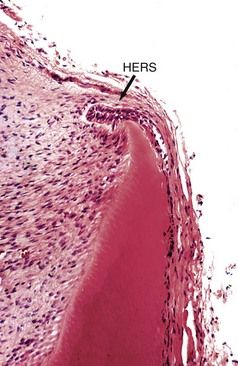
FIGURE 9-1 Remnants of the Hertwig epithelial root sheath (HERS) (arrow) in the developing root structure. This is a layer of inner and outer enamel epithelium responsible for root formation as it invaginates into the alveolar bone.

image

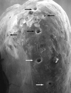
FIGURE 9-2 Palatal view of a mesial buccal root from a maxillary first molar, showing multiple foramina in the apical third of the root.

image

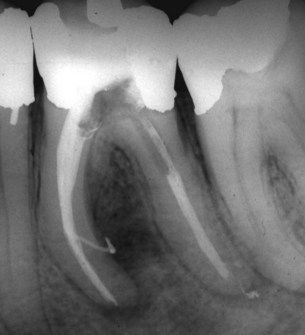
FIGURE 9-3 Large lateral canal leaving the mesial canals and exiting on the distal surface of the mesial root, as evidenced by filling during obturation. Note lateral lesion.

image

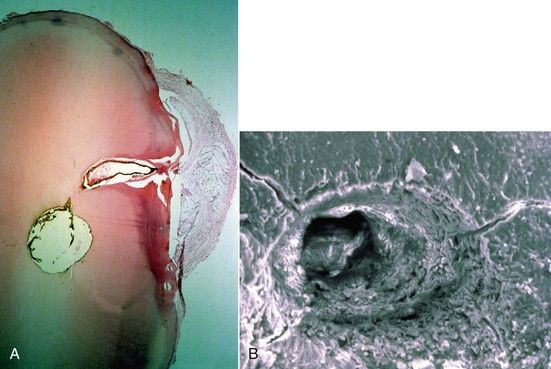
FIGURE 9-4 A, Histologic evidence of a large lateral canal in cross-section of the mandibular premolar. Note development of a cystic lesion on the lateral surface of the root. B, SEM (scanning electron microscopy) view of a lateral canal (×1800).

Extensive variability exists in the external apical root morphology of human permanent teeth with completed root apices (Fig. 9-5). In young teeth with incompletely formed apices, a funnel-shaped opening containing connective tissue (the dental papilla) is the typical appearance (Fig. 9-6). As the apex matures, the opening closes, and cementum is deposited on the apex continuing throughout life to compensate for loss of coronal tooth structure due to erosion, abrasion, or attrition. This gradual change in the morphology of the “normal” apical complex has been demonstrated,35 with further studies indicating that with increasing age, the center of the foramen deviates more and more from the vertex or apical center.55 Resorptive processes will also alter the morphology of the apical complex (Figs. 9-7 and 9-8; also see Chapters 3, 13, and 19). These resorptive processes may be the result of normal remodeling, orthodontic tooth movement, or inflammation of the pulp or periradicular tissues.

image

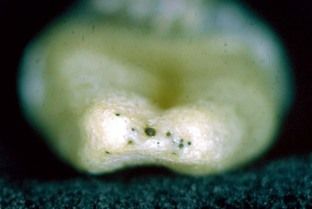
FIGURE 9-5 Multiple apical terminations pose clinical problems for working length determination, cleaning and shaping, disinfection, and obturation.

image

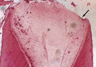
FIGURE 9-6 Overall view of an immature root that is forming shows a large dental papilla and advancing root sheath, left and right. Arrow indicates remnants of the epithelial sheath.

image

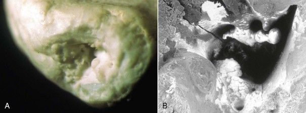
FIGURE 9-7 A, Resorbed root apex. B, SEM (scanning electron microscopy) of the same root apex shows significant irregularities. (×2000).

image

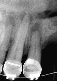
FIGURE 9-8 Radiograph showing apical resorption of the root apices of two teeth. Tooth on the left has an apical invaginating external resorptive defect that alters the position of the cemental-dentinal junction.

Internal morphology at the end of the root canal is determined by the odontoblasts responsible for development of the dentin. The transition from internal to external morphologic features occurs at the cementum-dentin junction (CDJ), delineated histologically by the odontoblasts. Coronal to this position, the tissue is classified as pulp tissue. However, the soft tissue contained within that portion of the canal apical to the CDJ is not dental pulp but a fibrous connective tissue that originates from the periodontal ligament and supplies the vessels and nerves leading to and from the pulp (Fig. 9-9).5 The walls of that portion of the canal as it enters the periodontal ligament (PDL) are covered with cementum. The root canal system tapers from the coronal end to its narrowest part, the constriction (minor foramen), which is usually but not necessarily within dentin. Early investigations indicated that the “pulp canal anatomy becomes extremely variable in the apical third.”5 Contemporarily, the internal morphology of the constriction has been classified into five main types: single constriction point, tapering constriction, multiple constriction, parallel constriction, and blocked (Fig. 9-10).17 Apical to the constriction, the root canal system diverges again to the major foramen that is within cementum. This hourglass shape dictates that canal cleaning, shaping, and obturation should be confined within dentin and not extend beyond the apical constriction or minor foramen.

image

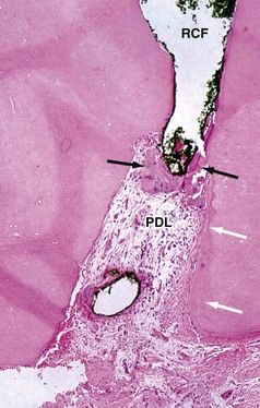
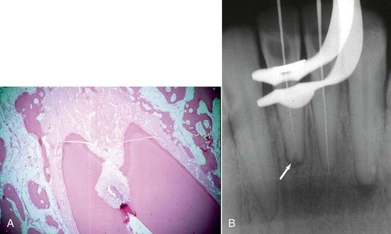
FIGURE 9-9 Root apex following root canal filling (RCF) short of the actual root length. Histologic evidence of hard-tissue formation (black arrows) that has formed from cells of the periodontal ligament (PDL) adjacent to root filling material. Note cementum formation (white arrows) on internal aspect of apical foramen.

image

FIGURE 9-10 Multiple possibilities for canal termination at the cemental-dentinal junction.

The apical portion of the root canal system presents the greatest number of ramifications, with 27.4% of teeth demonstrating accessory and lateral canals or an extensive arborization, also known as an apical delta (Fig. 9-11; also see Fig. 9-5).16 Their existence necessitates the use of adjuncts to mechanical cleaning and shaping to ensure thorough débridement of the root canal system (see Chapters 10 and 11). In all likelihood, the cleaning of these aberrant structures is all but impossible; in actuality, the tissues contained within these ramifications may contribute to ultimate apical healing.3,31

image

FIGURE 9-11 Apical delta formation in a demineralized and cleared tooth. Note presence of pulp stones in multiple small canals.

Studies have shown that the major foramina of most human teeth are distant from both the radiographic and anatomic apex9,36 (Fig. 9-12). Likewise, the major foramen is distant from the minor foramen or constriction by an average distance of 0.5 mm (see Fig. 9-12, B).9 All these anatomic variations directly affect clinical decisions made during root canal treatment, such as where shall the root filling end?30 It is easy to conjecture where this ideal position should be located, but more often than not, achieving this goal may elude the clinician. Ultimate clinical success and radiographic findings usually confirm or disprove this choice in treatment.41,47,53

image

FIGURE 9-12 A, Position of apical foramen is coronal to root apex. B, Histologic evidence and verification of this position (H&E stain ×4).

Apical Root Anatomy and Its Impact on Working Length

It is frequently impossible to know exactly where the apical foramen and apical constriction are located until after the canal has been obturated.30 However, knowledge of the possible three-dimensional variations (e.g., resorption or changes due to age, trauma, orthodontic movement, periradicular pathology, or periodontal pathosis) may prevent significant damage during working length determination and instrumentation to the cementum that has formed around the apical dentin and to the periapical tissues.18 In addition, extrusion of debris, medicaments used in root canal procedures, and obturation materials can be minimized, thereby preventing postoperative complications.10,14,31,37,60

The seventh edition of the Glossary of Endodontic Terms defines the working length of a tooth as the distance from a coronal reference point to the point at which canal preparation and obturation should terminate.21 To this purpose, the ideal apical terminus of the working length has been identified histologically as the CDJ. This junction is typified by a constriction or narrowing of the canal space (minor constriction) that provides an ideal point to prepare an apical seat in sound dentin. However, as previously mentioned, there can be vast variability in the nature of this constriction that will have an impact on any technique of working length determination.26,43,44 Furthermore, the constriction should not be confused with the apical foramen (major constriction), since the constriction is rarely if ever at the tip of the root.7,8,10,2729

The distance from the foramen to the constriction depends on a multitude of factors such as increased cemental deposition or radicular resorption (Fig. 9-13, A). Both processes are strongly influenced by multiple factors. Especially in periodontal disease states, the CDJ location has no predictable anatomic appearance or location, owing to resorptive processes or cemental depositions that may extend well into the root canal (see Fig. 9-13, B). The foreman and CDJ position on the root can be highly variable and exist anywhere from the direct radiographic apex up to 3 mm or more coronal to the radiographic apex, depending on a particular root morphology (Fig. 9-14). These potential anatomic variances have had a major impact on the precise region or location for determining the working length and termination of root canal instrumentation and obturation.

image

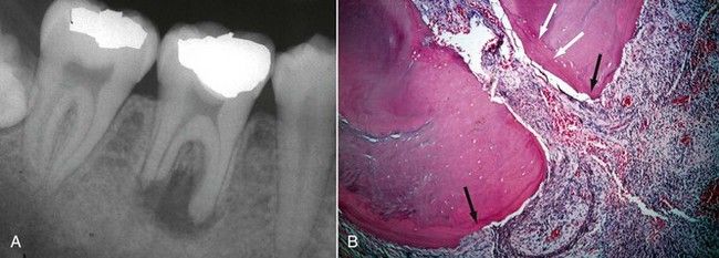
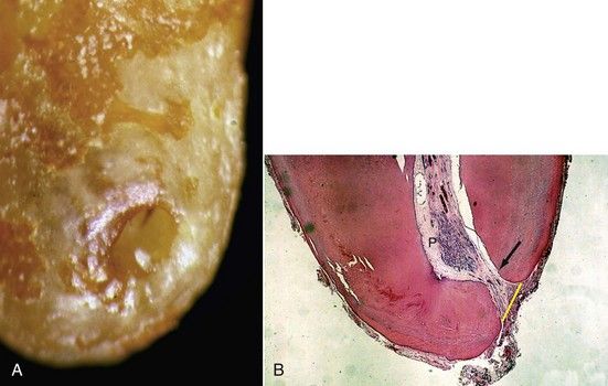
FIGURE 9-13 A, Mandibular molar with apical root resorption due to a necrotic, infected dental pulp that destroyed the natural cemental-dentinal junction. B, Histologic evidence of apical resorption on external cementum (black arrows) and layering of cementum (white arrows) into apical foramen (H&E stain ×10).

image

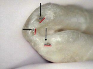
FIGURE 9-14 Apical view of tooth with a C-shaped root formation. Note root morphology around the canal exits as cementum invaginates into the foramen. K-files (arrows) are exiting from the canal long before they reach the actual root surface. Actual foramina are much larger than canal exits, as indicated by widths of the red lines. Working length determination to the root length in these cases would be destructive to periapical tissue.

Prior to establishing a definitive working length, coronal access to the pulp chamber must provide a straight-line avenue into the canal orifice, thereby facilitating subsequent canal penetration (see Chapter 8). In anterior teeth, failure to remove the lingual ledge or incisal edge often impedes this straight-line access, resulting in lack of depth penetration to the CDJ, failure to locate all canals present, or instrument penetration into the canal wall with ledge formation. In posterior teeth, primarily molars, or multirooted premolars, failure to remove cervical ledges or bulges results in missed canals or binding of the penetrating instrument in the coronal third of the canal with ledge formation (see Chapter 8). The ability to penetrate unimpeded to the CDJ is crucial to determining the working length of the root canal.

Current concepts of initial canal penetration recommend preflaring techniques for a coronal-to-apical approach to working length determination rather than immediate penetration to the apex region.39,54 Emphasis is placed on straight-line access to the radicular third of the canal, and considerable time and effort is spent preparing the coronal two-thirds of the root prior to apical penetration.39,54 This eliminates coronal impingements on the working length instrument and enhances penetration to the CDJ.

In curved canals, however, after obtaining straight-line access, the working length can change, especially if debris is packed around the curvature and not removed on a regular basis. Techniques have been advocated for this purpose, and cognizant use of them is recommended.38 If working length is obtained prior to straight-line access, it may be 1 mm less or even shorter after preparing the coronal two-thirds. Straight-line access eliminates the bend at the canal orifice and places the file in a more upright position closer to the reference point. A more accurate working length will be obtained after straight-line access in the canal is established (Fig. 9-15). This does not preclude obtaining a preliminary working length prior to preparing the coronal two-thirds if necessary to assist in overall knowledge of root shape, canal morphology, foramen patency, and to prevent canal blockage. Knowledge of average tooth length, tactile feel, or the use of digital radiography may provide tools for this purpose. However, once straight-line access is achieved, a more precise working length should be obtained.

image

FIGURE 9-15 When curves are present, as seen in the mesial buccal root, straight-line access is essential.

During access opening preparation, all caries, unsupported enamel, and faulty restorations are removed in an effort to secure stable reference points as aids in working length determination (see Chapter 8). This is especially helpful when more than one appointment is required to complete treatment. Typical reference points are those that are closest to the file and can be identified accurately as the cleaning and shaping process develops.18 If significant coronal destruction exists and extensive restorative procedures are anticipated, it is helpful to reduce unsupported tooth structure to prevent possible fracture between appointments, which may not only complicate the working length measurement already established but may also prevent associated periodontal and restorative problems should the fracture occur through the periodontal ligament (see Chapter 17).

Pathologic processes resulting in apical resorption can destroy the natural constriction of the CDJ (Fig. 9-16). This will create difficulty in locating a biologically acceptable position at which to establish the working length. The resorptive process generally produces a root end with an uneven, irregular radiographic appearance with few clues about where to prepare an apical stop. While the extent of proximal surface root end resorption may be discernible, the degree of buccal and lingual tooth loss is distinctly ambiguous. Buccal or lingual resorption cannot be discerned until 20% to 40% of the root structure has been demineralized and evidences some type of replacement resorption.1 If apical resorption presents radiographically with a scalloped or uneven proximal margin, significant three-dimensional resorption has already occurred, further complicating working length determination. Creation of an apical stop or enhancing an apical narrowing or constriction in these situations must rely on the clinician’s judgment, drawing on experience, tactile sensation, and reliable diagnostic radiographic techniques. If the root end is wide open from the resorptive destruction, electronic apex locators are unreliable and of little clinical value. Consequently, the coronal-most point on the root above the resorbed apex that exhibits sound radiodensity must be identified.28,52 This position is used as the new radiograph apex, and the working length is established 1 to 2 mm coronal to that point.28 In cases of extensive irregular apical resorption, the new working length can conceivably be 5 mm or more coronal from the original root apex.

image

FIGURE 9-16 A, Histologic demonstration of invasive apical resorption and how it destroys the cemental-dentinal junction. B, Working length established at the most narrow point in the canal when invasive apical resorption is present (arrow).

image


/>

Only gold members can continue reading. Log In or Register to continue

Stay updated, free dental videos. Join our Telegram channel

Jan 2, 2015 | Posted by in Endodontics | Comments Off on 9: Problem Solving in Working Length Determination

VIDEdental - Online dental courses

Get VIDEdental app for watching clinical videos