Dental pulp
Learning objectives
Key terms
Adventitia
Apical foramen
Basophils
Capillaries
Cell-rich zone
Cell-free zone (zone of Weil or Weil basal layer)
Coronal pulp
Denticles
Direct innervation theory
Endothelial cells
Eosinophils
Erythrocytes
False denticles
Formative
Free, attached, or embedded denticles
Gap junction
Hydrodynamic theory
Inductive
Intermediate junction
Intima
Leukocytes
Lymphocytes
Macrophages
Media
Nutritive
Odontogenic zone
Parietal layer of nerves
Pericytes
Plexus of Raschkow
Precapillaries
Protective
Radicular pulp
Reparative
Schwann cells
Terminal arterioles
Tight (zonula occludens) junction
Transduction theory
True denticles
Undifferentiated cells
Overview
Dental pulp is the soft, loose connective tissue located in the central portion of each tooth. It has a crown (coronal part) and a root (radicular part). Pulp is a delicate, specialized connective tissue containing thin-walled blood vessels, nerves, and nerve endings enclosed within dentin. Each pulp opens into the tissue surrounding the tooth, the periodontium, through the apex of the root canal. Accessory canals may be present at the apex of the tooth.
Pulp has a central zone and a peripheral zone, which are observed in both the coronal and radicular pulp. The central zone contains arterioles, veins, and nerve trunks that enter the pulp from the apical canal and proceed to the coronal pulp chamber. Fibroblasts are the preponderant cell, existing in an extracellular matrix of glycosaminoglycans and collagen fibers. Odontoblasts are the second most prevalent cell. The odontogenic zone in the periphery consists of odontoblasts and cell-free and cell-rich zones. Adjacent to the cell-rich zone is a parietal layer of nerves.
Odontoblasts form dentin throughout life, which causes the pulp to grow smaller with time. The terminal blood cells in the periphery are in thin-walled capillaries situated among the odontoblasts and are under local humoral control. Larger vessels with muscle cell support in their walls exist centrally and are under postganglionic sympathetic control. Several theories exist concerning pain conduction through dentin. The hydrodynamic theory is the most popular. It defines the movement of the odontoblast into contact with pulpal and intratubular nerve endings. Recent findings indicate, however, that odontoblasts are capable of receiving, conducting, and transmitting impulses to nerve endings in close proximity.
Pulp has several functions, such as initiative, formative, protective, nutritive, and reparative activities. All these clinical features are important to the production and maintenance of teeth.
Pulp may regress after trauma or with age and may contain diffuse areas of collagen fiber bundles and pulp stones. These pulp stones may be attached, embedded, or free in the pulp tissue. Pulp may also contain diffuse calcifications.
Anatomy of the pulp
Human beings have 52 pulps in their teeth, 20 in the primary dentition and 32 in the permanent dentition (Fig. 9-1). All pulps have similar morphologic characteristics, such as a soft, gelatinous consistency in a chamber surrounded by dentin, which contains the peripheral extensions of the pulpal odontoblasts. The total volume of the pulps of the permanent dentition is approximately 0.38 mL, and the mean volume of a single human tooth is 0.2 mL. The pulps of molar teeth are approximately four times larger than those of the incisors (see Fig. 9-1).
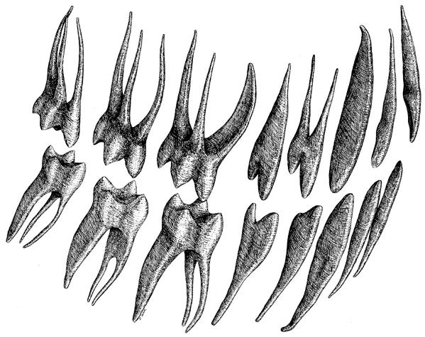
Upper row, Maxillary arch, left central incisor through third molar; lower row, mandibular arch, left central incisor through third molar.
Coronal pulp
The two forms of pulpal tissue are coronal and radicular (Figs. 9-2 and 9-3). Coronal pulp occupies the crown of the tooth. It is much larger than root pulp and has a structure different from the root tissue. In general, the coronal pulp follows the contour of the outer surface of the crown. Coronal pulp has six surfaces: mesial, distal, buccal, lingual, occlusal, and the floor. Coronal pulp has pulp horns, which are protrusions of pulp that extend into the cusps of the teeth. The number of pulp horns depends on the number of cusps (see Fig. 9-1). At the cervical region, the coronal pulp joins the root pulp. With age, the coronal pulp decreases in size because of continued dentin formation (see Fig. 9-2).
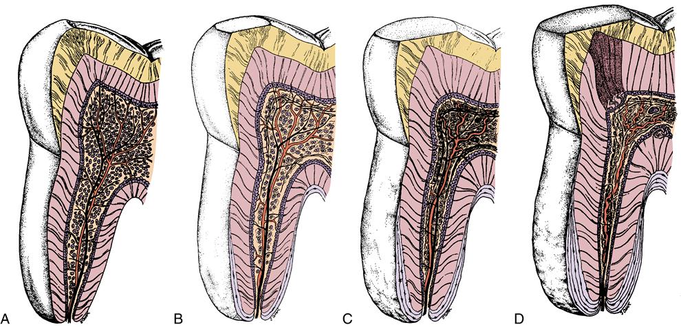
A, Young stage. B, After some attrition. C, At middle age. D, In old age. Pulp size and number of cells decrease, and fibrous tissue increases. Attrition also affects pulp horn with appearance of dead tracts and sclerotic dentin. (Modified from Bhaskar SN, editor: Orban’s oral histology and embryology, ed 11, St. Louis, 1991, Mosby.)
Radicular pulp
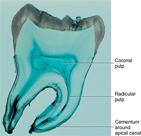
Pulpal root canals extend from the cervical region to the apex of the root. Radicular pulp of the anterior teeth is singular, whereas the posterior teeth have multiple root pulps. Radicular pulp is tapered or conical and, like coronal pulp, becomes smaller with age because of continued dentinogenesis (see Figs. 9-2 and 9-3). The apical foramen may become narrowed by cementum deposition.
 CONSIDER THE PATIENT
A patient calls a pink incisor to the dentist’s attention. He wants to know what causes this symptom.
 CLINICAL COMMENT
Radiographic knowledge of the pulp chamber’s shape and the extension of pulp horns into the overlying cusps is important in providing safe restorative dentistry. Pulp horns present a potential problem for pulp exposure.
Apical foramina and accessory canals
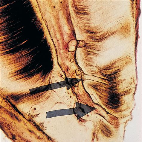
The apical foramen is the opening of root pulp into the periodontium. This opening varies from 0.3 to 0.6 mm, being slightly larger in the maxillary teeth than in the mandibular teeth. The apical foramen generally is centrally located in the newly formed root apex but becomes more eccentrically located with age (see Fig. 9-3 and Fig. 9-4). If several apical canals exist, the larger is designated the apical foramen, and the more lateral ones are called the accessory canals (see Fig. 9-4). Accessory canals may result from the presence of blood vessels obstructing dentin formation or from a break in the epithelial root sheath that induces initial root formation. The incidence of accessory canals is about 33% in permanent teeth. Accessory canals are located on the lateral sides of the apical region and may be found in the bifurcation area of multirooted teeth. Clinically, accessory canals are important because they represent contact of the pulp with the periodontal tissues and can contain bacteria and bacterial endotoxins which induce inflammation. If inflammation of the pulp is present, it can spread to the periodontium or vice versa.
 CLINICAL COMMENT
The presence of accessory pulp canals in an area where periodontal pathologic conditions exist may allow bacteria to spread into the pulp. If a pathologic condition exists in the pulp, on the other hand, it could be disseminated to the periodontium through such an accessory canal.
Histology of pulp
The pulp consists of coronal and root pulp. Coronal pulp is larger and contains many more elements than root pulp. Root pulp acts as a conducting tube to carry blood to and from the coronal area to the apical canal. Both pulp areas contain the same elements, although the cells, fibers, blood vessels, and nerves are more numerous in coronal pulp. Centrally, the pulp is composed of large veins, arteries, and nerve trunks surrounded by fibroblasts and collagen fibers embedded in an extracellular matrix (Fig. 9-5, A). Peripherally along the dentin in both coronal and radicular pulp are the formative cells of dentin, odontoblasts. The odontogenic zone includes these odontoblasts, the cell-free zone, and the cell-rich zone and the parietal plexus of nerves (Raschkow) (Fig. 9-5, B). The cell-free zone is known as the zone of Weil or the Weil basal layer. Adjacent to this zone is a zone of high cell density called the cell-rich zone, and pulpal to this zone is the parietal layer of nerves (Fig. 9-5, B). Thus the peripheral area of pulp is highly organized. The odontogenic zone appears most notably in coronal pulp and relates to the process of dentin formation, although the function of the cell-rich and cell-free zones in this process is still uncertain. In addition to the regions of the central and peripheral pulp is the area of the pulp horns. Here the odontoblasts are crowded and appear palisaded (pseudostratified) in contrast to their appearance in the remainder of the coronal area (Fig. 9-6). In the middle area, root pulp odontoblasts are short and cuboidal.
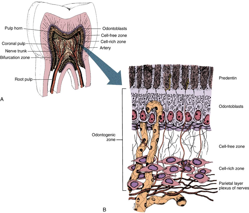
A, There appears high organization of the peripheral pulp and the appearance of centrally located nerve trunks (dark) and blood vessels (light). B, Odontogenic zone of pulp. Top to bottom: Predentin, odontoblasts, cell-free and cell-rich zones, and parietal layer of nerves. (Modified from Bhaskar SN, editor: Orban’s oral histology and embryology, ed 11, St. Louis, 1991, Mosby.)
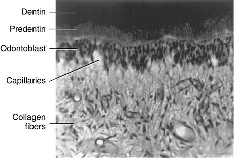
Pulpal capillaries are shown among these cells.
Odontoblasts
Odontoblasts line the perimeter of the pulp from the time they begin organizing to form dentin to the time they are quiescent and no longer producing dentin at a rapid rate. Odontoblasts are small and oval when they first differentiate but soon become columnar (Fig. 9-7). These cells then develop processes or extensions around which dentin forms. As the process lengthens, the amount of dentin thickens. Then the odontoblastic process develops many side branches that are contained within canaliculi. When these branches develop, space is provided in the dentin for them (Fig. 9-8). Odontoblasts are larger in coronal pulp than in the root and appear columnar in pulp horns (see Fig. 9-6). These tall, columnar ce/>
Stay updated, free dental videos. Join our Telegram channel
VIDEdental - Online dental courses


