Periodontium: Periodontal ligament
Learning objectives
■ Describe the location and structure of the periodontal ligament.
■ Explain the purpose of the periodontal ligament.
■ Describe the changes that occur in the periodontium as a result of the aging process.
Key terms
Aciniform
Alveolar crest fibers
Alveolar crest group
Apical fiber group
Attached gingival fibers
Cementoblasts
Circular or circumferential fibers
Dentoalveolar group
Fibroblasts
Free gingival fibers
Fundic alveolar bone
Gingival fibers
Gingival group
Horizontal fiber group
Interradicular fibers
Interstitial space
Macrophages
Oblique fiber group
Osteoblasts
Osteoclasts
Oxytalan fibers
Transseptal fibers
Overview
The periodontal ligament is a fibrous connective tissue between the alveolar bone proper and the cementum covering the root. This ligament covers the root of the tooth and connects with the tissue of the gingiva. The periodontal ligament occupies the periodontal space and is composed of fibers, cells, and intercellular substance. The latter consists of collagen fibers and ground substance, which in turn contains proteins and polysaccharides. The periodontium develops from dental follicular tissue that surrounds the tooth. The cells forming the ligament fibers, alveolar bone, and cementum develop from the follicle. The periodontium has a thickness of 0.15 to 0.38 mm, is thinnest in the midroot zone, and decreases slightly in thickness with age. The ligament is composed of collagen fiber bundles that attach the cementum to the alveolar bone proper. Interstitial spaces contain the blood vessels and nerve trunks, which communicate freely with vessels and nerves at the apex of the roots and the alveolar bone. This tissue is highly cellular, containing fibroblasts and vascular, neural, bone, and cemental cells. The primary function of the periodontal ligament is support for the teeth. The ligament also transmits neural input to the masticatory apparatus and has a nutritive function essential to maintaining the ligament’s health, which has important clinical implications.
Organization of the periodontal ligament
Two groups of principal fibers are named according to their location with respect to the teeth. The gingival group is located around the necks of the teeth, and the dentoalveolar group surrounds the roots of the teeth (Fig. 11-1). These principal fibers are bundles of collagen fibers strategically positioned at inclinations important to their functions along the root surface from the cervical region to the tooth’s apex (see parts 1 to 6 in Fig. 11-1). The collagen bundles are embedded in the cementum of the root and extend into the alveolar bone. Therefore, they act as a suspensor ligament for the teeth.
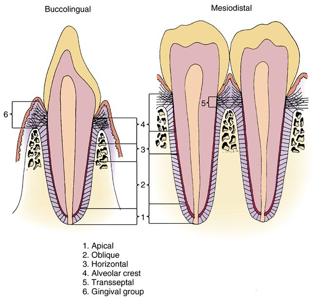
All fibers listed in the buccolingual plane are also present in the mesiodistal plane. The transseptal fiber group number 5, however, is seen only in mesiodistal plane, as these fibers are attached tooth to tooth. All other principal fibers are attached tooth to the gingiva or alveolar bone.
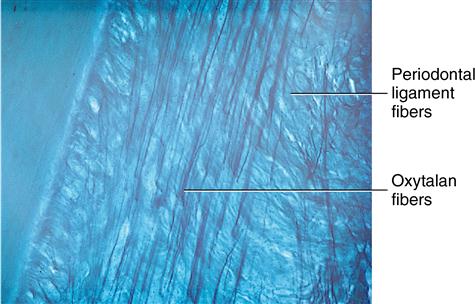
Between each group of fibers is a space termed the interstitial space (Fig. 11-2), which is not actually a space. Interstitial spaces contain a network of blood vessels, nerves, and lymphatics that maintain the vitality of the periodontal ligament and a network of finer fibers that interlace in the spaces, as well as support the dense collagen fiber bundles. The function of the interstitial space relates to the constant stretching and contraction of the fiber bundles during mastication. Most supporting fibers are collagenous, but a few have been described as elasticlike and of a structure different from that of collagen. These are termed oxytalan fibers (Fig. 11-3). Oxytalan fibers are small in diameter and appear to interface with the collagen bundles, supporting the collagen bundles and the blood vessel walls. These fine, elastic-like fibers stain with special stains that reveal their location to be almost longitudinal within the ligament when the fibers are viewed through a light microscope (see Fig. 11-3).
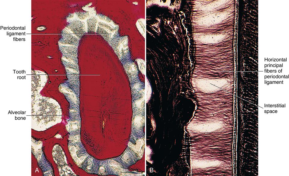
A, As they appear in cross-sectional plane. B, In plane longitudinal to tooth.
Gingival fiber group
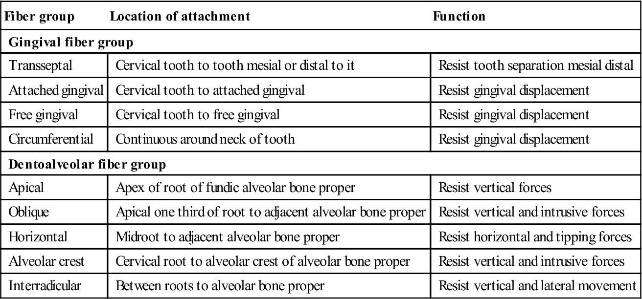
The principal fibers of the periodontal ligament in the gingival area are known as the gingival fibers. They consist of four groups of fibers, each having a different orientation and all supporting the gingiva (Fig. 11-4). The free gingival fibers arise from the surface of the cementum in the cervical region and pass into the free gingiva. The attached gingival fibers arise from the alveolar crest and pass into the attached gingiva. The circular or circumferential fibers are continuous around the neck of the tooth and resist gingival displacement. The alveolar crest fibers arise from the cementum at the neck of the tooth and terminate in the alveolar crest. Transseptal fibers originate in the cervical region of each crown and extend to similar locations on the mesial and distal surfaces of each adjacent tooth (Figs. 11-5 and 11-6). This fiber group functions in resistance to the separation of each tooth. Fig. 11-6 shows that transseptal fibers are found in the mesiodistal plane and are not present in the buccolingual plane. All these fiber groups are illustrated in Fig. 11-1 and listed in Table 11-1.
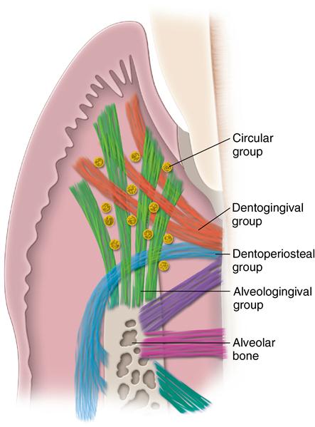
. Dentogingival fibers extend from the cervical cementum into free and attached gingiva. Alveologingival fibers extend from the alveolar crest into gingiva. Circular fibers surround the teeth, and the dentoperiosteal group extends from the cervical cementum into the alveolar crest. (From Nanci A: Ten Cate’s oral histology, ed 8, St. Louis, 2013, Mosby.)
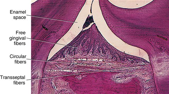
The transseptal fiber group extends from mesial of one tooth to distal of an adjacent tooth. Relationship of free gingival and circular fibers to transseptal fibers is shown.
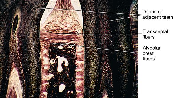
Periodontal fibers penetrate alveolar bone, and transseptal fibers extend from the tooth on the left to the right.
Table 11.1
< ?comst?>
| Fiber group | Location of attachment | Function |
| Gingival fiber group | ||
| Transseptal | Cervical tooth to tooth mesial or distal to it | Resist tooth separation mesial distal |
| Attached gingival | Cervical tooth to attached gingival | Resist gingival displacement |
| Free gingival | Cervical tooth to free gingival | Resist gingival displacement |
| Circumferential | Continuous around neck of tooth | Resist gingival displacement |
| Dentoalveolar fiber group | ||
| Apical | Apex of root of fundic alveolar bone proper | Resist vertical forces |
| Oblique | Apical one third of root to adjacent alveolar bone proper | Resist vertical and intrusive forces |
| Horizontal | Midroot to adjacent alveolar bone proper | Resist horizontal and tipping forces |
| Alveolar crest | Cervical root to alveolar crest of alveolar bone proper | Resist vertical and intrusive forces |
| Interradicular | Between roots to alveolar bone proper | Resist vertical and lateral movement |
< ?comen?>< ?comst1?>
< ?comst1?>
< ?come/>
Stay updated, free dental videos. Join our Telegram channel
VIDEdental - Online dental courses


