Salivary glands and tonsils
Learning objectives
■ Discuss the classification of the major and minor salivary glands.
■ Explain the composition and function of saliva.
■ Describe the location and purpose of salivary gland duct systems.
Key terms
Alveolus or acinus
Amylase
B cells
Buccal and labial glands
Epidermal growth factor (EGF)
Excretory portion
Exocytosis
Germinal centers
Glossopalatine glands
Intercalated ducts
Interlobular excretory ducts
Intralobular duct system
Lingual glands
Lingual tonsils
Lobes
Lobules
Major glands
Memory cells
Merocrine glands
Minor glands
Mixed glands
Mucin
Nerve growth factor
Palatine glands
Palatine tonsils
Parotid glands
Pharyngeal tonsil or adenoid
Pure mucous glands
Salivary corpuscles
Secretory end piece or intercalated duct
Secretory portion
Serous demilunes
Serous glands
Serous or mucous cells
Stensen duct
Striated duct
Sublingual glands
Submandibular glands
T cells
Waldeyer ring
Wharton duct
Zymogen granules
Overview
This chapter discusses the structure and function of the salivary glands, saliva, and tonsils. Despite different structures and functions, these soft tissues all contribute significantly to oral health. Saliva is a balanced secretion resulting from both the composition of the secretion and the location of the salivary gland secretions into the oral cavity. The two cell types are serous, which is high protein and low carbohydrate, and mucous, which is low protein and high carbohydrate. Glands of the lips, cheeks, and anterior floor of the mouth produce a watery mixture of a serous and mucous secretion, whereas other glands of the posterior palate, pharynx, and tongue contribute a viscous mucous solution that protects the membranes in those regions. The major salivary glands contribute 85% to 90% of the saliva into the more anterior area of the mouth. In addition to protein and carbohydrate, the parotid gland, which is the largest gland, secretes the enzyme amylase, which aids in digestion of carbohydrates. Therefore, the buffering ability of saliva results from ionic secretions by the salivary glands. These secretions are collected and modified through an elaborate secretory duct system.
Tonsils, like the salivary glands, have locations that maximally affect and protect the oral environment. These lymph node–like organs are positioned in the oropharynx at the entrance to the alimentary canal. They produce lymphocytes and, with the assistance of macrophages, protect against microbes, foreign cells, and cancer cells. Lymphocytes can recognize foreign cells and respond to them either by becoming T cells, which destroy the foreign cells directly, or by forming B cells, which transform into plasma cells that secrete antibodies to eliminate the foreign cells.
Classification of salivary glands
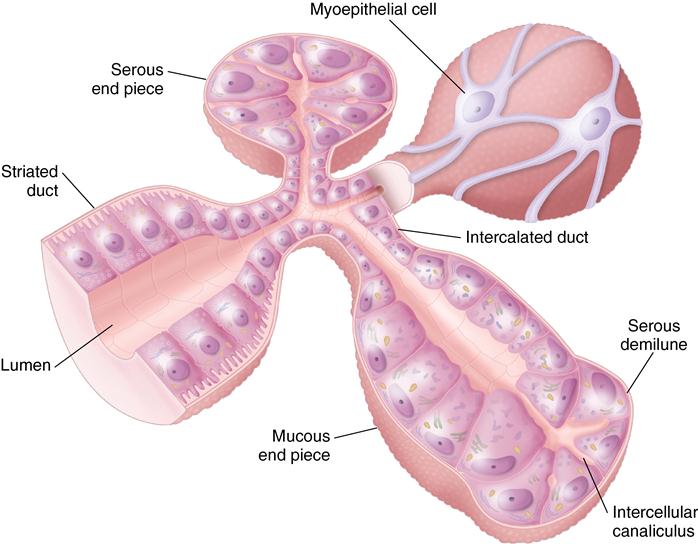
Salivary glands are classified as either major or minor depending on their size and the amount of their secretion. The major glands carry their secretion some distance to the oral cavity by means of a main duct. The smaller minor glands empty their products directly into the mouth by means of short ducts. Both are composed, however, of the same type of cells, either serous or mucous cells or a combination of the two called serous demilunes (Fig. 15-1).
 CLINICAL COMMENT
The salivary glands are also important for the production of growth factors including nerve growth factor (NGF) and epidermal growth factor (EGF), which belongs to a family of growth factors that stimulates cell growth, proliferation, and differentiation.
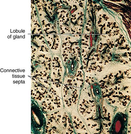
The functional unit of the salivary gland is the alveolus or acinus. An acinus is a cluster of pyramidal cells, either mucous or serous or a combination of the two, that secretes into a terminal collecting duct (see Fig. 15-1). The collecting duct is termed the secretory end piece or intercalated duct. Both the large and small glands are composed of many acini, although the larger glands contain more acini or units arranged in lobules and lobes (Fig. 15-2). Each cell type provides a different type of secretion. Serous cells secrete mostly proteins and small amounts of carbohydrates. Their secretion also contains zymogen granules, precursors of the enzyme amylase, which functions in the breakdown of carbohydrates. Serous cell secretion has a watery consistency. Mucous cells are high in carbohydrates and low in proteins and discharge a viscous product called mucin (Fig. 15-3). When mucin mixes with watery oral fluids, it becomes mucous, causing the saliva to be thick and viscous.
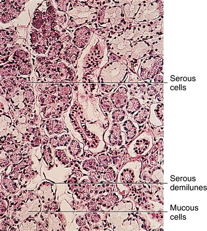
Serous cells are at the upper left, and mucous cells are at the lower left. A few mucous cells are capped with serous cells (serous demilunes).
Both types of acinar cells are pyramidal. The nucleus of the serous cell is oval to round, and that of the mucous cell is oval to spindle shaped (see Fig. 15-1). In each of these cell types, the nuclei appear in the basal part of the cell. The cytoplasm of the serous cell stains deeply because it is filled with albumin, whereas the mucous cell appears light and foamy because of the presence of carbohydrates in mucin (see Fig. 15-3 and Fig. 15-4).
Ultrastructurally, the serous cell is filled with secretory granules in the apical region, rough-surface endoplasmic reticulum, a Golgi apparatus, mitochondria, and an oval nucleus (Fig. 15-5). The mucous cells contain larger droplets of mucin apically and a prominent Golgi apparatus and rough endoplasmic reticulum around the flattened nucleus (Fig. 15-6). A third cell-type arrangement is a terminal alveolus of mucous cells with a cap of serous cells (see Fig. 15-4). This configuration is termed a serous demilune, with the secretion of the serous cells passing down a duct between the terminal mucous cells to the lumen of the alveolus (see Fig. 15-1).
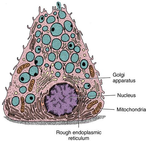
Note the round nucleus, Golgi apparatus, and rough endoplasmic reticulum that are characteristic of a protein-secreting cell. Arrows point to vesicles arising from the Golgi zone and migrating to the cell surface.
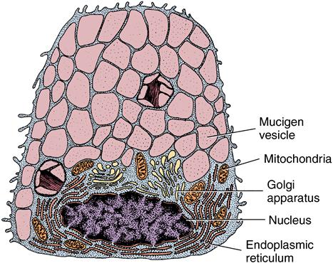
Compare the shape of this cell and its nucleus with those of the serous cell. Shown are the Golgi apparatus, rough-surface endoplasmic reticulum, and mucous accumulation (mucigen vesicles) in the cell.
Salivary glands are termed merocrine glands because the basic mode of product excretion is through membrane vesicles passing to the cell’s apex. These vesicles fuse with the cell plasma membrane and their contents are released by exocytosis, the process in which a cell can release substances to the extracellular milieu. (see Fig. 15-6).
 CLINICAL COMMENT
The serous and mucous cells of the major glands secrete 85% to 90% of saliva. Their combined secretions produce the viscosity as well as the important buffering actions of saliva. These properties result from the actions of protein, carbohydrate, carbonate, and phosphate that are contributed by the secretory ducts of the glands.
Major salivary glands
The major salivary glands are present as three bilateral pairs. The parotid glands are located on the sides of the face in front of the ears; the second pair, the submandibular glands, are inside the angle of the mandible; and the third pair, the sublingual glands, are on either side of the midline beneath the mucosa of the anterior floor of the mouth (Fig. 15-7 and Table 15-1).
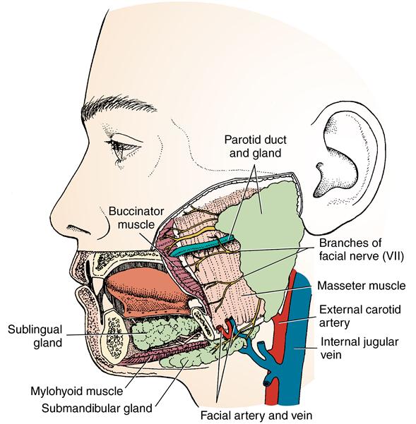
The diagram demonstrates the relationship of facial nerves and blood vessels to oral structures.
Table 15.1
Major salivary glands and contribution to saliva
< ?comst?>
| Gland | Size | Location | Type cells | Amount secretion (%) | Duct type: striated/intercalated |
| Parotid | Largest | Anterior ear | Serous | 25 | Long/long |
| Submandibular | Intermediate | Angle of mandible | Mixed serous demilune | 60 | Long/short |
| Sublingual | Smallest | Anterior floor of mouth | Mucous | 5 |
Stay updated, free dental videos. Join our Telegram channel
VIDEdental - Online dental courses


