Chapter 7
Surgical Endodontics
Aim
This chapter will describe the indications for, contraindications to and techniques of surgical endodontics.
Outcome
After reading this chapter you should know when surgical endodontics should be performed and the surgical techniques to be employed.
Introduction
The term surgical endodontics encompasses a number of procedures. This chapter will consider the following:
-
apicectomy
-
repair of a lateral perforation
-
root resection.
Apicectomy
Apicectomy means removing the apical portion of the root of a tooth. It is performed to remove atypical or complex apical foramina, together with abnormal, typically infected adjacent tissue.
It is important to establish at the outset that apicectomy is not an alternative to effective conventional endodontics. A simple explanation as to why apicectomy is inferior to good quality endodontics is that it is never performed under rubber dam. Other reasons include:
-
decreasing the crown:root ratio (Fig 7-1)
-
exposing accessory canals at the apex (Fig 7-1)
-
increasing the area of root filling material exposed at the apex (Fig 7-1)
-
iatrogenic damage to important structures (Fig 7-2).
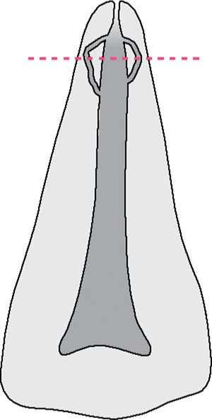
Fig 7-1 Removing the apex of a tooth can expose accessory canals and increase the area of root filling exposed.
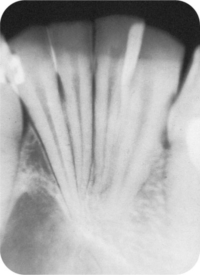
Fig 7-2 Performing apical surgery on one of these lower incisors could easily damage one of its neighbours.
Advances in endodontics, including the use of microscopes, have resulted in a reducing need for endodontic surgery. This is progress, as apicectomy is not an ideal treatment for a compromised tooth. The only thing worse is extraction, and this at least more or less guarantees a cure. Thus, apicectomy should be considered the last stop before the terminus of extraction. Not only is apicectomy a poor alternative to good conventional endodontics, it should not be considered if the existing root filling is less than ideal. Apicectomy should not be performed on a tooth with inadequate coronal restorations. This is because any coronal leakage will result in the failure of apical surgery. When bony support for the tooth is poor – for example, as a result of periodontal disease – surgery may worsen the support. Thus it is difficult to argue the case for surgical endodontics. In addition, if the patient’s general health is poor, surgery is contraindicated unless essential. Thus the contraindications to apicectomy are:
-
unsatisfactory endodontics
-
coronal leakage
-
poor bony support
-
where support will be insufficient following surgery
-
poor periodontal health
-
poor general health.
Notwithstanding the above, surgical endodontics is still performed. So there are times when this procedure is indicated. The indications for apicectomy are:
-
when it is impossible to clean, shape and fill root canals to within 1–2mm of their apical extension with conventional endodontics
-
fractured instrument
-
severe dilacerations
-
immovable post (of a well-fitting crown) (Fig 7-3)
-
-
when excess root canal material or a broken instrument is present through the apex and is giving rise to problems (Fig 7-4)
-
to remove a fractured apical portion
-
external root resorption (Fig 7-5)
-
when an apical biopsy is required.
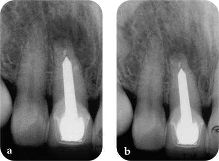
Fig 7-3 (a) A tooth with a well fitting crown with an apical area. (b) The area has healed following apical surgery.
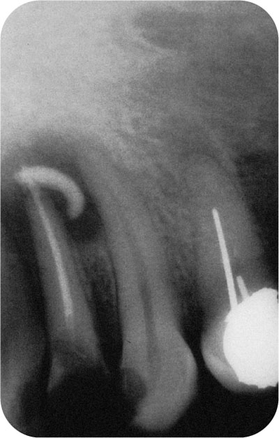
Fig 7-4 Excess root filling material at the apex of a tooth associated with apical infection. Apical surgery is indicated to remove this material.
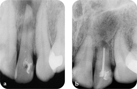
Fig 7-5 (a) Periapical pathology associated with external root resorption of an upper lateral incisor. (b) healing after apicectomy.
Radiographic Assessment
It is essential to have good radiographs before performing apical surgery. A long-cone paralleling technique periapical film showing at least 3mm of tissue beyond the apex of the appropriate tooth is ideal. If a large apical radiolucency is associated, then a sectional panoramic radiograph should also be taken.
Technique
The technique must allow:
-
access to the apex of the tooth
-
good visibility of the apex of the tooth
-
good healing.
Description of the technique is divided into:
-
anaesthesia
-
flap design
-
bone removal
-
apicectomy
-
curettage
-
apical seal
-
wound closure
The procedure is illustrated in Fig 7-6.
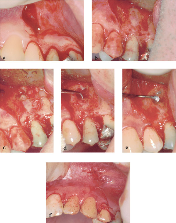
Fig 7-6 (a) Flap for apicectomy of upper second premolar showing vertical and horizontal incisions. (b) Flap raised to reveal bony defect at apex of upper second premolar. (c) Apicectomy of tooth revealing root canal. (d) Preparation of root canal for retrograde filling with ultrasonic tip. (e) Filling of retrograde cavity with IRM. (f) Wound closure.
Anaesthesia
Apical surgery is well suited to local anaesthesia. A combination of regional block and infiltration should suffice. The infraorbital nerve block in combination with infiltrations is particularly useful when dealing with upper anterior teeth. Good anaesthesia should be established before raising the flap. Once a flap is raised ooze of the anaesthetic solution can reduce efficacy and tastes awful.
Flap Design
A number of different flaps have been advocated for apical surgery (Fig 7-7). These can be described as:
-
those involving the gingival margin
-
two-sided (Figs 7-6a, b and 7-7a)
-
three-/>
-
Stay updated, free dental videos. Join our Telegram channel

VIDEdental - Online dental courses


