Chapter 10
Minor Oral Surgery and the Antrum
Aim
The aim of this chapter is to discuss the ways in which the maxillary antrum can be involved in the practice of minor oral surgery.
Outcome
After reading this chapter you should understand the techniques used to manage oro-antral communications.
Introduction
The antrum may become involved in the surgical treatment of the dentition in a number of ways. During tooth extraction an oro-antral communication (OAC) may be created. This may become epithelialised to form an oro-antral fistula (OAF). A root may be displaced into the antrum or the maxillary tuberosity may fracture. Closure of the antral defect may become necessary either immediately or later. The antrum may be involved in benign pathology such as cysts or be the site of a malignancy. The antrum is frequently involved in maxillofacial trauma and in infection, which may cause difficulty in diagnosis. Finally, prior to the placement of implants, bone grafting of the maxillary sinus floor may be required.
Anatomy
The maxillary antrum has been described as having:
-
a pyramidal shape
-
four walls
-
a floor – which is closely associated with the roots of the upper premolar and molar teeth
-
a roof – which is also the floor of the orbit
-
a respiratory epithelium lining – which is continuous with the nasal mucosa via the ostium.
Development
The antrum increases in size during childhood and continues to pneumatise with age.
Function
The maxillary antrum has a number of functions. These aid:
-
respiration – inspired air is warmed and humidified by circulation over the respiratory mucosa
-
speech – the paranasal air spaces of which the antrum is one, give resonation of speech
-
lightening of the skull – the weight of the head is limited by the presence of the air cells. This maxillary antrum may also help form a ‘crumple zone’ to absorb impact in maxillofacial trauma.
Drainage
It is important to appreciate the drainage pattern of the maxillary sinus. The antrum drains into the nose via the ostium, which is approximately half way up the medial wall. The ostium opens into the nose at the level of the middle meatus. Importantly the ciliated mucosa of the lining is extremely efficient and will normally move mucus from the base of the antrum to the ostium in around one hour. Thus drainage is away from the floor and not dependent upon gravity.
Surgical Involvement of the Antrum
The ways in which the antrum can impact on dental practice are described below.
Iatrogenic Injury
There are a number of occasions when the antrum can be involved during oral surgical procedures:
During extraction
This is the most likely way in which the dentist will encounter the antrum. The root apices of the upper teeth are closely associated with this structure. The maxillary sinus may extend as far forward as the upper canine (Fig 10-1). Any extractions in this area may cause a communication between the antrum and the mouth. The root most commonly associated is the palatal root of the upper first permanent molar. This is especially the case when there is evidence of apical pathology, which may cause resorption of the bone of the antral floor. The initial diagnosis is by suspicion (Table 10-1). Where there is any evidence to alert the clinician, the patient must be warned of the possibility of a communication and fistula formation. A conservative regimen may be started to allow healing to take place naturally (Table 10-2).
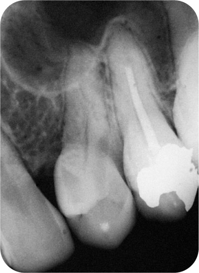
Fig 10-1 The antrum may extend anteriorly beyond the premolar teeth.
| INITIAL DIAGNOSIS OF ORO-ANTRAL COMMUNICATION (IMMEDIATELY AFTER EXTRACTION) |
| NOT BY FORCED NASAL EXPIRATION
NOT BY PROBING OBSERVATION SUSPICION RADIOGRAPH |
| CONSERVATIVE REGIMEN TO MANAGE AN ANTRAL COMMUNICATION |
| PATIENT INSTRUCTIONS – NO NOSE-BLOWING, ORAL HYGEINE INSTRUCTION, etc. |
| +/- |
| NASAL DECONGESTANTS – EPHEDRINE 0.5% |
| +/- |
| ANTIBIOTICS – BROAD SPECTRUM |
| REVIEW – UNTIL HEALED |
Patients should be warned not to increase antral pressure by blowing their nose and avoid sneezing. Such actions may cause a communication and possibly a prolapse of the antral lining (Fig 10-2). If the antral lining prolapses, it is unlikely to be easily reduced back into the antrum and will have to be excised. A buccal advanced flap is used to close the defect (see below).
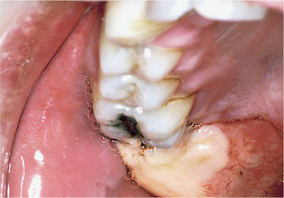
Fig 10-2 Antral prolapse following removal of upper third molar. The patient reported blowing his nose on the evening following extraction.
Oro-Antral Communication
An oro-antral communication may occur more frequently than one realises. The condition m/>
Stay updated, free dental videos. Join our Telegram channel
VIDEdental - Online dental courses


