7: Recognition and Management of Shock

Recognition and Management of Shock

Shock occurs when the cardiovascular system fails to perfuse vital organs. There are numerous clinical events that cause shock, such as severe hemorrhage, trauma, burns, sepsis, anaphylaxis, myocardial infarction, and pulmonary embolism. Despite the cause, shock is defined and ultimately diagnosed by the clinical evidence of inadequate tissue oxygenation and cellular death. Once cell death occurs, inflammation, free radical formation and edema can exacerbate hypoperfusion, creating a viscous cycle of irreversible tissue damage that causes further cardiovascular collapse, despite aggressive treatment. Therefore, it is essential that shock be recognized and treated early to prevent end-organ failure and death of the patient.

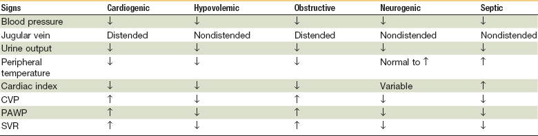
Treatment of shock varies depending on the type of shock. There are four major categories of shock—hypovolemic shock (hemorrhagic versus nonhemorrhagic), cardiogenic shock, obstructive shock, and distributive shock (septic, anaphylactic and neurogenic).1 Each category of shock may present with variable clinical signs and symptoms and requires different treatments. For example, aggressive volume replacement would be appropriate in a patient who is in hypovolemic shock, but may be detrimental in a patient with cardiogenic shock. Therefore, one must not only recognize shock but also be able to categorize it and implement the appropriate treatment based on the available clinical data. This chapter will give an overview of the cellular and systemic response to shock and then focus on its recognition, categorization, and current treatment philosophies.

Cellular Changes

Organ hypoperfusion creates a hypoxic environment that initiates cellular changes resulting in shock. At the cellular level, shock occurs when the delivery of oxygen is inadequate for cells to metabolize glucose for energy needed to run the cellular machinery. Cells depend on oxygen to carry out aerobic respiration via the mitochondria. Mitochondria are the power plants of cells that supply 36 adenosine triphosphate (ATP) molecules through the oxidation of each molecule of glucose. Without an adequate supply of oxygen, mitochondria switch to anaerobic respiration, yielding only two ATP molecules and lactate per glucose molecule. Lactate lowers the tissue pH and is released into the systemic system, becoming a useful marker for tissue hypoxia and acidosis. Not only is lactate toxic to cells, but a lack of ATP generation leads to free radical formation and damage to vital cell functions, most notably ATP-dependent ion channels that maintain normal membrane potentials. When ATP-dependent Na+, K+, and Ca2+ transport are disrupted, cellular membranes lose their electrical gradient, leading to intracellular bleb formation, cellular edema, and cell lysis. The tipping point for irreversible cell damage is not entirely understood but is thought to be a result of uncontrolled release of intracellular Ca2+ stores. Intracellular bleb formation and mitochondrial swelling are signs of irreversible cellular dysfunction that leads to cell lysis. When intracellular enzymes and cellular material are released into the surrounding tissue and circulation, further inflammation, edema, and tissue damage occur.2,3

Endothelium normally produces nitric oxide but when damaged by inflammation, nitric oxide synthase is overexpressed, producing toxic levels of nitric oxide and other oxygen-derived free radicals that are formed when tissue are reperfused with oxygen. This reperfusion injury creates free radicals and further activation of inflammatory mediators, notably interleukin-8 (IL-8), IL-1, and tumor necrosis factor (TNF). The combination of ATP depletion, tissue edema, pH changes, free radical formation, and inflammatory mediator formation leads to a complex vicious cycle of cell death and irreversible organ damage.3

Systemic Response

Different organ cells have extremely varying cellular oxygen demands. Two of the most metabolically active and oxygen-demanding tissues are the heart and brain. This is clearly evident in the systemic response to hypoperfusion. During shock, the cardiovascular system diverts blood flow from less vital structures to maintain adequate perfusion to the heart and brain at the expense of tissues such as skin, skeletal muscle, gastrointestinal (GI) tract, and kidneys. This diversion is made possible by differences in local and distant regulatory mechanisms. For example, the heart and brain at rest already exhibit a high sympathetic vascular tone and thus tightly autoregulate perfusion through local metabolites. In contrast, splanchnic blood flow is normally in excess of metabolic demand and its vasculature is influenced much more by increases or decreases in sympathetic tone. During prolonged shock, sympathetic-mediated vasoconstriction in the splanchnic vasculature leads to ischemia and necrosis, but the brain and cardiac blood flow are preserved because of metabolic autoregulation.

Cardiac output (CO) is the amount of blood pumped from each ventricle each minute. It can be represented by the following equation:

image

or

image

It is continuously regulated to meet tissue perfusion demands. For example, during strenuous exercise, CO will increase from 5.8 to 15 liters/min to meet the requirements of exercising skeletal muscle. CO is dependent on three variables:

Preload is the amount of blood that returns to the heart prior to contraction, the end-diastolic volume (EDV). Increasing preload is often referred to as priming the pump. More blood returning to the ventricles prior to systolic contraction not only increases the amount of blood ejected per heartbeat, but also increases the force of cardiac contractility as indicated by the Frank-Starling law of the heart. The result is an increase in the stroke volume and cardiac output.

Preload is determined by the pressure difference between the central venous system (great veins of the thorax and abdomen) and peripheral venous system. Combined, the venous system holds 60% to 70% of the circulating blood volume; most of this is stored in the peripheral venous pool of organs such as the skin, skeletal muscle, and splenic system. Veins are compliant because of their thin walls, allowing them to collapse or distend with small changes in pressure. This compliance enables the venous system to act as a reservoir of circulating blood volume, most of which is in the peripheral veins and does not contribute to the central venous pressure. During hypovolemia, volume loss is reflected in a loss in this venous reservoir as blood moves from the periphery into the central circulation. Also, an increase in circulating catecholamines—norepinephrine and epinephrine—causes further venous constriction. This venous compliance allows a net shift of blood from the peripheral venous pool to the central venous system, thus maintaining adequate blood return to the ventricles.

Afterload is the pressure or resistance to the forward movement of blood being pumped from the heart. As the heart contracts, it must overcome the forward pressure to propel blood through the circulatory system. Myocardial contractility is the force of the pump. A more forceful contraction allows for a greater percentage of blood entering the heart to be ejected into the arterial system. The ratio of blood entering and leaving the heart is represented by the ejection fraction (EF); EF = SV/EDV, which is normally 55% to 80% (mean, 67%). An EF of less than 55% indicates depressed myocardial contractility.

When mean arterial pressure falls below 60 mm Hg, tissue perfusion is compromised and shock ensues. Hypotension and hypoxia are sensed by chemoreceptors and baroreceptors, resulting in reduced vagal tone and the release of catecholamines (norepinephrine and epinephrine) by the neuroendocrine system. This causes an increase in heart rate, cardiac contractility, vascular tone, and total peripheral resistance to maintain blood flow to vital organs. It is the constriction of arterioles that decreases blood flow to the peripheral organs and increases the total peripheral resistance. Blood flow to the brain and heart are less affected by distant autonomic regulation and rely more on local control mechanisms, such as oxygen tension and metabolic byproducts. This allows for preservation of blood flow, despite significant reductions in cardiac output. In contrast, splanchnic blood flow is normally in excess of metabolic demands and is controlled more by circulating catecholamines. In times of shock, these organs can experience severe arteriolar constriction and early ischemia.

Vasoconstriction is vital for maintaining adequate perfusion and diverting blood flow in times of shock but, without volume replacement, prolonged vasoconstriction will lead to ischemia. The body can increase volume through mobilizing extravascular fluid into the circulation and by decreasing renal excretion.

Renin is released because of increased sympathetic stimulation and a decrease in blood flow sensed by the juxtaglomerular apparatus in the kidneys. Renin stimulates the formation of angiotensin I, which is converted to angiotensin II in the lungs. Angiotensin II is a potent vasoconstrictor. It also stimulates the release of aldosterone by the adrenal cortex and vasopressin, also known as antidiuretic hormone (ADH), by the posterior pituitary. Aldosterone expands the intravascular volume by increasing Na+ retention in the distal convoluted tubules and collecting ducts, and vasopressin retains water by increasing aquaporin channels in the collecting ducts. Overall, this causes a reduction in urinary output that can be measured to follow fluid status and resuscitation efforts. Generally, renal output of less than 0.5 mL/kg/hr in adults and 1 mL/kg/hr in children signifies renal hypoperfusion.

Not only does reducing the renal excretion of Na+ and water increase the intravascular volume, but the body is able to regulate changes in transcapillary fluid movement to improve intravascular volume. This occurs with increases in capillary oncotic pressure and a decrease in hydrostatic pressure. When arterioles constrict, blood flow and hydrostatic pressure through capillary beds is greatly reduced, causing a net shift of fluid into the vascular space. Also, larger molecules are unable to cross capillary fenestrations creating an oncotic pressure that drives extravascular fluid into capillaries. This oncotic pressure is magnified when catecholamines stimulate glycogenolysis and gluconeogenesis, causing an increase in the blood glucose concentration. These compensatory mechanisms are limited, and definitive treatment must be initiated quickly and will vary depending on the type and severity of shock.4

Major Categories of Shock

These are shown in Table 7-1.

Hypovolemic Shock

The most commonly observed form of shock, hypovolemic shock, results from a rapid loss of intravascular volume; this can be further subdivided into hemorrhagic and nonhemorrhagic types (Box 7-1). Trauma and/or GI bleeding are the most common sources of hemorrhagic shock, and exsanguination is responsible for 80% of deaths in the operating room and almost 50% of deaths in the first 24 hours after injury.5 Retroperitoneal accumulation is always a diagnostic challenge and the presence or amount of blood loss is frequently underestimated. Nonhemorrhagic shock results from increased noncompensated fluid loss (e.g., dehydration, vomiting, diarrhea, polyuria, fluid redistribution), as seen in burns and anaphylaxis.

Hemorrhage decreases intravascular volume. The average blood volume in a 70-kg person is approximately 5 liters (0.07 × weight in kilograms). The physiologic response is directly proportional to the volume of blood that is lost and can be classified by the percentage exsanguinated. Class I hemorrhage is a blood volume loss up to 15%. In uncomplicated situations, there are minimal clinical signs. Patients with class I hemorrhage normally compensate by baroreceptor-mediated reflex vasoconstriction, with a concurrent increased level of catecholamines from baroreceptor stimulation and increased renin levels from the juxtaglomerular system. Intravascular volume is regenerated by osmotic shifts from the extravascular fluid and through fluid absorption from the GI tract. Vascular resistance increases secondary to the vasoconstriction.

Class II hemorrhage is a blood volume loss between 15% and 30%. Clinical symptoms include tachycardia, tachypnea, and a decrease in pulse pressure. These patients are normally stabilized and usually respond to expansion of the intravascular volume with crystalloids, but some patients may require blood transfusion.1 Patients with class II blood loss can usually sustain normal pressures while supine but may be orthostatically dependent.6

Class III hemorrhage is a blood volume loss between 30% and 40%. These patients will have obvious signs of inadequate perfusion, including significant tachycardia and tachypnea, and are in need of blood products. In class III blood loss, patients are at great risk if resuscitation with restoration of intravascular volume is not performed.

Class IV hemorrhage is a blood volume loss of more than 40% and is an imminent life-threatening situation. Symptoms include a significant depression in systolic blood pressure and a very narrow pulse pressure. Urinary output is negligible and mental status is depressed.1 These patients exhibit an impaired level of consciousness and will experience a precipitous death if intravascular volume is not quickly restored (Table 7-2).

Stay updated, free dental videos. Join our Telegram channel

Jan 14, 2015 | Posted by in Oral and Maxillofacial Surgery | Comments Off on 7: Recognition and Management of Shock

VIDEdental - Online dental courses

Get VIDEdental app for watching clinical videos