18: Ophthalmic Consequences of Maxillofacial Injuries

Ophthalmic Consequences of Maxillofacial Injuries

Outline

The globe is protected from injury by a number of structures and mechanisms, including the prominence of the bones of the orbit and the natural reflexes of self-protection—namely, blinking, averting the head, and protecting the eye with the hand or forearm. Despite these factors, the eye may sustain injury, but the resilient structure of the globe allows it to withstand blows of considerable force without rupture.

Both prospective and retrospective studies of patients who have sustained midfacial fractures indicate that as many as 40% may sustain serious ocular injury that warrants ophthalmologic referral.18 A recent study9 has demonstrated that up to 91% of patients with orbital fractures who had an ophthalmic evaluation within 1 week of their injury sustained some form of ocular injury. Many of these were classified as mild but 45% were deemed to be moderate or severe injuries.

Some ophthalmic injuries may be clearly apparent. However, other potentially blinding complications can easily be missed unless they are actively sought. Inadequate care can result in blindness, with its attendant social and medicolegal implications. Examination of the eyes is mandatory for every patient who has sustained midfacial trauma severe enough to cause a fracture. This chapter reviews methods of ophthalmic examination and the ophthalmic consequences of injury and provides guidelines for ophthalmologic referral.

Ophthalmic Assessment

The assessment comprises the history, evaluation of visual function, and examination for structural disorders.

Clinical Examination

Assessment of Visual Function

At the time of initial assessment, visual acuity, which is a measure of the resolving power of the eye, is determined in every case of fracture of the midface if possible. Distance acuity is assessed with the patient at 6 m (20 feet) from a Snellen chart. The test letters are constructed so that the edges of the lines composing the letter subtend a visual angle of 1 minute of arc when they are a certain specified distance away. The complete test letter subtends a total angle of 5 minutes of arc at the eye, for an eye with 6/6 (20/20) vision.

Visual acuity is recorded as a fraction. The numerator denotes the distance of the patient from the chart and the denominator the line that he or she sees at this distance. For example, the top letter of the Snellen chart subtends 5 minutes of arc at the eye when read from 60 m (200 feet). If the patient can read only the top letter of the chart, the visual acuity is 6/60 (20/200).

One eye must be fully covered while acuity is determined. If the patient cannot read at 6/6 (20/20) and yet does not have glasses for distance, acuity is measured with the patient looking through a pinhole; a device for this purpose can be easily improvised with a card and a pin. If acuity improves, the most likely cause of the poor acuity is a refractive error. Occasionally, acuity improves in patients with cataracts or opacities in the vitreous.

When visual acuity is less than 6/60 (20/200), the distance at which the top letter can be read is recorded (e.g., 3/60 or 10/200). When the chart cannot be read, the patient is asked to count fingers (CF), and the distance at which this task is achieved is documented (e.g., CF, 0.5 m). If acuity is less than this, the perception of hand movements is recorded as HM or the perception of light only as PL.

In some patients with multiple injuries, it may be possible to assess only the visual acuity for near vision. The reduced Snellen letters subtend the same angle at the eye at 0.33 m as the full Snellen letters at 6 m.

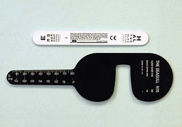
For convenience, the clinician can carry a means of assessing visual acuity for near vision in her or his pocket. For older patients, near acuity must be determined with the use of reading glasses or a pinhole (Fig. 18-1) because the eye’s ability to accommodate declines with age. If a formal means of visual acuity assessment is not available, the clinician can estimate visual acuity using a newspaper or paper currency.

Visual Fields

Visual fields are assessed in patients who have sustained severe head trauma, in those who are aware of a defect in their vision, and in those whose behavior indicates that a visual field defect may be present. Confrontation methods of visual assessment are most commonly used to screen for a visual field defect. However, more sensitive methods must be used if a minor defect is to be detected. We recommend the following strategy.

Binocular Visual Field Testing by Quadrants.

This method is used to test for homonymous visual field defects. The examiner sits opposite the patient at a distance of 1 m (3 feet). The patient is asked to look at the examiner’s eyes. Both hands are placed in the lower outer quadrants and then in the upper outer quadrants. The patient is asked to identify a small movement of the extended forefinger of each hand. The examiner moves each finger in turn and then moves both fingers together. The patient is asked to point at the moving finger (or fingers). A patient with a left homonymous hemianopia, in which the left field of vision in each eye is deficient, will not point to the moving fingers on the left. A patient with a visual inattention defect in that area will not perceive movement when the finger is moved in the outer half of the visual field at the same time. Inattention hemianopia is indicative of unilateral diffuse occipital pathologic conditions.

Assessment of the Central Visual Field to Confrontation.

Traumatic damage to the visual pathways is more likely to cause impairment of the central 30 degrees of the visual field than of the periphery. Therefore, a small target, such as a small red pin, should be used to screen for such defects. The examiner sits opposite the patient and closes one eye. The examiner asks the patient to cover her or his corresponding eye with the palm of the hand and to fixate on the examiner’s open eye. The red target is introduced from the periphery to the center along a coronal plane halfway between the examiner and the patient. The patient is instructed to say “now” as soon as he or she becomes aware of the head of the pin. The examiner is specifically looking for a quadrantic field loss; therefore, the pin is introduced into the fields along the oblique meridians (if the examiner tests only in the horizontal and vertical meridians, she or he may miss the field defect).

To determine the sensitivity of the technique, the examiner checks the position and dimensions of the blind spot by placing the target in his or her own blind spot. The examiner’s blind spot should correspond approximately to that of the patient.

Subjective Visual Field Assessment.

Occasionally, all these tests may be normal, but the patient still complains of impaired vision. The examiner sits opposite the patient. The examiner closes one eye and covers the corresponding eye of the patient. The patient is asked to fixate on the examiner’s pupil, and the examiner places the red pin close to the patient’s face in each quadrant of the patient’s visual field adjacent to the examiner’s eye. The patient is asked to compare the colors in each position. In particular, in cases of traumatic chiasmatic damage, the patient is aware of color desaturation in the upper temporal fields, but no other detectable visual field defect may be noted with any of the other methods used.

When a visual field defect is detected, more accurate charting of the defect by perimetry may be necessary to determine the pattern and extent of the defect.

Pupils

If the patient’s visual acuity is reduced and shows no improvement with use of the pinhole, the pupils are tested to seek evidence of an afferent pupillary defect. This occurs in defects in the visual pathway anterior to the chiasm, gross retinal detachment, and traumatic optic neuropathy.

Swinging Flashlight Test.

This test is used to detect a subtle defect caused, for example, by incomplete optic nerve damage. The pupils are illuminated in the same manner, but on this occasion the light is shined into each eye for about 2 seconds and then swung rapidly to illuminate the other eye. For an incomplete right afferent pupillary defect, when the light is shined into the right eye, both pupils constrict. When the light is swung to the left eye, both pupils constrict further. When the light reilluminates the right eye, the pupils return to their previous resting position and dilate slightly. This technique can be used even in the presence of a unilateral third nerve palsy, in which one pupil is poorly reactive or nonreactive. The swinging flashlight test is performed and the size of the contralateral pupil is determined for both its direct and its consensual reflexes. Any difference in size indicates a relative afferent pupillary defect.

For example, a fracture at the right orbital apex may damage the right oculomotor and optic nerves. The right pupil would, therefore, not react directly or consensually because of the oculomotor nerve damage. However, the diameter of the left pupil will be smaller for its direct response than for the consensual response from illuminating the right eye.

Examination for Structural Disorders

Ophthalmoscopy Through Dilated Pupils

This test is indicated for all patients with reduced visual acuity. Tropicamide 1% produces rapid pupillary dilation with little effect on accommodation and with a return to normal within 3 hours. The addition of phenylephrine 10% may be necessary for those patients with pigmented irides the examiner must check first for any history of cardiac dysrhythmia or systemically administered monoamine oxidase inhibitors.

The contraindications to pupillary dilation are as follows:

Although the optic disc can be assessed without dilating the pupil, the central and surrounding retina cannot be adequately examined.

The reader will no doubt be conversant with the normal appearance of the retina (Fig. 18-2) and the use of the direct ophthalmoscope. The following hints may, however, be of value:
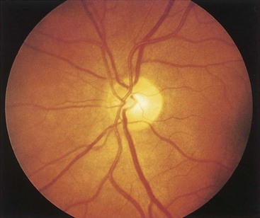
1. The examiner looks through the ophthalmoscope from a 0.33-m (1-foot) distance, examining the red reflex initially. By this means, the examiner can identify any opacities in the media—for example, vitreous hemorrhage or traumatic cataract.

2. The patient is asked to fixate into the distance with the other eye. If the patient focuses for near vision, the examiner will have difficulty in focusing the ophthalmoscope.

3. If a bright light reflex gets in the way, the light is reflecting from the cornea. If the ophthalmoscope is rotated very slightly, the light reflex will diminish or disappear, because it will no longer be reflected back along the examiner’s visual pathway.

4. To observe a scene through a keyhole, the eye needs to be placed close to the keyhole. The same principle applies to ophthalmoscopy—the closer to the patient’s eye is the examiner, the wider the angle of view.

5. To examine the fovea, the patient is asked to look at the light.

6. To examine the peripheral retina, the patient is asked to move her or his eyes in sequence in different directions. When the patient looks up, the examiner is looking at the upper retina as it is brought down into view; the same principle applies to the other positions of gaze.

Examination of Eye Movements

Eye movements are commonly impaired following facial and head injury. It must be remembered, however, that an antecedent squint is not uncommon. Moreover, ptosis, blurred vision as a result of the eye injury, amblyopia, and a history of patching of the eye in childhood all may prevent the patient from experiencing double vision. Eye movements are therefore objectively assessed in all patients who have sustained an injury likely to be complicated by a motility disorder (e.g., a blowout fracture). Figure 18-3 indicates the primary directions of action of each of the extraocular muscles. The eye movements into each of these positions of gaze are examined.

The assessment of eye movements is a skilled procedure. The following strategy is suggested as a means of identifying patients who warrant referral.

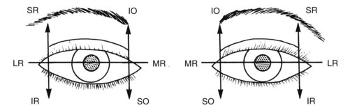
The examiner sits directly opposite the patient and uses a penlight to examine the eye movements. The penlight is moved in a manner similar to that for peripheral visual field testing. The light is held at approximately 0.33 m (1-foot) from the patient. The examiner observes the exact position of the light reflexes on the cornea with respect to the pupil. The patient is asked to follow the light. The light is moved in an arc into each position of gaze, with the light constantly directed at the eyes. The symmetry of the light reflexes and symmetry of the positions of gaze are closely examined (see Fig. 18-3). The skilled observer is able to detect most motility disorders.

The cover-uncover test is performed while the patient fixates on the light in the primary position of gaze and in the positions of gaze in which double vision is experienced and a motility disorder has been detected. An eye occluder or a piece of a card is used. The examiner watches one eye and covers the other one. The eye that the examiner is watching should not move. If the eye does move to look at the light, it is a squinting or deviated eye. The procedure is repeated for the other eye. This method provides an objective means of validating and quantifying the patient’s subjective double vision.

Forced Duction Test.

This test can be performed on a patient with a motility disorder in whom the differential diagnosis between entrapment and muscle weakness is in doubt. Topical local anesthetic (e.g., benoxinate) is instilled into both eyes. The conjunctiva in line with the muscle in question is grasped just adjacent to the corneoscleral junction (the limbus) with a pair of fine-toothed forceps and the globe is gently rotated. The procedure is repeated for the other eye to allow a comparison between both eyes to be made. The force required for rotating the globe is estimated in relation to the normal contralateral eye. Tethering of the globe is indicative of entrapment.

An alternative means of rotating the globe is to use a cotton swab (cotton-tipped applicator) soaked in local anesthetic and to rotate the globe by pressing the swab onto the eye and applying a tangential force. With practice, this method can be equally sensitive and is less likely to cause subconjunctival hemorrhage. However, it should be noted that in our experience, not all patients will tolerate the forced duction test.

Position of the Globe

In every case of facial fracture, the position of the eye should be carefully examined. The eyes may be displaced in any one of three dimensions.

Minor Eye Injuries

Subconjunctival Hemorrhage and Bruised Eyelids

Subconjunctival hemorrhage (Fig. 18-4) with bruised eyelids commonly follows midfacial injury. Blood may track forward from an orbital injury, or bleeding may take place locally. A clear demarcation line to the bruising of the eyelid suggests orbital hemorrhage. Such bruising is usually benign, but it may be related to severe ocular injury. Careful examination of the eye is required in every case.
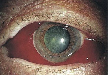
Corneal Abrasion or Corneal Foreign Body

This injury causes severe pain, blurring of vision, photophobia, and lacrimation, except in the presence of corneal anesthesia. Loss of the corneal epithelium may be caused by direct injury to the eye or inadequate eyelid closure as a result of facial palsy, eyelid laceration, or injury during surgery. Alcohol-based skin preparations, incomplete eyelid closure, and accidental injury to the cornea during surgery all must be carefully avoided. When such a lesion is suspected, fluorescein stain allows a diagnosis to be made.

Treatment

After the administration of one drop of topical local anesthetic, which allows clinical examination and gives temporary pain relief, a medium-acting cycloplegic agent such as cyclopentolate (24 hours), which alleviates pain caused by ciliary spasm, and a topical antibiotic such as chloramphenicol are instilled. If a foreign body is present, it is removed. A corneal foreign body is removed with great care, preferably using binocular magnification. The physician can usually lift off a foreign body on the surface of the cornea by using a hypodermic needle held tangentially to the corneal surface, ensuring throughout the procedure that the patient is unable to move forward toward the needle. One drop of a topical nonsteroidal anti-inflammatory preparation is then instilled to alleviate pain.10

Nonperforating Eye Injuries

A blunt injury severe enough to cause an orbital fracture may also damage the eye. Depending on the nature, direction, and force of the injury, any anatomic component of the globe may be disrupted. The effects of blunt injury can be divided into those resulting from distortion and those resulting from concussion. Both types of injury are commonly seen in the same eye.

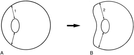
A high-speed anteroposterior force results in marked distortion of the globe (Fig. 18-5). The eye is transiently deformed, with notable distention in the coronal plane and shortening of the anteroposterior dimension. The sclera is inelastic and the aqueous and vitreous cannot be compressed. The iris, ciliary body, zonule of the lens, and peripheral retina may be torn from their insertions and, in severe cases, the sclera may rupture. Distortion of the posterior segment of the eye can result in a tear of the choroid associated with subretinal hemorrhage and, in the most severe case, avulsion of the optic nerve from the globe.

The concussional component of the injury results from a coup-contrecoup effect. The cells of the cornea, lens, retina, and choroid all are susceptible to such injury and may transiently or permanently cease to function.

In this section, the results of injury to each component of the eye are discussed separately. However, almost any combination of injuries can occur, which can occasionally result in disorganization of the structures of the globe (Fig. 18-6).
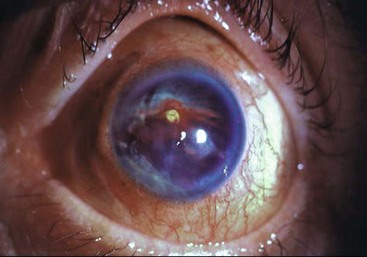
Conjunctiva and Cornea

Swelling of the conjunctiva (chemosis) is common in association with subconjunctival hemorrhage and resolves spontaneously. A tear of the conjunctiva is suggestive of a more severe blunt injury. In every case, internal injury to the globe must be sought.

Loss of the corneal epithelium (Fig. 18-7) is fairly common and causes the same signs and symptoms as a corneal erosion. The corneal endothelium is comprised of a monolayer of cells that probably do not replicate following injury. Their function is to maintain the clarity of the cornea by pumping water out of the cornea and into the anterior chamber. Damage to the corneal endothelium results from a combination of contusion, reactive inflammation, and raised intraocular pressure.11 This condition may culminate in permanent edema if the endothelial cell population is reduced below a critical level. Recovery of corneal clarity can take place in some patients after a number of/>

Only gold members can continue reading. Log In or Register to continue

Stay updated, free dental videos. Join our Telegram channel

Jan 14, 2015 | Posted by in Oral and Maxillofacial Surgery | Comments Off on 18: Ophthalmic Consequences of Maxillofacial Injuries

VIDEdental - Online dental courses

Get VIDEdental app for watching clinical videos