Overview of the Cell
Learning Objectives
• Define and pronounce the key terms in this chapter.
• Outline cell division and describe the phases of mitosis that are involved.
• Describe the extracellular materials surrounding the cell and its intercellular junctions.
New Key Terms
Anaphase (an-ah-faz)
Cell, membrane (sen-tree-ols)
Centrioles (sen-tree-ols)
Centromere (sen-tro-mere)
Centrosome (sen-tro-some)
Chromatids (kro-mah-tids)
Chromatin (kro-mah-tin)
Chromosomes (kro-mah-somes)
Cytoplasm (cy-to-plazm)
Cytoskeleton (site-oh-skel-it-on)
Desmosome (des-mo-som)
Endocytosis (en-do-sigh-toe-sis)
Endoplasmic reticulum (en-do-plas-mik rey-tik-u-lum)
Exocytosis (ek-so-sigh-toe-sis)
Golgi complex (gol-jee)
Hemidesmosome (hem-eye-des-mo-som)
Histology (his-tol-oh-jee)
Inclusions (in-kloo-zhins)
Intercellular junctions (fil-ah-ments)
Intercellular substance (fil-ah-ments)
Intermediate filaments (fil-ah-ments)
Interphase (in-ter-faz)
Lysosomes (li-sah-somes)
Metaphase (met-ah-faz)
Mitochondria (mite-ah-kon-dree-ah)
Mitosis (my-toe-sis)
Microfilaments (my-kroh-fil-ah-ments)
Microtubules (my-kroh-too-bules)
Nuclear envelope (noo-kle-er), pores
Nucleolus (noo-kle-oh-lis)
Nucleoplasm (noo-kle-ah-plazm)
Nucleus (noo-kle-is) (plural, nuclei, noo-kle-eye)
Organ (or-gah-nels)
Organelles (or-gah-nels)
Phagocytosis (fag-oh-sigh-toe-sis)
Prophase (pro-faz)
Ribosomes (ry-bo-somes)
System (tel-oh- faz)
Telophase (tel-oh- faz)
Tissue, fluid (ton-oh-fil-ah-ments)
Tonofilaments (ton-oh-fil-ah-ments)
Vacuoles (vak-you-oles)
The Cell
As an introduction to Unit III, the organization of the body’s histology is discussed in this chapter. Histology is the study of the microscopic structure and function of cells and associated tissue. Another term for histology is microanatomy because the dimensions of the anatomical structures studied are on a microscopic scale (see Appendix B). A dental professional must have a clear understanding of the basic unit of the body, the cell and its components, as well as understanding the larger concepts involved in the histology of tissue, such as those found in the oral cavity. This chapter gives an overview of the cell and its various components, and then Chapter 8 presents a review of basic tissue types in the body. A discussion of the histology of each of the tissue types in the oral cavity follows in later chapters of Unit III.
Anatomy of the Cell
The smallest living unit of organization in the body is the cell, because each cell is capable of performing any necessary functions without the aid of other cells (Figures 7-1 and 7-2, Table 7-1). Each cell has a cell membrane, cytoplasm, organelles, and inclusions. Thus, every cell is a world unto itself, like a gated small city, surrounded by a boundary, having “factories” and other “industries” that make it almost self-sufficient.
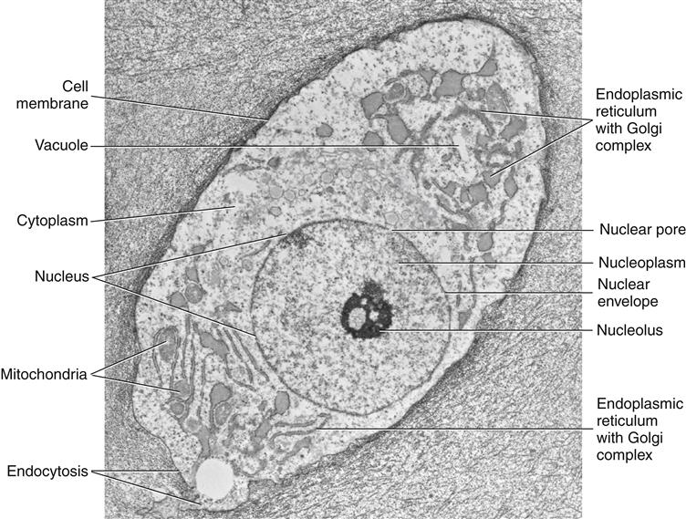
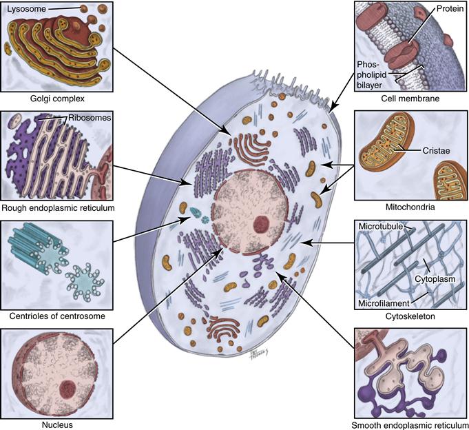
TABLE 7-1
Components of the Body and Examples
| Cell | Smallest living unit of organization: epithelial cell, neuron, myofiber, chondrocyte, osteocyte, fibroblast, erythrocyte, macrophage, sperm |
| Tissue | Collection of similarly specialized cells: epithelium, nervous tissue, muscle, cartilage, bone, connective tissue, blood |
| Organ | Independent body part formed from tissue: skin, brain, heart, liver |
| System | Organs functioning together: central nervous system, respiratory system, immune system, cardiovascular system |
Cells also interact with one another as a city reacts with other cities. Cells with similar characteristics of form and function are grouped together to form a tissue (see Table 7-1), as countries are formed from cities for a common goal. Thus, a tissue is a collection of similarly specialized cells, most often surrounded by extracellular materials. Various tissue types are then bonded together to form an organ, a somewhat independent body part that performs a specific function or functions, similar to organizations formed from countries. Organs can function together as a system.
Cells in a tissue undergo cell division (or mitosis) to reproduce and replace the dead tissue cells. As a result of the division process, two daughter cells that are identical to each other and to the original parent cell are formed. This process consists of different phases, which are discussed later with regard to different components of the cell.
However, cells also interact with the extracellular environment in many ways. Cells can perform exocytosis, which is an active transport of material from a vesicle within the cell out into the extracellular environment. Exocytosis occurs when there is fusion of a vesicle membrane with the cell membrane and subsequent expulsion of the contained material.
The uptake of materials from the extracellular environment into the cell is endocytosis. E/>
Stay updated, free dental videos. Join our Telegram channel
VIDEdental - Online dental courses


