Head and Neck Structures
Learning Objectives
• Define and pronounce the key terms in this chapter.
New Key Terms
Acinus (plural, acini) (as-i-nus, as-i-ny): serous (sere-us), mucoserous (mu-ko-sere-us), mucous (mu-kis)
Capsule (kap-sule)
Cells: goblet, mucous (mu-kis), myoepithelial (my-oh-epee-thee-lee-al), secretory (sek-kre-tory)
Colloid (kol-oid)
Duct: excretory (ex-kreh-tor-ee), intercalated (in-turk-ah-lay-ted), striated (stri-ate-ed), thyroglossal (thy-ro-gloss-al)
Erectile tissue (e-rek-tile)
Follicles (fol-i-kls)
Germinal center (jurm-i-nil)
Gland: endocrine (en-dah-krin), exocrine (ek-sah-krin)
Goiter (goy-ter)
Hilus (hi-lus)
Hyposalivation (hi-po-sal-i-vay-shen)
Lobes (lob-ules)
Lobules (lob-ules)
Lumen (loo-men)
Lymph (limf)
Lymphadenopathy (lim-fad-uh-nop-ah-thee)
Lymphatic ducts (lim-fat-ik), nodules (nah-jools), vessels
Lymphatics (lim-fat-iks)
Mucosa: olfactory (ol-fak-tor-e), respiratory
Mucocele (mu-kah-sele)
Nasal cavity (nay-zil kav-it-ee), conchae (kong-kay)
Nicotinic stomatitis (nik-ah-tin-ik sto-mah-ti-tis)
Paranasal sinuses (pare-ah-na-zil sy-nuses)
Pharyngeal tonsils (fah-rin-je-il)
Ranula (ran-u-lah)
Saliva (sah-li-vah)
Salivary glands (sal-i-ver-ee): major, minor
Septum (plural, septa) (sep-tum, sep-tah)
Serous cells (sere-us), demilune (dem-ee-lune)
Sinusitis (sy-nu-si-tis)
Thyroxine (thy-rok-sin)
Tonsillar tissue (trah-bek-u-lay)
Trabeculae (trah-bek-u-lay)
Vessels: afferent (af-er-int), efferent (ef-er-ent)
von Ebner’s salivary glands (von eeb-ners sal-i-ver-ee)
Xerostomia (zer-oh-sto-me-ah)
Head and Neck Structures
Dental professionals must have a clear understanding of the histology and prenatal development concerning not only the oral cavity but also of the associated head and neck structures. The clinical functioning of the head and neck structures is related to the underlying histology. In addition, many pathological lesions that are encountered in the oral cavity can be associated with changes in these associated structures of the head and neck, and thus are reflected in changes in their underlying histology. The head and neck structures to be discussed include the salivary glands, thyroid gland, lymphatics, nasal cavity, and paranasal sinuses.
Glands
A gland is a structure that produces a secretion necessary for normal body functioning. An exocrine gland is a gland having a duct associated with it. A duct is a passageway that allows the glandular secretion to be emptied directly into the location where the secretion is to be used. An endocrine gland is a ductless gland with its secretions conveyed directly into the blood, and then carried to some distant location to be used. Motor nerves associated with both types of glands help regulate the flow of the secretion. Sensory nerves are also present in the gland.
Salivary Glands
The salivary glands produce saliva, or “spit.” Saliva contains immunoglobulins (secretory IgA), minerals, electrolytes, buffers, enzymes, and metabolic wastes. The secretion by these glands is controlled by the autonomic nervous system. Saliva lubricates and cleanses the oral mucosa, protecting it from dryness and potential carcinogens. This secretory product also helps in digestion of food by enzymatic activity. Additionally, it serves as a buffer, protecting the oral mucosa against acids from food and dental biofilm; it is also involved in antibacterial activity.
Finally, saliva helps maintain tooth integrity, because it is involved in remineralization of the tooth surface. However, because it contributes to the formation of the pellicle on the tooth and mucosal surfaces, saliva is also involved in the first step in dental biofilm formation. It also supplies the minerals for supragingival calculus formation.
Salivary glands are classified as either major or minor, depending on their size, but both types have similar histological features. Further, both the major and minor salivary glands are exocrine glands, and thus have associated ducts that help convey the saliva directly into the oral cavity, where it is used.
Histology of Salivary Glands
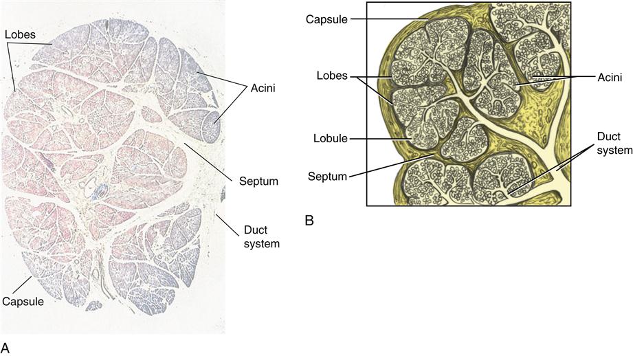
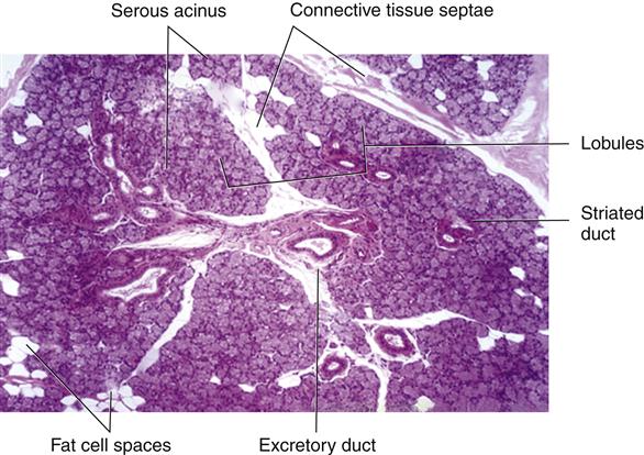
Both major and minor salivary glands are composed of both epithelium and connective tissue (Figure 11-1). Epithelial cells both line the ducts and produce the saliva. Connective tissue surrounds the epithelium, protecting and supporting the gland. The connective tissue of the gland is divided into the capsule, which surrounds the outer part of the entire gland, and the septa. Each septum (plural, septa) helps divide the inner part of the gland into the larger lobes and smaller lobules. Both the capsule and septa carry nerves and blood vessels that serve the gland.
Secretory Cells and Acini
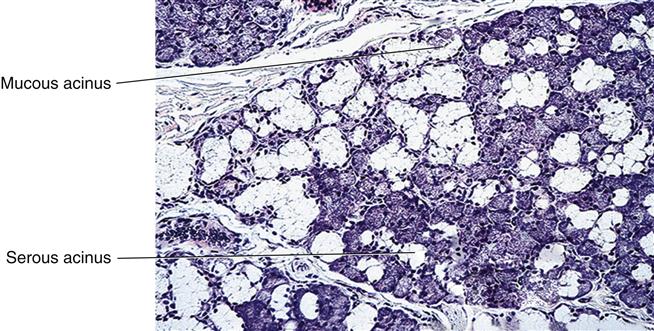
Epithelial cells that produce the saliva are the secretory cells (Figure 11-2). The two types of secretory cells are classified as either mucous or serous cells, depending on the type of secretion produced. Mucous cells have a cloudier-looking cytoplasm and produce mucous secretory product. In contrast, serous cells have a clear cytoplasm and produce serous secretory product. A combination of secretory cells present in the gland can produce a mixed secretory product. In some glands, one type of cell predominates so that the product is either more mucous or more serous than if it had a range of both cell types.
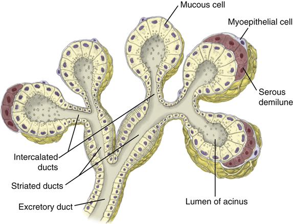
Secretory cells are found in a group, or acinus (plural, acini), which resembles a cluster of grapes. Each acinus is located at the terminal part of the gland connected to the ductal system, with many acini within each lobule of the gland. Each acinus consists of a single layer of cuboidal epithelial cells surrounding a lumen, a central opening where the saliva is deposited after being produced by the secretory cells.
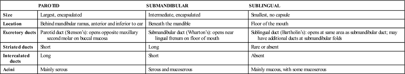
The three forms of acini are classified in terms of the type of epithelial cell present and the secretory product being produced. The major and minor salivary glands have different types of acini (Table 11-1). However, the different types of acini are often difficult to classify on histological sections of the glands.
TABLE 11-1
Comparison of Major Salivary Glands
| PAROTID | SUBMANDIBULAR | SUBLINGUAL | |
| Size | Largest, encapsulated | Intermediate, encapsulated | Smallest, no capsule |
| Location | Behind mandibular ramus, anterior and inferior to ear | Beneath the mandible | Floor of the mouth |
| Excretory ducts | Parotid duct (Stenson’s): opens opposite maxillary second molar on buccal mucosa | Submandibular duct (Wharton’s): opens near lingual frenum on floor of mouth | Sublingual duct (Bartholin’s): opens at same area as submandibular duct; may have additional ducts at submandibular folds |
| Striated ducts | Short | Long | Rare or absent |
| Intercalated ducts | Long | Short | Absent |
| Acini | Mainly serous | Serous and mucoserous | Mainly mucous, with some mucoserous |
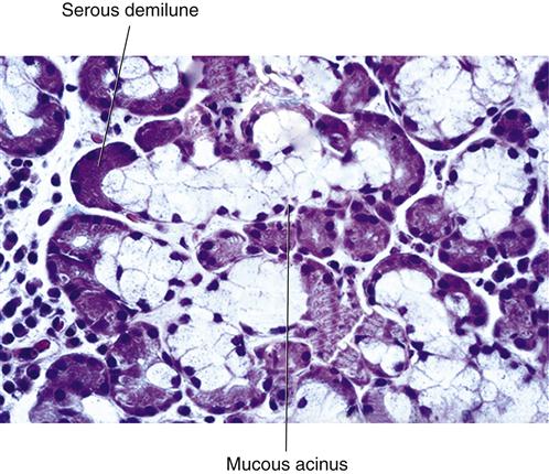
Serous acini are composed of serous cells producing serous secretory product and have a narrow lumen (Figure 11-3). In contrast, mucous acini are composed of mucous cells producing mucous secretory product, and their lumen is wider. Mucoserous acini have both a group of mucous cells surrounding the lumen and a serous demilune, or “bonnet” of serous cells superficial to the group of mucous secretory cells (Figures 11-4 and 11-5). Because the mucoserous acini contain both types of secretory cells, they produce a mixed secretory product. However, the major distinctions between serous and mucous cells have become less important with additional studies of their cellular functioning.
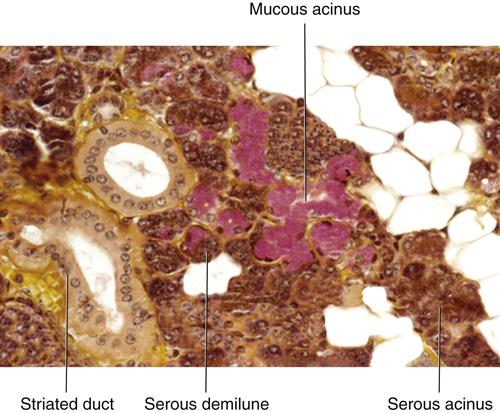
To facilitate the flow of saliva out of each lumen into the connecting ducts, myoepithelial cells are located on the surface of some of the acini, as well as on parts of the ductal system, the intercalated ducts (Figure 11-6). Each myoepithelial cell consists of a cell body with four to eight cytoplasmic processes radiating outward. They are specialized cells of epithelium that resemble an octopus on a rock; they are situated on the surface of the acini and have a contractile nature.
When these cells contract, they squeeze the acinus, forcing the saliva out of the lumen and into the connecting duct. When />
Stay updated, free dental videos. Join our Telegram channel
VIDEdental - Online dental courses


