Chapter 7
Management of Oral Precancer
Having established both a clinical and then a histopathological diagnosis for an oral potentially malignant disorder in a patient, one of the principal difficulties in determining a management strategy is the lack of agreed and defined treatment protocols. Treatments are rarely evidence-based and are strongly influenced by individual clinician preferences and skill. There is a lack of clinically relevant randomised controlled trials, and most evidence is based upon cohort or observational studies.
Similarly, there are no universally agreed objectives of treatment interventions, although it is sensible to assume that there is a consensus view that prevention of oral cancer is a fundamental priority. The natural history of oral precancer remains ill defined and unpredictable, however, so that uncertainty inevitably haunts clinical management decisions. It is thus important for us to define pertinent management goals in attempting to treat oral precancer and a proposed summary of these are listed in Box 1.1. We have already seen that accurate diagnosis may not always be straightforward, but the ability to recognise the presence of early malignant change within lesions, together with the removal of dysplastic tissue and the prevention of further precancer disease, should be regarded as essential prerequisites for a successful management technique.
As detailed in Chapter 6, it is very important to ensure that we obtain as accurate and definitive a histopathological diagnosis for precancer patients as possible and it now seems clear that excision of precancer lesions in their entirety is necessary to establish this. Small, incisional biopsies for obvious reasons may not always be truly representative of the entire potentially malignant disorder [1, 2].
Whilst formal surgical excision is carried out in order to remove all clinically apparent dysplastic tissue, it is extremely important to note that in nearly 10% of excised precancer lesions, microinvasive or frankly early invasive carcinomas are also identified unexpectedly on histological examination, presumably at a pathological stage when otherwise clinically undetectable [3]. It is thus obvious that, whilst the principal dilemma in oral precancer management strategies has always been whether to observe patients or to intervene and offer treatment, the recognition that a truly definitive diagnosis requires excision biopsy of the entire visible lesion places the emphasis in modern treatment protocols firmly upon surgical intervention.
In this chapter we will review the management techniques that have been proposed for oral precancer treatment (Box 7.2). We will also discuss in some detail our preferred option of interventional laser surgery, which appears to be both an appropriate diagnostic tool and an efficacious treatment modality.
Box 7.1 Management goals in treating oral precancer.
- Accurate diagnosis
- Early recognition of malignancy
- Removal of dysplastic mucosa
- Prevention of recurrent or further disease
- Prevention of malignant transformation
- Minimal patient morbidity
Box 7.2 Treatment modalities for oral precancer.
- Risk factor identification and elimination
- Clinical observation
- Medical treatment
- Surgical excision
It is difficult to obtain meaningful figures for malignant transformation rates for precancers in the current literature, with quoted figures for the development of oral squamous cell carcinoma varying between 0.13% and 36.4% [3]. For leukoplakia, which is worldwide the commonest oral potentially malignant disorder, a figure of around 1% has been quoted as an overall annual transformation rate [1]. Widely disparate data such as these clearly have very limited use in discussing prognosis and behaviour with individual patients and certainly do not help in dealing with the myriad of potentially malignant lesions that present in the clinical situation.
Of more pragmatic benefit, therefore, is to try to identify which patients are at ‘high risk’ of malignant disease compared with those more likely to be ‘low-risk’ individuals. High-risk lesions, in other words those demonstrating severe dysplasia or carcinoma in situ, have been quoted to progress to cancer in up to 80% of cases. Low-risk lesions, showing at worst only mild dysplasia, tend to progress in less than 15% of cases.
As we have seen, however, such risk stratification is more difficult than might appear obvious at first sight. This is because a ‘high-risk’ patient is not just simply an older individual with a long history of smoking and alcohol use presenting, for example, with severely dysplastic leukoplakia in the floor of the mouth. An unpredictable ‘high risk’ may also affect young patients, particularly those with no demonstrable risk factor behaviour and can apply equally to mucosal lesions demonstrating sometimes quite minimal dysplasia.
There remains a surprising level of ignorance in the general population regarding mouth cancer and its causes. Any management protocol must therefore begin with education of patients about the aetiology of oral cancer and the importance of both recognising and then eliminating risk factors. In the vast majority of cases, of course, this relates primarily to tobacco and alcohol use, and it is disheartening how often patients fail to stop smoking or reduce their alcohol intake following diagnosis and treatment [4, 5].
Smoking Cessation
Tobacco smoking, primarily of cigarettes, is well documented as the principal aetiological agent in oral cancer development and there is a recognised dose–response relationship between increasing cancer risk and the amount and duration of tobacco use. Patients can usually estimate the number of cigarettes or amount of tobacco they smoke daily, although it is likely that they may underestimate this, either consciously or quite often subconsciously, when self-reporting to clinicians.
There is evidence that brief health care interventions can be effective in educating patients about the risks of tobacco smoking, and we have clearly shown that formal stopping smoking regimes can be very useful in precancer patients. However, these interventions are most effective when dedicated smoking cessation advisers attend clinics as members of the specialist dysplasia team [4]. Patients who try to stop smoking without formal cessation advice, who do not use nicotine replacement therapy to prevent acute withdrawal symptoms, or who have no regular contact with a dedicated cessation advisor are statistically highly unlikely to achieve long-term quitting.
It is salutary to note that nearly three-quarters of smoking patients treated for potentially malignant disorders remain as persistent smokers 5 years after their initial diagnoses. This not only risks further precancerous lesion development but also puts them at increased risk of malignant transformation [4].
In addition to tobacco smoking, as we have already seen, the use of smokeless tobacco is also an important risk factor but varies considerably worldwide, being particularly prominent in India and the southern states of the USA. In some parts of Sweden and Norway it is used as snus, which is a moist tobacco that is held under the upper lip and then sucked. In the UK smokeless tobacco is primarily seen as the classic betel quid in Asian communities and is composed of chewing tobacco mixed with areca nut, slaked lime and betel. People who use smokeless tobacco absorb similar amounts of nicotine and are as equally dependent as smokers. Advice on stopping the use of smokeless tobacco, behavioural therapy and the treatment of nicotine withdrawal symptoms are all thus equally important as treatment modalities in these patients as in their smoking counterparts [6].
Alcohol Counselling
Assessing and then attempting to modify risk factor behaviour is integral to any management protocol for potentially malignant disorders. In relation to alcohol use it is complicated by patients’ subjective and potentially unreliable estimates of self-reported intake and is also, of course, confounded by the concomitant use of tobacco [5]. Whilst it is undoubtedly true that most patients presenting with oral potentially malignant lesions are either current or ex-smokers, there is a small minority who have never smoked.
Controversy exists about whether alcohol alone increases precancerous transformation, but there is clear evidence that alcohol availability and consumption are both rising in most populations worldwide. It is probably within cohorts of patients who have never smoked that the role of alcohol is most significant. This effect of alcohol, however, is probably most likely to affect patients who consume alcohol both regularly and excessively. It is most likely that it is the overall amount that is drunk rather than the use of any individual type of alcohol that is most relevant.
Separating the complex interactions and observed synergistic effects between tobacco and alcohol use in oral precancer patients is probably impossible and the reality is such that the majority of patients usually consume both.
Attempts to establish more objective estimates of alcohol use may, however, be of value in precancer management. We investigated a cohort of 54 smokers presenting with new, single dysplastic oral lesions and demonstrated that an objective mean corpuscular red blood cell volume greater than 100 fl, as recorded in routine preoperative blood tests, corresponded with both a patient self-reporting of alcohol intake in excess of 28 units per week, and also an increased severity of dysplasia seen in their presenting oral lesions [5]. How successful individual patient counselling and advice to reduce alcohol intake is in influencing oral precancer outcomes is difficult to assess. However, it was disappointing to note that all 54 patients in our cohort study continued to drink alcohol following interventional laser treatment. Indeed, we also observed a significantly increased risk of further precancer disease developing in patients who continued with a high level of alcohol intake post-surgery [5].
Nonetheless, identification of patients with excessive alcohol intake offers important opportunities for education and health care interventions and should be encouraged in all potentially malignant disorder cases.
Modification of Other Risk Factors
In non-smoking patients and those with low alcohol consumption, the importance and relevance of other risk factors may require further elucidation. The type of risk factors requiring attention here include deficiencies in dietary intake of fresh fruit and vegetables, a possible influence for human papillomavirus (HPV) and oral sexual behaviour, together with a number of background medical conditions, such as immunosuppression, all of which may influence both dysplasia development and disease progression.
It may not always be possible to reduce the significance or alter the course of these rather disparate disease influences, but it is certainly important to be able to identify them and recognise their relevance to individual patient care pathways.
Observation Versus Intervention
Traditionally, clinical observation protocols – which essentially comprised initial lesion recognition and diagnosis, photography, routine oral inspection and patient monitoring – have been the mainstay of oral potentially malignant disorder management. Unfortunately this has led in many cases to, effectively, a passive observation of cancer development in a previously identified precancer patient. This seems both self-defeating and highly inadequate as a 21st century patient care pathway. Thus, it is hardly surprising that interventional management protocols have developed to try to prevent such disastrous transformations and most authors now recommend active treatment, rather than clinical observation, for all oral precancer lesions [1].
A period of clinical observation may, however, be appropriate for patients with low-grade dysplasia who stop smoking, who are prepared to address other relevant risk factor behaviours, and who are willing and able to attend for regular clinical review. It is recognised that in many cases lesions may improve and regress following smoking cessation, but it is important that this is carefully monitored.
Failure of precancer lesions to resolve may mean that the patient has actually been unable to stop smoking or, perhaps of more significance, the recognition of persistent disease in the absence of smoking may indicate irreversible dysplastic change. As a result this may well be an important patient subgroup in which to reconsider surgical intervention.
In the absence of appropriate randomised controlled clinical trials we retrospectively reviewed two of our patient cohorts over a 3-year period. One cohort was a laser excision group of 78 patients exhibiting high-grade single dysplastic lesions and the other an observed group of 39 patients with low-grade dysplasia. During the follow-up period 64% of laser patients showed complete clinical resolution of their disease whilst, perhaps unsurprisingly, only 23% of observed only lesions resolved [7].
Whilst the above is a useful comparative, observational ‘snap shot’, there is a clear need for proper randomised controlled trials with long-term patient follow up to investigate such treatment decisions and resultant clinical outcomes in more detail.
A variety of medical interventions have been attempted through the years to treat precancer lesions, utilising both local and systemic chemopreventive agents. These have included the use of carotenoids, vitamins A, C and E, bleomycin and more recently cyclo-oxygenase inhibitors. The types of drugs that have been utilised, their proposed mechanisms of action and any recognised side effects consequent upon their use are summarised in Table 7.1.
Table 7.1 Medical treatments in oral precancer management.
| Agent | Mode of action | Side effects |
| Carotenoids | ||
| Beta carotene (vitamin A precursor) | Antioxidant | Yellow skin discolouration, headaches, muscle pain |
| Lycopene | Antioxidant | None |
| Vitamins | ||
| Ascorbic acid (vitamin C) | Antioxidant | None |
| Alpha tocoferol (vitamin E) | Antioxidant | None |
| Retinoic acid (vitamin A): 13-cis-retinoic acid | Keratin production; epithelial cell growth and differentiation; collagen matrix production | Dermatitis, teratogenic, headaches, muscle pain, xerostomia, dizziness |
| Isotretinoin | ||
| Tretinoin | ||
| Fenretinide (vitamin A analogue) | Apoptosis induction | None |
| Bleomycin | Cytotoxic antibiotic | Stomatitis, alopecia, skin pruritus and vesiculation |
| Ketorolac | NSAID | Gastroduodenal irritation |
| Celecoxib | Selective COX-2 inhibitor | Cardiovascular thrombosis |
| Tea/green tea | Anti-angiogenesis | Insomnia, nervousness |
| NSAID, non-steroidal anti-inflammatory drug. | ||
Systematic reviews of medical interventions for oral precancer have, unfortunately, proved unhelpful in determining realistic management strategies and very few relevant randomised controlled trials exist in the literature [8]. It is also disheartening to note that very little benefit has ever been shown from such medical treatments. There is no evidence, for example, of any effective long-term clinical resolution, no significant prevention of malignant transformation of lesions and no reduction in either disease incidence or recurrence compared with observation alone or placebo treatment [8–10].
Furthermore, many of the clinical trials undertaken were carried out on small numbers of patients over short study durations, usually with cases followed up for only a few months rather than years post treatment, which is necessary to identify relevant malignant transformation risks. Where clinical improvement was noted during the trial, lesions often worsened again on cessation of therapy and, because no clear definitions of treatment goals or end points were established, the overall significance of many of these treatments remains unclear. Of similar concern was the observation that numerous side effects of medical treatments were common (Table 7.1). These, to a greater or lesser extent, affected the majority of study participants, sometimes interfering with compliance and prompting subjects to withdraw from treatments.
Finally, and perhaps of most significance, is the fact that all pre-treatment diagnoses relied entirely upon incisional biopsy histopathological classification, which we now recognise, of course, as only a provisional diagnosis. As whole lesion examination is deemed necessary for definitive histopathological diagnosis, it is not unreasonable to view medical treatments as fundamentally flawed therapies applied to diseased mucosa in the absence of definitive pathological diagnoses [1, 2].
Photodynamic therapy (PDT) is a particularly specialised and emerging form of medical intervention, which relies on cellular destruction by a cold photochemical reaction following activation of a photosensitising drug, such as aminolevulinic acid or temoporfin, by low-power visible light. This technique has been advocated by some as a non-invasive oral precancer treatment although it remains primarily a tool for palliative treatment of advanced squamous cell carcinoma of the head and neck. A number of observational studies have reported variable success rates using PDT to treat oral leukoplakia. However, there has been inconsistent reporting regarding recurrent disease and to date only limited, between 3 and 6 months, or no follow-up data [10].
There thus remains a fundamental lack of randomised controlled trials for non-surgical treatment of oral potentially malignant disorders. Those that do exist in the literature show no satisfactory evidence of effective treatment of individual precancer lesions or prevention of oral malignancy.
One feature that is agreed by most authors, and which is really central to oral precancer management, is that regular patient follow up and repeated detailed clinical examination remains mandatory for all potentially malignant disorder patients irrespective of the mode of proposed treatment [10].
Surgical treatment of oral potentially malignant disorders is clearly designed to prevent malignant transformation by removing lesions and thus freeing patients from the risk of cancer at affected sites. Critics of surgical intervention observe that not only is there little evidence to support the above hypothesis, but that no randomised controlled trials of surgical intervention, particularly one that includes a no treatment or placebo arm, have ever been carried out [8]. Nonetheless, surgery remains the first choice in oral precancer management by most specialists, especially for lesions exhibiting high-grade dysplasia [11, 12].
Initial surgical interventions relied upon scalpel excision and primary closure of defects and mucosal or split skin grafting for larger defects. Neither proved popular with patients or clinicians, primarily due to localised postoperative contracture and deformity and the frequent failure of skin grafts to take in the oral environment. Management of widespread, multifocal disease with such techniques was virtually impossible.
Cryotherapy is a specialised technique involving the localised destruction of diseased tissue by the surgical application of extreme cold usually via liquid nitrogen. This has been popular in the past for treating oral lesions but is less commonly used today and is actually a highly unsatisfactory technique which is not recommended for precancer or cancer treatment. Not only does significant postoperative pain and swelling result from the tissue damage consequent upon cryotherapy, but also potentially malignant lesions are rarely successfully or completely destroyed and, of course, no excision biopsy is undertaken to establish a definitive diagnosis. This potentially leaves partially treated dysplastic tissue in situ, which may then be ‘stimulated’, presumably by extensive tissue damage, to more aggressive clinical behaviour. Interestingly, both high recurrence and increased malignant transformation rates have been reported following the use of cryotherapy for precancer, which supports these concerns [9].
Interventional Laser Surgery
Interventional laser therapies have evolved following demonstrable failure of observational or medical therapies and the limitations of conventional surgery in treating oral precancer disease. A number of cohort studies have demonstrated an important role for laser surgery as both a diagnostic tool and a treatment modality in the management of potentially malignant disorders [2, 13, 14].
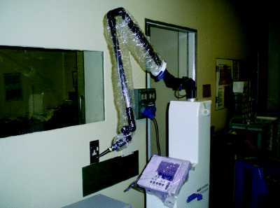
The term laser is an acronym for ‘light amplification by stimulated emission of radiation’. The laser device essentially emits a monochromatic, coherent wave of light energy that is then delivered to the target tissue via fibreoptic systems, hollow waveguides or a series of articulated arms and mirrors. Figure 7.1 illustrates the laser device and articulated arm system of a standard carbon dioxide (CO2) surgical laser, as seen prepared for use in the operating theatre. This type of laser uses sealed CO2 gas as the active medium to generate laser light in the mid-infrared range at 10 600 nm. As this is near the spectroscopic absorption peak for water, all oral soft tissues successfully interact with the CO2 laser beam.
Figure 7.1 Carbon dioxide laser prepared for use in the operating theatre, demonstrating the articulated arm arrangement linked by freely moveable joints and containing precisely aligned mirrors that maintain the laser beam in a central position until it reaches the handpiece.
A photothermal reaction occurs when laser light interacts with the target tissue. Between 60 and 100°C, this produces coagulation which facilitates either localised haemostasis or tissue necrosis. At and above 100°C, when water boils, there is vapourisation which allows the surgeon to incise tissue, and to either resect or ablate lesions.
Safety regulations govern the use of surgical lasers and include wearing wavelength-specific protective eyewear, using a high-volume smoke evacuation system and restricting access to the laser surgery area.
Introduction of the CO2 laser to oral surgery practice in the 1970s revolutionised interventional therapy allowing convenient, effective and reproducible treatments. Box 7.3 lists a number of specific advantages of laser surgery. With preserved function of oral tissues and the ability to re-apply laser therapy to previously treated areas, CO2 laser surgery is now the preferred treatment in many medical institutes.
Box 7.3 Advantages of interventional laser surgery.
- Rapid, precise tissue dissection and lesion excision
- Minimal damage to adjacent normal tissue
- Haemostasis
- Postoperative analgesia
- Definitive histopathological diagnosis
- Low morbidity and minimal swelling with low infection rates
- Rapid healing
- Reduced oral mucosal scarring and contracture
- Excellent patient acceptance
- Facilitates multiple or repeated treatment
In recent />
Stay updated, free dental videos. Join our Telegram channel
VIDEdental - Online dental courses


