Enamel
Learning objectives
■ Discuss how these affect the permeability of enamel.
■ Discuss the surface characteristics and the etching of enamel.
Key terms
Enamelin
Gnarled enamel
Hunter-Schreger bands
Hydroxyapatite
Imbrication lines
Lines of Retzius
Microlamellae
Neonatal line
Perikymata
Prismless enamel
Spindles
Striae of Retzius
Tufts
Overview
Enamel, the hard protective substance that covers the crown of the tooth, is the hardest biologic tissue in the body. It consequently is able to resist fractures during the stress of mastication. Enamel provides shape and contour to the crowns of teeth and covers the part of the tooth that is exposed to the oral environment.
Enamel is composed of interlocking rods that resist masticatory forces. Enamel rods are deposited in a keyhole shape by the formative ameloblastic cells. Groups of ameloblasts migrate peripherally from the dentinoenamel junction as they form these rods. Ameloblasts take variable paths, which produces a bending of the rods. These cells maintain a relationship as they travel in different directions and produce adjacent rods. The enamel rod configuration viewed in incidental light appears as light and dark bands of rod groups termed Hunter-Schreger bands. Because these rods bend in an exaggerated, twisted manner at the cusp tips, they are called gnarled enamel.
All enamel rods are deposited at a daily appositional rate or increment of 4 μm. Such increments are noticeable, like rings in a cross section of a tree, and appear as dark lines known as striae of Retzius or lines of Retzius. The growth lines become apparent on the surface of enamel as ridges, known as perikymata. Two structures are noticeable at the dentinoenamel junction: spindles, the termination of the dentinal tubules in enamel, and tufts, hypocalcified zones caused by the bending of adjacent groups of rods.
Because enamel is composed of bending rods, which in turn are composed of crystals, minute spaces or gaps exist where crystals did not form between rods. This feature causes enamel to be variable in its density and hardness. Therefore some areas of enamel may be more prone to penetration by small particles. This characteristic leads to tooth destruction by dental caries. After enamel is completely formed, no more enamel can be deposited.
 CLINICAL COMMENT
Perikymata are surface manifestations of the incremental lines usually found at the cervix of the crown. Some perikymata are more prominent and present difficulties to the novice clinician, who may confuse them with calculus.
Physical properties
Because enamel is very hard, it is also brittle and subject to fracture. Fracture is especially likely to occur if the underlying dentin is carious and has weakened the enamel’s foundation.
Enamel is about 96% inorganic mineral in the form of hydroxyapatite and 4% water and organic matter. Hydroxyapatite is a crystalline calcium phosphate that is also found in bone, dentin, and cementum. The organic component of enamel is the protein enamelin, which is similar to the protein keratin that is found in the skin. The distribution of enamelin between and on the crystals aids enamel permeability. Enamel is grayish white but appears slightly yellow because it is translucent, and the underlying dentin is yellowish. Enamel ranges in thickness from a knifelike edge at its cervical margin to about 2.5 mm maximum thickness over the occlusal incisal surface.
 CLINICAL COMMENT
Although enamel is the hardest tissue in the human body, it is permeable to some fluids, bacteria, and bacterial products of the oral cavity. Enamel exhibits cracks, checks, and microscopic spaces within and between rods and crystals, allowing penetration.
Rod structure
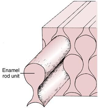
Enamel is composed of rods that extend from their site of origin, at the dentinoenamel junction, to the enamel outer surface (Fig. 7-1). Each rod is formed by four ameloblasts. One ameloblast forms the rod head; a part of two ameloblasts forms the neck; and the tail is formed by a fourth ameloblast. Fig. 7-2 shows the six-sided design that is the shape of the ameloblast in contact with the forming keyhole- or racquet-shaped rod, which is columnar in its long axis. The head of the enamel rod is the broadest part at 5 μm wide, and the elongated thinner portion, or tail, is about 1 μm wide. The rod, including both head and tail, is 9 μm long. The enamel rod is about the same size as a red blood cell (Fig. 7-3).
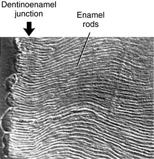
This figure is possible because the section is etched and viewed with a scanning electron microscope. (From Avery JK: Oral development and histology, ed 3, Stuttgart, 2002, Thieme Medical.)
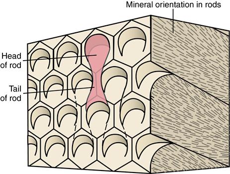
Parts of four cells form each enamel rod. Crystal orientation of three rods can be seen on the right side of the model. (From Avery JK: Oral development and histology, ed 3, Stuttgart, 2002, Thieme Medical.)
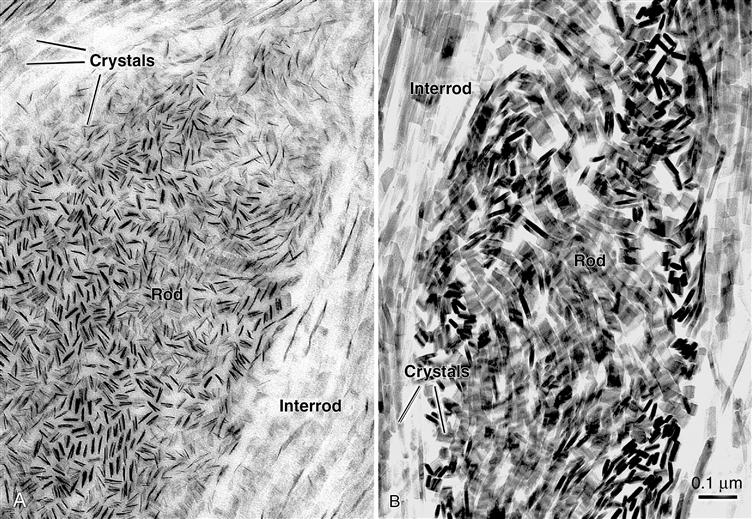
Each rod is filled with crystals. Those in the head follow the long axis of the rod, and those in the tail lie in the cross axis to the head (Figs. 7-4 and 7-5). The upper right rod head of Fig. 7-4 indicates how the mineral is oriented during the rod’s development, which forms the rod head and tail as seen on the left side of the figure. The architecture of the mineral orientation is complex, especially when viewed in any direction other than cross section (see Fig. 7-5).
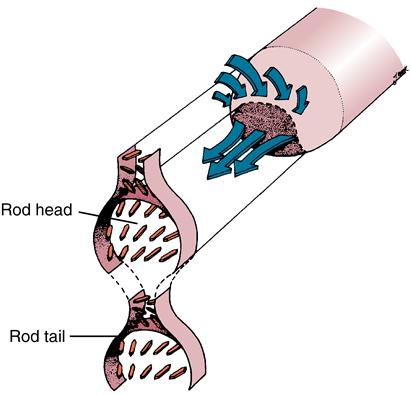
The right part shows how forming crystals pack in the rod from the cell complex (arrows).
 CLINICAL COMMENT
Enamel rods interlock to prevent fracture and splitting of the tooth. Enamel rod groups also intertwine, thereby preventing separation. The rod direction in the crown is normally perpendicular to the incisal surface, which provides additional support in preventing fracture.
Rods form nearly perpendicular to the dentinoenamel junction and curve slightly toward the cusp tip. This unique rod arrangement also undulates throughout the enamel to the surface. Each rod interdigitates with its neighbor, the head of one rod nestling against the necks of the rods to its left and right (see Fig. 7-3). The rods run almost perpendicular to the enamel />
Stay updated, free dental videos. Join our Telegram channel
VIDEdental - Online dental courses


