7: Dental Anomalies

Dental Anomalies

An anomaly is defined as something that is noticeably different or that deviates from the ordinary or normal. Dental anomalies are deviations of dental tissue origin and therefore are derived from the dental tissues enamel, dentin, or cementum. Anomalies can be extreme variations or just slight deviations. They can be caused by a multitude of things or by just one small variation in the environment.

Some abnormalities result from intrinsic factors such as heredity, metabolic dysfunction, or mutations; other causes are extrinsic such as physical or chemical trauma, biologic agents, nutritional deficiencies, stress, habits, or environmental conditions. In many instances anomalies result from a combination of intrinsic and extrinsic factors.

If a condition occurs because of an individual’s genetic makeup, the condition is termed hereditary. If the condition occurs at or before birth, it is termed congenital. A congenital condition is sometimes the result of heredity, and sometimes a hereditary condition does not become evident until years after birth. If a condition exhibits some evidence of an inherited tendency but such evidence is inconclusive, it is often referred to as a familial tendency. If a condition results during the formation and development of a dental structure, it is referred to as a developmental anomaly.

CLASSIFICATION OF DENTAL ANOMALIES

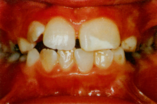
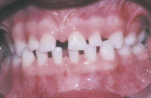
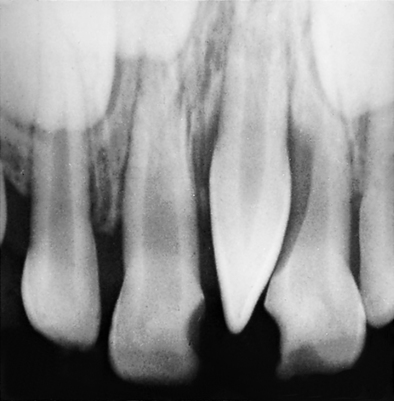
Anomalies resulting in a variation in the size of teeth are called macrodontia, which is when teeth are too large, (Fig. 7-1) and microdontia, which is when teeth are too small (Fig. 7-2).

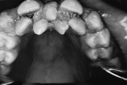
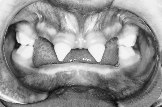
Anomalies resulting in a variation in the number of teeth are hyperdontia (multiple or extra teeth) (Fig. 7-3) and anodontia (too few teeth) (Fig. 7-4). Total anodontia exists if no teeth are present at all, and partial anodontia is if less than the normal number of teeth are present. True anodontia is the congenital absence of teeth. It may involve the permanent dentition, the primary dentition, or both. If the primary teeth are congenitally missing, their permanent replacements will also be absent.

The most commonly missing permanent teeth are the third molars, and the maxillary thirds are absent more often than the mandibular. The next most likely teeth to be missing are the permanent maxillary lateral incisors. Between 1% and 2% of the population are missing at least one permanent maxillary lateral incisor. The third most commonly missing tooth is the permanent mandibular second premolar. About 1% of the population are affected. The least likely permanent teeth to be missing are the canines.

Hyperdontia is not an uncommon anomaly. It has been reported that from 0.1% to 3.6% of individuals from various populations have too many teeth. These extra teeth are referred to as supernumeraries. Supernumerary teeth are most commonly located in the midline and molar regions of the maxillae, followed by the premolar region of the mandible, whereas other sites are only rarely involved. Maxillary supernumerary teeth outnumber mandibular nine to one.

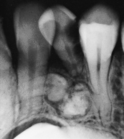
Supernumerary teeth arising in the midline of the maxillae are termed mesiodens (Fig. 7-5) and are the most common supernumeraries. The maxillary distomolars are the next most common supernumerary teeth. These distomolars are also called fourth molars and are located distal to the maxillary third molars. Mandibular distomolars do occur but not nearly as often as in the maxilla. A supernumerary tooth situated buccally or lingually to a molar is called a paramolar; they are usually small and rudimentary. The next most likely location for supernumeraries is the premolar area of the mandible. Only 10% of all supernumeraries occur in the mandible.

If a supernumerary resembles a regular tooth, it is termed supplemental; if it is cone shaped, it is called conical; and if it is very small, it is called tubercle. Supernumerary teeth are much more common in the permanent than in the primary dentition.

ANOMALIES IN SHAPE

Odontoma

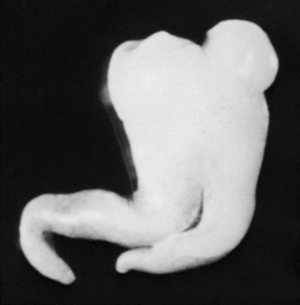
An odontoma (Fig. 7-6) is a tumorous anomaly of calcified dental tissues. The two types of odontoma are complex, which consists of a single mass of dentin, cementum, and enamel in a large blob or unspecified shape; and compound, which consists of several small masses that more or less resemble rudimentary teeth. Although compound odontomas may sometimes resemble multiple mesoderms because they are similar to small, supplemental teeth, they are smaller.

Dens in Dente

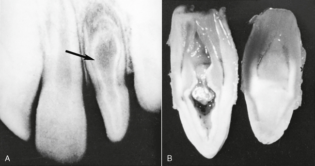
Dens in dente (Fig. 7-7) is a developmental variation thought to occur when the outer surface of the tooth crown invaginates or turns itself inward before mineralization. The term, dens in dente means tooth within a tooth. An x-ray image of a dens in dente shows what appears to be a tooth actually within a tooth (see Fig. 7-7, A). This invagination allows communication between the oral cavity and the inner enamel-lined cavity, which could be considered an extremely deep pit. Permanent maxillary lateral incisors are the teeth most often affected by dens in dente.

Dilaceration

A dilacerated tooth (Fig. 7-8) is a tooth that has a sharp bend or curve in the root or crown. It appears as though the tooth suddenly bent under pressure. The term dilaceration should only be used to describe teeth with very sharp bends.

Only gold members can continue reading. Log In or Register to continue

Stay updated, free dental videos. Join our Telegram channel

Jan 4, 2015 | Posted by in General Dentistry | Comments Off on 7: Dental Anomalies

VIDEdental - Online dental courses

Get VIDEdental app for watching clinical videos