6: Occlusion

Occlusion

POSITION AND SEQUENCE OF ERUPTION

The preceding chapter described eruption patterns of teeth. The eruption schedule helps permanent teeth emerge in their proper position. The loss of certain deciduous teeth at the particular times allows the permanent teeth to move into key positions.

What about the deciduous teeth? What enables them to take their position for alignment? When examining the deciduous teeth, they not only appear in a certain position that is normal for each tooth but are also arranged in a row, which is referred to as being in alignment.

Normally, the eruption schedule helps the deciduous teeth take their proper position. For example, the central incisors come into position anterior to the lateral incisors because the centrals erupt before the lateral incisors. Facial development and growth encourages the teeth to erupt properly. The anterior teeth are not covered by as much bone, and therefore the tooth buds begin their formation earlier than those of the posterior teeth; the result is that most of the anterior teeth erupt before the posterior teeth. Some posterior teeth must actually wait until growth has occurred in the mandible because they are initially trapped under the ramus of the mandible. Thus the eruption pattern, facial development, and the sequence in which tooth buds begin forming all contribute to the eventual relationship of the teeth and jaws.

The development of occlusion begins with the eruption of the primary teeth (see Fig. 5-7, A). Usually the first teeth to erupt are the central incisors with the mandibular teeth erupting slightly before the maxillary. The eruption of the lateral incisors, which occurs next, follows the same sequence.

At 16 months the primary molars erupt, which is an important event because the primary molars establish the vertical height of the primary occlusion. Primary molars also establish intercuspation, the mesial-distal and buccal-lingual relationships that determine how the upper teeth will touch, hit, and interlock with the lower teeth. The upper primary molars also help establish the anteroposterior (mesial-distal) relationship of the remaining deciduous teeth because their presence prompts the canines and second deciduous molars to erupt around them.

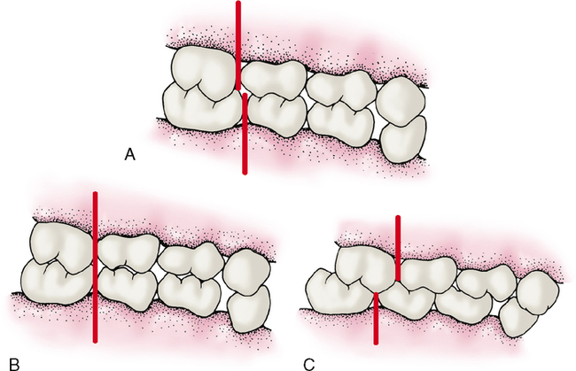
The primary dentition, which is usually complete by about 2.5 years of age, erupts in a more upright position than secondary teeth replacements. The average overjet of primary teeth is 3.0 mm, and the average overbite is 2.5 mm. The primary occlusion has one of three possible anteroposterior molar relationships called steps or planes. The majority of children has a mesial step between the distal surfaces of the second primary molars (Fig. 6-1, A). The mandibular molars are situated more mesially than their maxillary counterparts, thereby forming a mesial step. A smaller but still large group of children exhibits a flush terminal plane, with the distal surfaces of the deciduous second molars even with each other (Fig. 6-1, B). A still smaller minority has a distal step (Fig. 6-1, C). How would you describe a distal step in comparison with a mesial step or flush terminal plane? Note the large diastema or space in the mandibular arch between the canine and first molar.

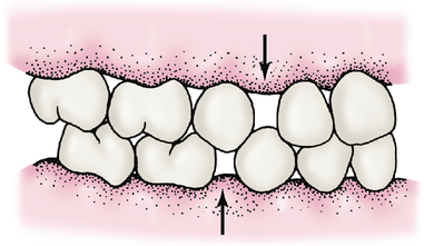
As a child grows in height and weight, so too do the jaws. This growth of the mandible and maxilla results in horizontal and vertical growth of the dental arches. The teeth, however, remain the same size. Thus as the arches grow, spaces called diastemas form between the teeth. The largest spaces are often found mesial to the maxillary primary canines and distal to the mandibular canines. These spaces are called primate spaces, and although not always present, they are characteristic of all primates, including man (Fig. 6-2). As growth continues, diastemas also develop between the incisors.

DEVELOPMENT OF THE MESIAL STEP

The permanent molars erupt and eventually touch the distal surfaces of the deciduous molars. As the permanent molars push up against the deciduous molars, they cause a chain reaction that pushes all of the spaces between the teeth closed. A mesial step occurs in the majority of individuals because closing the primary space allows room for the lower molars to move mesially.

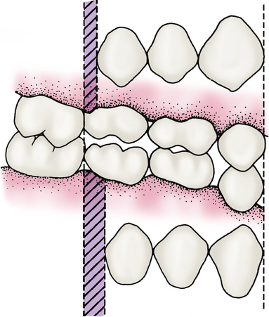
A mesial step is further enhanced as the deciduous molars exfoliate and are replaced by the narrower permanent premolars. Extra space called leeway space is gained from this exchange of the second premolars. The earlier eruption cycle of the mandibular teeth allows them to capitalize on this exchange before the maxillary teeth, which further helps establish the mesial step (Fig. 6-3).

Finally the heads of the condyles of the mandible continue to grow later than the maxilla, allowing even further mesial mandibular advancement and ensuring in most instances a mesial step heading the patient toward a class I relationship. Further growth of the condyle head could push the patient into an extreme mesial step resulting in a class III relationship. A class II relationship could result if the mandible does not continue to grow or if the maxilla outgrows the mandible. It is possible for this type of relationship to occur on just one side while the other side maintains a class I relationship.

A deep bite could result if the condyle head is displaced distally in the glenoid fossa, if the posterior teeth do not erupt enough, if the muscles of mastication are so hyperactive that they prevent the eruption of the posterior teeth, or if the condyle grows at an angle that causes the jaw to develop in a less mesial direction.

The development of the occlusion is further influenced by hereditary factors such as congenitally missing teeth, impacted teeth, or the size and shape of muscle and bone. Controllable factors that also affect occlusal development include the premature loss of deciduous teeth, decayed teeth that were not restored, and harmful habits.

HORIZONTAL ALIGNMENT

After the teeth erupt into the oral cavity, the tongue acts as a huge internal force, pushing the teeth toward the lips and cheeks. Conversely, resistance from the muscles that form the cheeks and lips is the controlling factor that prevents the teeth from moving too far facially. The balance or relative equilibrium between the tongue and the facial muscles allows the teeth to be brought into proper alignment and to be maintained in their proper positions once they have erupted.

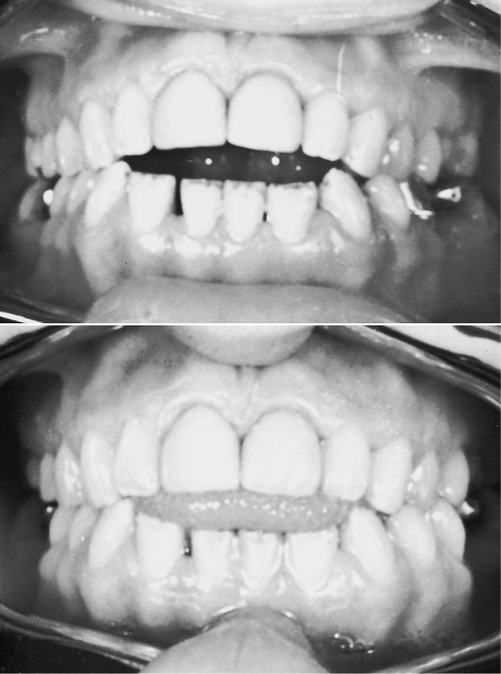
If this balance of forces is disturbed, a malocclusion, or an abnormal alignment of the teeth within the dental arches, can result. Abnormal forward thrusting of the tongue against the anterior teeth can cause such an imbalanced state (Fig. 6-4). Tongue thrusting causes the maxillary anterior teeth to protrude labially out of the mouth. This is especially true if an underdeveloped upper lip is evident along with the tongue thrust.

An opposite situation can also occur if the lower lip is overdeveloped; the retrusion of the lower anterior teeth occurs. The patient is constantly tightening the lower lip against the lower anterior teeth. These lip muscles are so strong that the lower teeth will be pushed back into the mouth by this overdeveloped lower lip.

The lip, tongue, and cheek muscles and their relationship to one another are not the only factors that determine the alignment of the teeth. The intercuspation of the teeth helps prevent tooth deviations in a buccal or lingual direction. The maxillary posterior teeth have buccal and lingual cusps, and when the jaws are closed, the buccal cusps of the mandibular posterior teeth are interlocked between the buccal and lingual cusps of the maxillary teeth. This interlocking is similar to the interlocking of two gears.

The alignment of previously erupted teeth in turn affects the alignment of successive teeth. Adequate space between teeth allows for complete and unhindered eruption of more teeth. If a tooth does not have room enough to erupt, it will deflect off the obstructing tooth and erupt out of alignment. It could also be blocked entirely by the obstruction and never erupt.

Other factors also influence the alignment of teeth. Mesial drift could account for the closure or loss of space necessary for tooth eruption. The size and shape of the jaws, the shape of the teeth, and the amount of lingual convergence of each tooth affect not only the alignment of the teeth but also the curvature of the dental arch and the spacing necessary for incoming teeth.

CURVE OF SPEE, CURVE OF WILSON, AND SPHERE OF MONSON

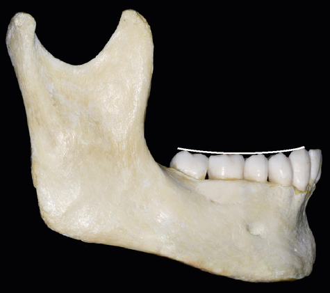
Usually the buccal cusp tips of posterior teeth, seen in alignment from a lateral view (see Fig. 6-6), conform to a fairly even curve in an anterior to posterior direction. This curve is known as the curve of Spee.

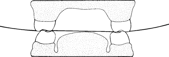
An occlusal curve exists for posterior teeth in a direction from right to left as seen from a posterior view (Fig. 6-5). This transverse occlusal curve is called the curve of Wilson.
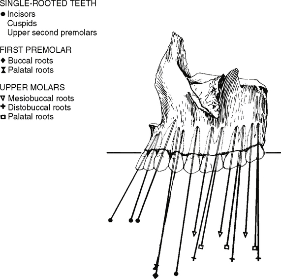
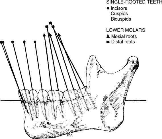
The occlusal surfaces of the natural dentition are thought to be aligned in such a way that a spherical curve 8 inches in diameter could rest on the buccal cusp tips of the mandibular posterior teeth. The curves of Wilson and Spee, when studied simultaneously in three-dimensional alignment, demonstrate an illusion of the cusp tips of the mandibular posterior teeth resting on a sphere known as the sphere of Monson. This theory has yet to be proved.

VERTICAL ALIGNMENT

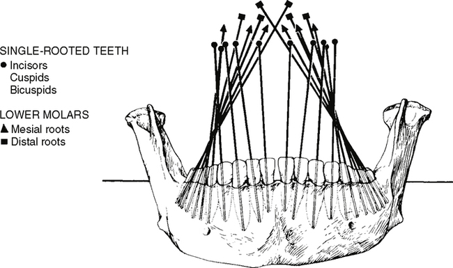
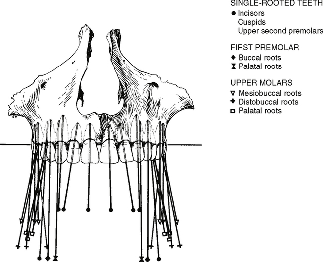
Teeth are often thought to be vertically straight, but this is not true. The teeth are not positioned straight up and down in the mouth. The mandibular posterior teeth have a tendency to tip their crowns lingually and their roots laterally (Fig. 6-7). The maxillary posterior teeth tend to keep the crown straighter but with a slight buccal inclination and as a lingual inclination of the root (Fig. 6-8). From a lateral view, all the teeth, maxillary and mandibular, anterior and posterior, show a slight mesial inclination, with the possible exception of the maxillary third molar. Notice that the anterior teeth (Figs. 6-9 and 6-10) have a slight labial protrusion (a condition of being tipped forward), and from a frontal view, their crowns seem to incline laterally. In other words, the anterior teeth tip out to the side and toward the front (see Figs. 6-7 and 6-8).

OCCLUSION

Occlusion is the term used to describe the relationship of the mandibular and maxillary teeth when the teeth are closed together or during excursive movements when the teeth are touching. When the jaws are completely closed together, two possible relationships occur—a relationship of the upper jaw to the lower jaw (centric relation) or a relationship of the upper teeth to the lower teeth (centric occlusion).

Stay updated, free dental videos. Join our Telegram channel

Jan 4, 2015 | Posted by in General Dentistry | Comments Off on 6: Occlusion

VIDEdental - Online dental courses

Get VIDEdental app for watching clinical videos