Chapter 6
OCCLUSION
6.1 Introduction to occlusion
Working impression
Casting working model
occlusion
Occlusion is the subject that is concerned with how the teeth and associated bones, joints and muscles function together.
The natural dentition
When you put your teeth together, the occlusal surfaces meet in the same position each time (Figure 6.1.1). This position is called intercuspal position (ICP) and is used extensively in dentistry. ICP is a relationship between the maxilla and mandible when the teeth are in maximum intercuspation or maximum meshing. Other terms used for ICP are centric occlusion or habit bite.
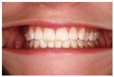
In the ICP, the occlusal load is distributed through the molars. You can feel this if you squeeze your teeth together very hard; there should be little or no pressure on your anterior teeth. The molars are well suited to distribute this load as the roots have a large surface area with which to transmit the load to the bone.
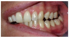
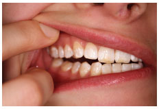
From ICP, move your mandible to the left with your teeth in contact. In most cases you will find that the canines are the only teeth contacting or working together (Figure 6.1.2). In this excursion or movement from ICP, the left side is called the working side. The teeth on the right should not be contacting or not working (Figure 6.1.3), which is why this side is termed the non-working side.
Of course when moving your mandible to the right from ICP, you should find the right canines contact, therefore the right is now the working side and the left teeth have space between them as this is the non-working side.
In these lateral excursions, only the canines contact and take the occlusal load, while the other teeth are separated. The canines are better suited than the posterior teeth to distribute these sideways forces for several reasons.
- Their shape is strong enough to resist force unlike molar teeth that have fissures causing weaknesses.
- They have long roots that prevent them from moving or tipping.
- They are further from the mandibular hinge (the temporomandibular joint (TMJ)) and therefore the muscles cannot exert such high forces.
- They are also more highly innervated or sensitive than other teeth and can easily detect light contact. This stimulus informs the brain, which in turn reduces the load on the tooth. You can try this for yourself, squeeze together in ICP and feel your masseter muscle on the angle of your mandible. Now move you jaw sideways such that only the canines on one side touch and squeeze in this position. You should feel that the brain has limited the activity of the muscle.
When the canine dictates the sideways movement of the mandible, the occlusal scheme is considered to be canine guided. This means that the canine is the only tooth guiding the sideways movement of the jaw.
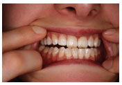
The anterior teeth act in the same way during forward movements of the mandible, dictating is pathway and causing the posterior teeth to separate (Figure 6.1.4).
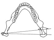
The two TMJs also dictate the mandibular movement at the posterior end. As the jaw moves to the left, the left or working side condyle rotates on an axis through the head as shown in Figure 6.1.5. This working side condyle may also be termed the rotating condyle.
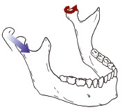
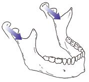
The right side condyle moves forwards, down the articular eminence (the bony slope of the joint) and towards the left at the same time. This non-working side condylar pathway is guided by the articular eminence (Figure 6.1.6). Both condyles may translate (slide) forward in the protrusive movement of the jaw (Figure 6.1.7).
Centric relation
The mandible can be related to the maxilla in one more relationship, this time determined by the position of the condyles rather than the teeth. The condyles need to be located in their optimal position at the top of the articular eminence (Figure 6.1.8). Here the condyles can distribute any load through the bony structures of the skull rather than using muscles to hold the condyle on the slope of the articular eminence.
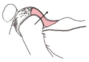
When the condyles are in this position the mandible can hinge open and closed by approximately 20 mm without any forward movement. This range of movement is called centric relation (CR) and again is used extensively in dentistry. You should assume that in an ideal situation ICP coincides with the CR arc of closing, therefore when the mandible is closed in CR, the teeth meet in ICP. This is the basic arrangement and function of the teeth in the idea/>
Stay updated, free dental videos. Join our Telegram channel

VIDEdental - Online dental courses


