5
The respiratory system
Chapter contents
5.1 Introduction
Oxygen derived from the air is essential for providing energy to drive the metabolic processes in cells and tissues. Air is drawn into and expelled from the body through the respiratory system by the process of ventilation. Within the respiratory system, gaseous exchange takes place between air and blood in the lungs. This is respiration in its true sense; oxygen enters the blood and carbon dioxide leaves it. The activities of the respiratory system must be regulated to ensure adequate oxygen supplies and clearance of carbon dioxide to meet the functional demands of the body. The respiratory and cardiovascular systems work in concert to maintain homeostasis and share several control mechanisms. The respiratory system also provides the driving force for production of speech and modifying sounds during speech.
Anatomically, the respiratory system consists of a series of air passages that terminate in the lungs where gaseous exchange takes place across the thin walls of individual alveoli within them. The air passages are supported by bone or cartilage to prevent them from collapsing when air pressure is reduced. A schematic diagram of the respiratory tract is shown in Figure 5.1. In succession, the nose, pharynx, larynx, trachea, and bronchial tree constitute the conducting portion of air passages and the lung alveoli form the respiratory portion where gaseous exchange takes place. Clinically, the air passages as far as the larynx are known as the upper respiratory tract (URT) and the passages below the larynx and the lungs are the lower respiratory tract (LRT).
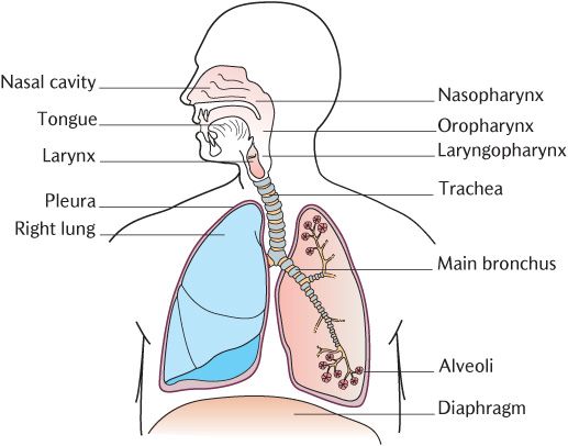
Fig. 5.1 A diagrammatic representation of the respiratory system.
5.2 The upper respiratory tract
Air is drawn into the body through the nose. The nose is more than a simple air passage; it has important functions in cleaning, warming, and moistening air. Air is filtered by hairs at the entrance to the nose, warmed by heat exchange with the abundant blood vessels in the mucosa of the nasal cavities, and humidified by fluid evaporating from mucus secreted by the lining mucosa. Figure 5.2A shows how bone in the lateral walls of the nasal cavities is folded to increase the surface area available and thus increase their efficiency of heating and humidification. The mucosa lining the respiratory portion has an outer covering known as respiratory epithelium although its full description, pseudostratified ciliated columnar epithelium with goblet cells, is more informative. Its structure is shown diagrammatically in Figure 5.2B. ‘Pseudostratified’ means that there appear to be several layers (strata) of cells when in fact, there is only a single layer of columnar cells; ‘ciliated’ indicates that the cells are covered by cilia, tiny hair-like structures, on their outer surface and goblet cells which are mucus-secreting cells embedded between the ciliated cells. Aggregates of mucus cells also form mucous glands in the connective tissue below the epithelium to add to the volume of mucus. As well as its function in humidifying the air, mucus traps tiny particles that may be harmful to the LRT. The cilia are in constant wave motion to move the mucus across the epithelium. In the nasal cavities, the cilia are beating back towards the pharynx. In the air passages below the pharynx, the cilia are beating in an upward direction. Thus mucus containing potentially harmful material is moved towards the pharynx from where it can be swallowed and rendered harmless.
Notice in Figure 5.2A, the large holes in the bones forming the lateral walls of the nasal cavity. These are two of the paranasal air sinuses that occupy the middle of some of the skull bones surrounding the nasal cavity. The air sinuses connect with the nasal cavity and are lined with respiratory epithelium. They probably function to provide an additional/>
Stay updated, free dental videos. Join our Telegram channel
VIDEdental - Online dental courses


