4
The interface between epithelium and connective tissue
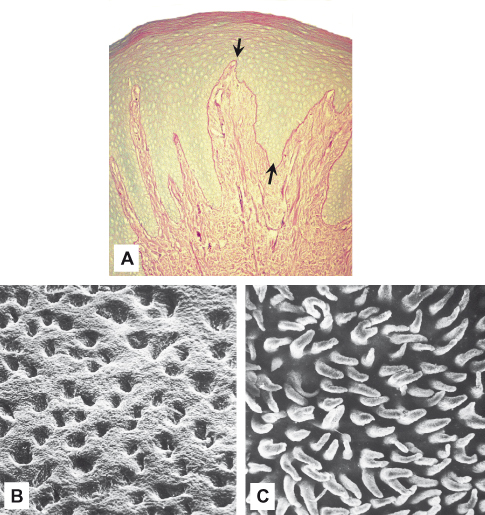
The junction between the connective tissue and the overlying oral epithelium is seen in two-dimensional histological sections as an undulating interface at which papillae of the connective tissue interdigitate with the epithelial ridges (Fig. 4.1A). In three dimensions, the interface is seen to consist of connective tissue ridges, conical papillae, or both, projecting into the epithelium (Fig. 4.1B,C). The undulations increase the surface area of the interface and may provide better attachment and permit forces applied at the surface of the epithelium to be dispersed over a greater area of connective tissue. In this regard, it is notable that masticatory mucosa has the greatest number of papillae per unit area of mucosa; in lining mucosa, the papillae are fewer and shorter. The junction also represents an increased interface for metabolic exchange between the epithelium and the vasculature of the connective tissue, for there are no blood vessels in the epithelium.
Figure 4.1 (A) Gingival epithelium stained by the PAS method shows the basement membrane (arrows) and extensive interdigitation between epithelium and connective tissue. Scanning electron micrographs of gingival epithelium separated at this interface shows the circular orifices in the underside of the epithelium (B) into which fit the cone-shaped papillae of connective tissue (C). ((B) and (C) from Klein-Szanto and Schroeder, 1977, with permission from Wiley-Blackwell.)

4.1 ORGANIZATION OF THE NORMAL INTERFACE
In routine hematoxylin and eosin stained histological sections of oral mucosa, the basement membrane between the epithelium and connective tissue appears as a structureless band, but it stains brightly with reagents such as periodic acid-Schiff (PAS) which detects glycogen, neutral mucosubstances, glycolipids, and phospholipids (Fig. 4.1A). Ultrastructurally, this region is described as the basal lamina or basal complex and is highly organized (Fig. 4.2). It consists of a layer of finely granular or filamentous material about 50 nm thick, the lamina densa, which runs parallel to the basal cell membranes of the epithelial cells but is separated from them by an apparently clear zone some 45 nm wide, the lamina lucida. Hemidesmosomes represent condensations of the proteins bullous pemphigoid antigen and intermediate filament-associated protein on />
Stay updated, free dental videos. Join our Telegram channel

VIDEdental - Online dental courses


