4: Oral and Maxillofacial Infections

Oral and Maxillofacial Infections

Odontogenic and nonodontogenic maxillofacial infections are among the oldest disease processes treated by oral and maxillofacial surgeons. They commonly present to the office, or in severe cases to the hospital emergency department. Although the majority of infections can be treated in a nonemergent fashion, early recognition and correct management of severe infections can be lifesaving. Knowledge of the surgical anatomy and path of spread of infections in the head and neck is fundamental to correct diagnosis and treatment. Severe infections of the sublingual, submandibular, and parapharyngeal spaces can cause airway compromise, cavernous sinus thrombosis, and possibly mediastinal spread of infection, resulting in significant mortality and morbidity, especially in the medically compromised patient who presents late in the disease process.

Despite the availability of a wide spectrum of antimicrobial agents and increasing knowledge of microbiology, the treatment of odontogenic infections remains primarily surgical. Removal of the source of infection and establishment of adequate drainage for elimination of the purulent material is the mainstay treatment. However, adequate antibiotic coverage is important and should not be overlooked.

The response to treatment can be monitored by several clinical (swelling, erythema, pain, interincisal opening) and laboratory (white blood cell [WBC] count, C-reactive protein) parameters. The measurement of temperature is an ancient method of monitoring the response to an infectious process. Fever occurs secondary to the production of endogenous pyrogens (cytokines, interleukins, tumor necrosis factor), which affect the hypothalamus and medulla to increase the temperature set point. The definition of fever is arbitrary, and there is considerable variability in “normal temperature” for a population of healthy adults. A range of definitions is acceptable (37.5° to 38.5°C), depending on how sensitive an indicator the surgeon wants to use. The lower the temperature used to define fever, the more sensitive the indicator is for detecting an infectious process, but the less specific it will be. Normal body temperature is generally considered to be 37°C and varies according to circadian rhythm and menstrual cycle. Many different variables can influence temperature such as exercise and environmental factors.

Chills occur in response to the elevation in temperature set point during the initiation of fever. This is often accompanied by the need for increased insulation and decreased exposure of skin. Shivering is also seen, contributing to the increase in temperature.

The differential is a ratio of the different types of white blood cells present (polymorphonuclear neutrophils [PMNs], lymphocytes, monocytes, eosinophils, and basophils). With a rise in the WBC count, as is seen during an acute infection, the predominant increase occurs in the PMNs. Chemotactic factors contribute to the recruitment of PMNs to the site of infection (or injury), with a subsequent increase in production of neutrophils by the bone marrow. Production of the precursor forms of the PMN (myelocytes and promyelocytes) increases, and these cells are released into the circulating blood. The movement toward circulation of immature forms is termed a shift to the left and is usually seen during acute infections.

In this chapter we present four teaching cases of infections that have an odontogenic etiology. We also present a case of osteomyelitis of the mandible.

Ludwig’s Angina

HPI

The patient presented to the emergency department with a 3-day history of progressive swelling and pain in his neck. He reports a 3-week history of severe, intermittent pain in his lower right third molar. Ten days ago, his general dentist prescribed him amoxicillin for a periapical abscess and pericoronitis associated with his right mandibular third molar, which was partially impacted and decayed. Despite compliance with antibiotics, he progressively developed persistent swelling and a foul-tasting drainage around the tooth. He began to have swelling under the right side of his tongue (right sublingual space), which spread to the contralateral side (there is no anatomic barrier between the right and left sublingual spaces). Simultaneous with the floor of the mouth swelling, the neck began to swell on the right side, and the swelling spread to the other side. The patient reports having subjective fevers and chills (signs of systemic inflammatory involvement), in addition to dysphagia (difficulty swallowing) and odynophagia (painful swallowing). He states that he has not been able to eat or drink in the past 48 hours (causing dehydration). He denies dysphonia (difficulty speaking, seen with edema of the vocal cords and upper airway) or any chest discomfort (seen with advanced mediastinal involvement).

Examination

General. The patient is sitting upright, appears very restless, and is unable to tolerate his oral secretions (evidenced by constant use of a Yankauer suction and drooling of saliva). He appears to be in mild respiratory distress, but there is no evidence of stridor (a high-pitched, crowing noise due to partial upper airway obstruction).

Airway. The airway is stable on examination. The trachea is difficult to palpate due to edema but appears to be in the midline. Fiberoptic nasopharyngoscopy can be performed to further evaluate the patency of the upper airway and the amount of edema of the surrounding soft tissue (see Emergent Surgical Airway in Chapter 3). Alternatively, computed tomography (CT) scans of the neck can delineate neck and airway swelling.

Vital signs. The patient’s blood pressure is 138/89 mm Hg, heart rate 110 bpm (tachycardia), respirations 28 per minute (tachypnea), temperature 40°C (febrile), and oxygen saturation 96% on room air.

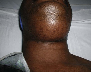
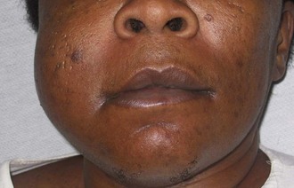
Maxillofacial. There is obvious moderate to severe facial swelling over the lower third of the face. Brawny and painful induration of the submandibular and submental spaces is noted bilaterally (Figure 4-1). There is erythema over the anterior neck extending down to the clavicles. However, subcutaneous crepitus (indicative of subcutaneous air from gas-producing organisms) is not present. No cervical lymphadenopathy or fluctuance was palpated (lymphadenopathy would be difficult to assess in the presence of neck edema or induration).

Intraoral. The patient’s mouth opening is limited, with a maximal interincisal opening of 20 mm (trismus indicates masticator space involvement or guarding secondary to pain). The floor of the mouth and tongue are elevated and edematous (sublingual space). The oropharynx is not clearly visualized due to the limited mouth opening and elevated tongue (positive predictors of difficult laryngoscopy and endotracheal intubation).

Cardiovascular. The patient is tachycardic, without rubs, murmurs, or gallops (tachycardia and friction rubs can be indicative of mediastinitis). He is negative for Homan’s sign (crepitus heard with a stethoscope during systole, indicative of mediastinitis).

Pulmonary. Lung fields are clear to auscultation bilaterally, without rales, bronchi, or wheezes (aspiration of saliva or exudates can be seen with advanced cases).

Imaging

Before obtaining any imaging studies, the surgeon needs to decide on the urgency of the infectious process compromising the airway. If the airway is deemed stable, imaging studies should be obtained to guide surgical treatment. However, any possibility of acute airway embarrassment should not delay direct transfer to the operating room for advanced airway interventions. Once the airway is stabilized, imaging studies can be safely obtained.

A panoramic radiograph is the initial screening study of choice. It provides an excellent overview of the dentition, identifying any odontogenic sources of the infection. CT scans of the neck with contrast material are indicated when dealing with deep neck space infections (chest CT should be included if there is a suspicion of descending mediastinitis). This study can help determine the anatomic spaces involved, localize any fluid collections (loculations of purulence), and determine whether the airway is deviated or compromised. CT is also helpful in surgical planning for incision and drainage. When a chest CT is deemed unnecessary, chest radiographs (posteroanterior and lateral views) can be an important screening tool to detect a widened mediastinum, which may be indicative of descending mediastinitis.

In the current patient, the panoramic radiograph revealed a carious right mandibular third molar with a large periapical radiolucent lesion. The CT scan of the patient’s neck revealed a rim-enhancing fluid collection involving the bilateral submandibular, submental, and right sublingual spaces (Figure 4-2). In addition, there was diffuse soft tissue edema consistent with cellulitis in the involved spaces. No subcutaneous emphysema was seen in the cervical tissues (subcutaneous gas collection is considered a hallmark of cervical necrotizing fasciitis and is seen in up to 46% to 67% of cases). The patient’s airway was patent and midline. A chest CT was ordered due to the erythema tracking down the anterior neck. No mediastinal involvement was observed.
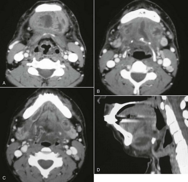
Labs

A complete blood count (CBC) and complete metabolic panel (CMP) are indicated during the work-up of severe odontogenic infections. The presenting WBC count is a marker of the severity of infection, and this value should be followed during the course of treatment. C-reactive protein (CRP) is an acute-phase reactant that is released in response to inflammation, and it can be used to monitor the response to therapy. Studies have also suggested that a very high CRP level at the time of admission is a predictor of a complicated hospital course. Electrolyte disturbances (sodium, potassium, magnesium, calcium) are common among patients with severe head and neck infections, especially when the patient is not able to tolerate oral intake due to swelling or pain. Blood urea nitrogen (BUN) and creatinine levels are useful for evaluating for prerenal azotemia due to hypovolemia. Blood cultures are indicated in the patient with persistent fever. An electrocardiogram (ECG) should be obtained with suspicion of mediastinitis. Arterial blood gas (ABG) measurement is warranted in the critically ill patient presenting with septic shock.

The current patient presented with these lab values: WBC count 21,000 cells/mm3 with a 35% bandemia, BUN 30 mg/dl (normal range, 7 to 18 mg/dl), and creatinine 1.2 mg/dl (normal range, 0.6 to 1.2 mg/dl). The BUN/creatinine ratio was 25 (a ratio greater than 20 is indicative of prerenal azotemia). The remainder of his electrolyte values were within normal limits.

Assessment

Ludwig’s angina secondary to carious right mandibular third molar (odontogenic source of infection accounts for 70% to 90% of cases, the vast majority arising from second or third molars).

Ludwig’s angina was first described by Karl Friedrich Wilhelm von Ludwig in 1836 as a rapidly progressing, gangrenous cellulitis originating in the region of the submandibular area that extends without any tendency to form abscesses. Ludwig’s angina is now known as an aggressively spreading cellulitis that simultaneously affects the bilateral submandibular, sublingual, and submental spaces. Although Ludwig’s angina is classically described as a cellulitis, progression to abscess formation within the involved spaces is most often the case, and to this date, clinicians still use the term “Ludwig’s angina” when describing a bilateral submandibular, sublingual, and submental space infection. Grodinsky and Holyoke’s criteria for Ludwig’s angina may no longer have any useful clinical application. The term “angina” is a misleading term, because any chest discomfort seen with this is from descending mediastinitis and is not related to ischemic heart disease.

Treatment

Treatment begins with evaluation of the patient’s airway and appropriate management to prevent acute airway embarrassment (see Emergent Surgical Airway in Chapter 3). The airway is first evaluated by the general appearance of the patient (a distressed patient with stridorous respirations is assumed to have an airway compromise until proved otherwise). The oral cavity should be examined to evaluate the amount of tongue, floor of the mouth, soft palate, and pharyngeal wall edema (many times an oral examination is very limited due to the patient’s inability to open). A fiberoptic nasopharyngoscopy can be performed in the emergency department to further assess the airway, including the vocal cords. Intravenous dexamethasone can be given to reduce the airway edema in patients with impending upper respiratory obstruction. An emergent cricothyroidotomy should be performed if the patient loses the airway before arrival in the operating room. An awake tracheotomy or an awake fiberoptic nasal intubation can be performed in the operating room if the situation is less acute (oral intubation by direct laryngoscopy may also be possible in less severe cases). There is support in the current literature for the assumption that a tracheotomy may be indicated in patients with Ludwig’s angina (see Complications).

Supportive measures should be initiated while arrangements are made with the operating room. This should include fluid resuscitation and initiation of broad-spectrum empiric antibiotic therapy. Fluid resuscitation is commonly needed because patients present with hypovolemia due to lack of oral intake (insensible losses are accelerated by fever) and/or some degree of sepsis or septic shock. Adequacy of fluid resuscitation should be continuously monitored (heart rate, blood pressure, urine output, and BUN/creatinine). Vasopressive therapy may be indicated in patients presenting with septic shock. Tight glycemic control (blood glucose 90 to 110 mg/dl) is desirable, especially in the critically ill patient.

Empiric antimicrobial therapy should be promptly initiated to cover the mixed aerobic-anaerobic polymicrobial organisms (gram positive, gram negative, aerobic, and anaerobic) commonly involved in these infections. Penicillin G at an adult dose of 4 million to 30 million units per day, divided and given every 4 to 6 hours, in combination with metronidazole, is an appropriate regimen. Other recommendations include clindamycin 900 mg given intravenously every 8 hours; ticarcillin clavulanate 3.1 g given intravenously every 6 hours; ampicillin sulbactam 3 g given intravenously every 6 hours; and piperacillin tazobactam 3.375 g given intravenously every 6 hours. Chow in 1992 recommended high-dose intravenous penicillin G combined with clindamycin, metronidazole, or cefoxitin. When available, the antibiotic regimen should be guided by cultures and sensitivity studies. The CRP has been shown to be an excellent marker for the severity of the infection and the patient’s response to surgical and antibiotic therapy.

Aggressive surgical drainage and debridement, along with elimination of the source of infection, are necessary for definitive treatment. Delay in taking the patient to the operating room for surgical treatment is associated with a worse outcome. Cultures should be taken either via aspiration techniques or with a culturette swab. Most cases of Ludwig’s angina can be managed using small incisions in the submandibular and submental regions (larger cervical hockey-stick or apron incisions may be indicated when the condition is complicated by necrotizing fasciitis). Blunt dissection is carried out to explore all the involved spaces. Subperiosteal dissection and debridement are important in the area around the source of infection, and any offending teeth should be extracted. The intraoral and extraoral dissections can be dissected to freely communicate, allowing for dependent extraoral drainage. Therefore the abscess is decompressed, the necrotic debris is debrided, and the wounds are copiously irrigated. Red rubber catheters and/or Penrose drains can be used to facilitate postoperative wound irrigation and to allow dependent drainage. Drains can be slowly advanced out of the wound postoperatively or removed when purulent drainage ceases. Repeat drainage and lavage procedures in the operating room should be considered, especially in more severe infections that are refractory to treatment. Bouloux and associates evaluated the efficacy of irrigating surgical drains on postoperative odontogenic infections. They found that nonirrigating drains (Penrose drains) appear to be equally efficacious as irrigating drains (red rubber catheter).

The current patient was given 16 mg of dexamethasone intravenously in the emergency department; intravenous fluid resuscitation was initiated; and empiric intravenous antibiotics were administered. Antibiotic therapy consisted of ampicillin-sulbactam (Unasyn) 3 g every 6 hours and clindamycin 900 mg every 8 hours. The patient was urgently taken to the operating room for incision and drainage of the involved anatomic spaces of the neck and extraction of the right mandibular third molar. The patient was intubated successfully via an awake nasal fiberoptic endotracheal intubation. An 18-gauge needle was used to aspirate purulent exudate from the submandibular space, which was sent for Gram stain, aerobic and anaerobic cultures, and antibiotic sensitivity studies. The surgical drainage consisted of three incisions of 1.5 to 2 cm in length, 2 cm below the inferior border at the angle of the mandible bilaterally and anteriorly in the submental area. Consideration should be given to placement of the incisions to allow dependent drainage. Blunt dissection with a hemostat and a Kelly clamp was carried out to explore all involved spaces. Copious amounts of purulence and necrotic tissue were expressed from the surgical sites. The right mandibular third molar was elevated and extracted. The gingival cuff was elevated, and subperiosteal dissection was carried out along the lingual plate to enter the sublingual and submandibular spaces. All the incisions were connected to each other in the subplatysmal and subperiosteal planes. Irrigation drains were placed in the submandibular, sublingual, and submental spaces. All drains were irrigated with copious amounts of antibiotic irrigation and/or normal saline irrigation. The patient was left intubated for 3 days postoperatively due to surgical and airway edema. After significant resolution of the infection and edema, he had a positive cuff leak test and was extubated over an Eschmann tube, which was left in place for several hours. He did not experience any postextubation airway compromise and was transferred to the ward the following day.

Complications

The most feared complication associated with Ludwig’s angina is death due to airway compromise. Loss of airway from upper airway obstruction can occur at any time during the perioperative period, before arrival at the operating room, during an attempted intubation, after an accidental or self-extubation in the intensive care unit (ICU), or after a planned extubation (see Emergent Surgical Airway in Chapter 3). Potter and colleagues in 2002 reported a 3% incidence of loss of airway for patients who received a tracheotomy versus 6% for patients maintained with endotracheal intubation. They reported two deaths (4% mortality rate) secondary to loss of airway, and both deaths occurred in the endotracheal intubation group (one occurred after a planned extubation and the other occurred after an unplanned extubation). The tracheotomy group had shorter ICU stay (1.1 versus 3.1 days) and shorter overall hospital stay (4.9 versus 5.9 days). Patients with Ludwig’s angina or a retropharyngeal space abscess have a significant need for tracheotomy. Har-El et al’s review of 110 patients showed that 4 of 8 patients meeting their criteria for severe infection who did not receive a tracheostomy developed upper airway obstruction necessitating an emergent surgical airway (50% incidence of airway loss in the endotracheal intubation group). They concluded that tracheotomy is indicated in patients with Ludwig’s angina. In 1985, Loughnan and Allen reported successful endotracheal intubation in 9 of 10 patients with Ludwig’s angina using an inhalational induction technique and direct laryngoscopy, but they did not report on the postoperative morbidity and mortality. If postoperative endotracheal intubation is planned, adequate sedation, four-point restraints, and a secured tube (taped around the head or wired to the teeth) are paramount to prevent unanticipated self or iatrogenic extubation. Upon extubation, a cuff leak test should be performed and an Eschmann tube should be left in place to facilitate reintubation if needed (postextubation laryngeal edema may cause loss of airway despite having a good cuff leak test result).

Before the advent of antibiotics, the mortality rate from Ludwig’s angina was greater than 50%. Fortunately, the prevalence and mortality rates have significantly decreased due to better access to dental care and antibiotic therapy. When the condition is complicated by descending mediastinitis and thoracic empyema, the mortality rate remains as high as 38% to 60% despite antibiotic therapy (Figure 4-3). When the condition in complicated by cervical necrotizing fasciitis, the more recent reported mortality rate is 18% to 22% (any delay in surgical treatment increases mortality). Tung-Yiu and colleagues reported that an immunocompromised state (e.g., diabetes mellitus) increases the risk of an odontogenic infection developing into cervical necrotizing fasciitis. Of their series of 11 cases, seven patients were immunocompromised (four with diabetes mellitus), which accounted for all major complications, including two deaths. Others have reported a mortality rate as high as 67% with severe odontogenic infections associated with diabetes mellitus. Currently there is no evidence to suggest that HIV/AIDS status increases the risk of developing Ludwig’s angina and its associated complications.

Other potential complications include aspiration, ventilator-acquired pneumonia, septic shock, and acute renal failure.

Discussion

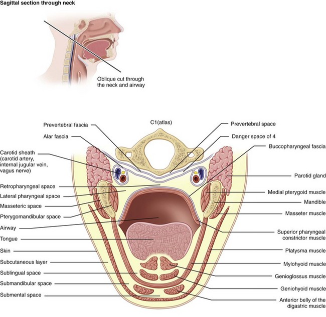
Ludwig’s angina is defined by the involvement of specific anatomic spaces (bilateral submandibular, sublingual, and submental spaces). The sublingual spaces are bounded anteriorly and laterally by the mandible, superiorly by the floor of the mouth and tongue, and inferiorly by the mylohyoid muscle. There is no anatomic barrier between the left and right sublingual spaces. The submandibular space is separated from the sublingual space by the mylohyoid muscle, thus forming the roof of the submandibular space. The hyoglossus and styloglossus muscles form the medial border, and the body of the mandible forms the lateral border. The skin, superficial fascia, platysma, and superficial layer of the deep cervical fascia form the superficial boundary. The anterior bellies of the digastric muscles form the lateral borders of the submental space. The roof is formed by the mylohyoid muscle. The symphysis of the mandible and the hyoid bone form its anterior and posterior borders, respectively. The sublingual and submandibular spaces posteriorly communicate freely with each other and with the medial masticator and lateral pharyngeal spaces, which in turn is contiguous with the retropharyngeal space. Extension of the infection along the carotid sheath (contained within the posterior compartment of the lateral pharyngeal space [LPS]) or retropharyngeal space can lead to descent into the superior mediastinum. The alar fascia separates the retropharyngeal space from the “danger space” (space 4 of Grodinksy and Holyoke), which extends to the diaphragm and the posterior mediastinum. The anterior paratracheal spaces provide anterior access to the superior mediastinum (Figure 4-4).

Ludwig’s angina most commonly has an odontogenic etiology (70% to 90%). A periapical abscess from the second or third mandibular molars is the most common cause. The roots of these teeth are commonly below the attachment level of the mylohyoid muscle to the internal oblique ridge. The periapical abscess perforates the lingual cortex, with spread into the sublingual (if the root is above the mylohyoid attachment) or submandibular (if the root is below the mylohyoid attachment) space. The infection can then rapidly spread to adjacent continuous or contiguous spaces. Other causes include peritonsillar or parapharyngeal abscesses, oral lacerations, mandibular fractures, and submandibular sialadenitis.

The bacteriologic profile of Ludwig’s angina is usually polymicrobial and includes aerobes and anaerobes. The most common organisms are Streptococcus viridans, β-hemolytic streptococci, staphylococci, Klebsiella pneumoniae, anaerobic Bacteroides organisms, and Peptostreptococcus organisms. S. viridans is one of the most commonly isolated organisms. This is consistent with previous reports associating this organism with odontogenic infections. K. pneumoniae is another commonly isolated organism that has a higher incidence in patients with diabetes mellitus.

Bibliography

Allen, D, Loughnan, TE, Ord, RA. A re-evaluation of the role of tracheostomy in Ludwig’s angina. J Oral Maxillofac Surg. 1985; 43:436–439.

Barsamian, JG, Scheffer, RB. Spontaneous pneumothorax: an unusual occurrence in a patient with Ludwig’s angina. J Oral Maxillofac Surg. 1987; 45:161–168.

Bouloux, GF, Wallace, J, Xue, W. Irrigating drains for severe odontogenic infections do not improve outcome. J Oral Maxillofac Surg. 2013; 71:42–46.

Chidzonga, MM. Necrotizing fasciitis of the cervical region in an AIDS patient: report of a case. J Oral Maxillofac Surg. 2005; 63:855–859.

Chow, AW. Life-threatening infections of the head and neck. Clin Infect Dis. 1992; 14:991.

Dugan, MJ, Lazow, SK, Berger, JR. Thoracic empyema resulting from direct extension of Ludwig’s angina: a case report. J Oral Maxillofac Surg. 1998; 56:968–971.

Fischmann, GE, Graham, BS. Ludwig’s angina resulting from infection of an oral malignancy. J Oral Maxillofac Surg. 1985; 43:795–796.

Har-El, G, Aroesty, JH, Shana, A, et al. A retrospective study of 110 patients. Oral Surg Oral Med Oral Pathol. 1994; 77(5):446–450.

Loughnan, TE, Allen, DE. Ludwig’s angina: the anaesthetic management of nine cases. Anaesthesia. 1985; 40:295–297.

Mihos, P, Potaris, K, Gakidis, I, et al. Management of descending necrotizing mediastinitis. J Oral Maxillofac Surg. 2004; 62:966–972.

Potter, JK, Herford, AS, Ellis, E. Tracheotomy versus endotracheal intubation for airway management in deep neck space infections. J Oral Maxillofac Surg. 2002; 60:349–354.

Steiner, M, Gau, MJ, Wilson, DL, et al. Odontogenic infection leading to cervical emphysema and fatal mediastinitis. J Oral Maxillofac Surg. 1982; 40:600–604.

Sugata, T, Fujita, Y, Myoken, Y, et al. Cervical cellulitis with mediastinitis from an odontogenic infection complicated by diabetes mellitus: report of a case. J Oral Maxillofac Surg. 1997; 55:864–869.

Tsuji, T, Shimono, M, Yamane, G, et al. Ludwig’s angina as a complication of ameloblastoma of the mandible. J Oral Maxillofac Surg. 1984; 42:815–819.

Tsunoda, R, Suda, S, Fukaya, T, et al. Descending necrotizing mediastinitis caused by an odontogenic infection: a case report. J Oral Maxillofac Surg. 2000; 58:240–242.

Tung-Yiu, W, Jehn-Shyun, H, Ching-Hung, C, et al. Cervical necrotizing fasciitis of odontogenic origin: a report of 11 cases. J Oral Maxillofac Surg. 2000; 58:1347–1352.

Ylijoki, S, Suuronen, R, Jousimies-Somer, H, et al. Differences between patients with or without the need for intensive care due to severe odontogenic infections. J Oral Maxillofac Surg. 2001; 59:867–872.

Zachariades, N, Mezitis, M, Stavrinidis, P, et al. Mediastinitis, thoracic empyema, and pericarditis as complications of a dental abscess: report of a case. J Oral Maxillofac Surg. 1988; 46:493–495.

Buccal and Vestibular Space Abscess

HPI

The patient has not received any dental care for the past several years (risk factor for odontogenic infections). Two weeks earlier, she noticed that a segment of a restoration broke off the right maxillary second molar. At the time, there was an acute, localized pain in the right posterior maxillary molar with subsequent development of swelling in the right buccal vestibule (buccal vestibular space infection). The patient decided to take over-the-counter medications, and her pain subsided but the swelling persisted. Two days before presentation, she developed acute onset of right-side swelling of the cheek associated with mild pain and discomfort that have progressively exacerbated. There is no history of trismus, visual changes, dysphagia, swelling of the floor of the mouth, or difficulty breathing (all of which are signs of more severe facial space involvement, such as masticator space infection, periorbital extension, and parapharyngeal or sublingual infections; these signs are not seen with pure buccal and vestibular space involvement).

Examination

General. The patient was a well-developed and well-nourished woman in mild distress (patients with buccal space infections are frequently very concerned due to the extent of swelling and pain).

Vital signs. Stable and WNL. Temperature of 98.8°F (an elevated temperature is not always seen, especially in well-localized infections, unless there is surrounding cellulitis or systemic dissemination of the infection).

Maxillofacial. Significant right-side facial edema extending from the inferior border of the mandible superiorly to the level of the zygoma (Figure 4-5). The swelling is soft and fluctuant and with no apparent intraoral or extraoral drainage (untreated buccal space infections may spontaneously drain, providing some relief or spread into other fascial spaces). There is tender right submandibular lymphadenopathy (due to active infection).

Intraoral. The maximal interincisal opening is 35 mm (trismus is not seen with vestibular space or buccal space infections, because it does not involve the muscles of mastication, unless the infection has spread from buccal space to the submasseteric space posteriorly, pterygomandibular space inferiorly, and infratemporal space superiorly). Bimanual examination of the right posterior buccal vestibule and right cheek reveals fluctuance within the right maxillary vestibule extending to the depth of the mandibular vestibule. The right />

Stay updated, free dental videos. Join our Telegram channel

Jan 12, 2015 | Posted by in Oral and Maxillofacial Surgery | Comments Off on 4: Oral and Maxillofacial Infections

VIDEdental - Online dental courses

Get VIDEdental app for watching clinical videos