11: Oral Cancer

Oral Cancer

Malignant diseases of the oral cavity include a spectrum of neoplastic disorders that can emerge from the cellular structures present in the oral cavity or, less frequently, from metastatic disease to the area. Primary oral squamous cell carcinoma accounts for more than 90% of all head and neck malignancies. Malignant diseases of the salivary glands, and ductal epithelium account for the majority of the remaining cases. A greater proportion of oral cancer is diagnosed and referred for treatment by dental professionals than by general medical practitioners. Therefore, recognition and knowledge of the diagnostic tools necessary to identify these disorders are the minimum requirements for general dental practitioners and oral and maxillofacial surgeons. Today an increasing number of oral and maxillofacial surgeons are additionally trained in the treatment of head and neck malignancies. Surgery remains the primary treatment modality for head and neck cancer.

Oral cancer poses a significant challenge to our specialty. As the future unfolds, advances in molecular biology, cell signaling, immunomodulation, and angiogenesis will result in novel targeted therapies that will allow patients with cancer to live longer and healthier lives. Therapies will be tailored to the biologic behavior of the tumor, whether benign or malignant, not just to the histologic diagnosis. This behavior, in turn, will be determined by pretreatment genetic, molecular, and proteomic assessment so that the prescribed cure matches the patient’s disease.

In this chapter we present three cases representing the most commonly encountered oral cavity malignancies. One case discusses oral squamous cell carcinoma, and another case discusses verrucous carcinoma; these cancers originate from the epithelium. The third case discusses three salivary gland malignancies—acinic cell carcinoma, adenoid cystic carcinoma, and mucoepidermoid carcinoma. A new section discusses neck dissection, a procedure that is an important aspect of head and neck cancer treatment.

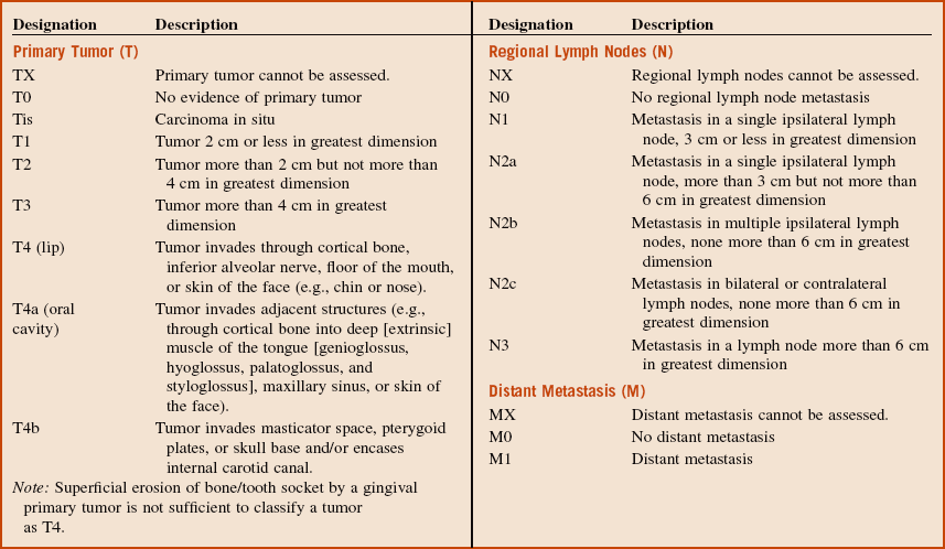
Box 11-1 outlines the TNM staging classification used for lip and oral cavity cancers; Box 11-2 looks at the staging of squamous cell carcinoma.

Box 11-1

TNM Staging for Lip and Oral Cavity Cancers*


*Staging system of the American Joint Committee on Cancer.

From Sobin L, Gospodarowicz M, Wittekind C: International Union Against Cancer (UICC) TNM classification of malignant tumors, ed 7, West Sussex UK, 2010, Wiley-Blackwell.

Box 11-2

Staging of Oral Squamous Cell Carcinoma

From Sobin L, Gospodarowicz M, Wittekind C: International Union Against Cancer (UICC) TNM classification of malignant tumors, ed 7, West Sussex UK, 2010, Wiley-Blackwell.

Squamous Cell Carcinoma*

HPI

The patient recently visited his general dentist for evaluation of his loose upper dentures. Upon examination, a red and white, fungating mass of the right lateral border of his tongue was noted (SCCa until proved otherwise). The patient was otherwise asymptomatic. (Early mucosal lesions of oral cancer are usually asymptomatic; painful ulcers would be more suggestive of an inflammatory or infectious etiology). Within 2 weeks he had an incisional biopsy of the lesion by a local oral surgeon, with a subsequent diagnosis of an invasive SCCa.

The biopsy report from the previous surgeon was requested and reviewed (it is important to confirm the diagnosis before definitive treatment). The histopathology report described a loss of normal maturation of the epithelial cells, with invasion of abnormal cells beyond the basement membrane into the underlying subcutaneous tissues and muscle layers (indicative of invasive tumor). Stranding and islands of keratin-like material are also noted (keratin is indicative of greater cellular differentiation). The abnormal cells appeared pleomorphic (having many different shapes) with an increased nuclear-to-cytoplasmic ratio and occasional mitotic figures (signs of cellular malignant transformation). Generalized inflammatory infiltration was noted at the deepest portion of the specimen. The diagnosis of a grade III (discussed later in this section) invasive SCCa was made.

PMHX/PDHX/Medications/Allergies/SH/FH

The patient has a 45 pack-year history of tobacco use. In addition, he regularly consumes alcoholic beverages on weekends and occasionally on weekdays (tobacco and alcohol are both risk factors for the development of oral SCCa; see Discussion). He does not receive routine medical or dental treatment.

The strong association between SCCa and tobacco use is well established. The risk of SCCa developing in a smoker is approximately five to nine times greater than in a nonsmoker. It is also postulated that smoking is responsible for approximately 90% of oral cavity tumors in men and 61% of those in women. Chewing tobacco is associated with an increased risk of oral SCCa. Alcohol use alone and in conjunction with tobacco use has been shown to pose an increased risk of oral SCCa. In studies controlled for smoking, those who consumed moderate to heavy amounts of alcohol were found to have a 3 to 9 times greater risk of the development of SCCa. When alcohol and smoking are combined, alcohol is considered to be a promoter and a possible co-carcinogen to tobacco, with some studies showing a 100-fold increased risk.

Examination

The examination of a patient with the diagnosis of SCCa should entail a complete head and neck examination to search for neck metastasis (the most common areas of distant metastasis are the lungs), synchronous primary tumors or, in cases of presenting neck disease, occult primary tumors. Particular attention is given to the status of the lymph nodes and the size of the presenting lesion. Previous studies have shown that on initial examination of a known primary tumor, there is a 3% to 7% incidence of a synchronous tumor in the upper aerodigestive tract. A nasopharyngoscopic examination is indicated to evaluate the subepiglottic and supraepiglottic regions, posterior oropharynx, and nasopharynx.

General. The patient is a well-developed and well-nourished African American man who appears his stated age, with no signs of cachexia (seen with advanced disease).

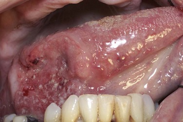
Maxillofacial. There is a 3.5-cm red and white, fungating mass on the right lateral border of tongue with central ulceration (a nonhealing ulcer in the oral cavity is considered to be SCCa until proved otherwise) (Figure 11-1). There is no pain or bleeding noted on palpation of the lesion (although ulcers from SCCa may occasionally bleed, they are usually painless). Some patients may also complain of ear pain if the lesions are deep and involve the lingual nerve. When ear pain is present, perineural invasion cannot be ruled out until final pathology. Examination of the remaining oral cavity, including the buccal mucosa, hard and soft palate, parotid and submandibular glands, oropharynx, and nasopharynx, reveals no other abnormalities. Nasopharyngoscopy reveals no abnormal tissues in the posterior oropharynx, subglottic or supraglottic regions, or nasopharynx (nasopharyngoscopy should be performed as part of the head and neck evaluation of tongue SCCa).

Neck. No cervical or submandibular lymphadenopathy is noted (cancers of the tongue usually metastasize to the level I and II nodes). There is no pain on palpation of the neck (lymphadenopathy from cancer is usually painless).

The presence of occult neck disease in the N0 neck is related to the tumor’s stage, size, and depth of invasion; perineural invasion; and histologic grade. Lesions greater than 4 mm in depth, along with a high-grade histology, have a greater than 20% risk of neck disease in the N0 neck.

Imaging

The initial imaging modalities for the evaluation of patients with SCCa begin with a panoramic radiograph. This is a useful screening tool to evaluate for the presence of bony infiltration associated with the tumor. It also provides valuable information regarding the long-term prognosis of the remaining dentition, because some patients may require extraction of carious or periodontally involved teeth before radiotherapy.

A computed tomography (CT) scan of the head and neck is the commonly used imaging study of choice to delineate the lesion and assess the neck for cervical lymphadenopathy (nodes greater than 1.5 cm, with central necrosis, an ovoid shape, and fat stranding are indicative of nodal metastasis). Additional tests, such as magnetic resonance imaging (MRI) and ultrasonography, can be used to assess the status of the cervical nodes.

Anteroposterior and lateral chest radiographs are used to screen for underlying pulmonary disease and evaluate for pulmonary metastasis, because the lungs are the most common areas of metastasis for this tumor. Positron emission tomography (PET) scans are becoming a common modality for the evaluation of distant metastasis. This technology uses an 18F-fluorodeoxyglucose (FDG) marker to examine sites of increased glucose uptake, which are seen with metabolically active cancer cells. This imaging modality is commonly used to rule out distant disease, and it is also helpful for clinically staging the tumor. Several studies have demonstrated that a standardized uptake valve (SUV) of greater than 3 correlates with hypermetabolism suggestive of a pathologic process. Clinical staging is helpful, because a treatment plan can be worked up for the patient and adjuvant modalities recommended.

In the current patient, axial and coronal CT images of the head and neck, with and without contrast, revealed a 3.5-cm, well-circumscribed lesion of the right lateral border of the tongue musculature. No evidence of cervical lymphadenopathy was noted. The PET scan performed with 18F-FDG showed a hypermetabolic area in the right tongue coinciding with the clinical lesion. No other abnormal uptake in the neck or chest was noted. The panoramic and chest radiographs revealed no abnormalities.

Treatment

Treatment of SCCa of the tongue begins with a complete history and physical examination, including nasopharyngoscopy. This is followed by appropriate tests, including CBC with differentials, electrolytes, liver function tests, chest radiographs, and CT with contrast. The role of PET scanning for occult metastasis continues to evolve.

The treatment of SCCa is site specific; surgical ablation with minimum 1 to 1.5-cm margins is the main modality of treatment. Most oral cavity tumors are approached intraorally; however, some tumors may need to be accessed extraorally via a transfacial approach. When the tumor is located in the mandible, the inferior border can be preserved (marginal mandibulectomy), depending on the degree of infiltration. However, when the cancellous portion of the mandible is invaded, segmental resection is required to maintain oncologic safety.

A common procedure that accompanies the removal of the tumor is the removal of the fibrofatty contents of the neck, for treatment of cervical lymphatic metastases and for complete staging of the cancerous process (see the section Neck Dissections later in this chapter).

Reconstruction and rehabilitation. Depending on the defect, the reconstructive surgery can be divided into soft tissue and/or bony reconstruction. Closing the defect primarily is ideal if it can be accomplished. Soft tissue surgical procedures include closure by secondary intention, skin grafts, local flaps, or microvascular free flaps. Simultaneous bony reconstruction can be accomplished using vascularized free flaps from the iliac crest, scapula, or fibula when needed. When large ablative and reconstructive procedures are performed, they can be performed simultaneously (see Chapter 12). Depending on the amount of healing and dysfunction anticipated, a percutaneous endoscopic gastrostomy tube and elective tracheostomy can be performed to secure the airway and aid in the nutritional support of the patient during the postoperative period.

Radiation therapy. Radiation therapy can be used as a primary or an adjuvant therapy. Primary radiotherapy is usually reserved for patients with significant comorbidities or when the primary tumor or the patient is not amenable to surgery. This is not a primary indication for early-stage SCCa because of the associated morbidity, including dysphagia and xerostomia. Another significant risk is the development of metachronous lesions after radiation therapy.

Postoperative radiation therapy is commonly used as a part of the comprehensive treatment. The indications for its use include positive or near margins, significant perineural or perivascular invasion, bone involvement, multiple nodal involvement, extracapsular spread, or stage III or stage IV disease. Typically, about 6,000 cGy in divided doses is administered, and treatment is initiated soon after healing from the initial surgery is complete. Surgery combined with radiation therapy and chemotherapy has increased the 5-year survival rates for stage III and stage IV cancers by 10%.

In the current patient, a right partial glossectomy via a transoral approach was performed with 1.5-cm margins. The status of the margins was evaluated using frozen section microscopy, which demonstrated negative margins. An ipsilateral supraomohyoid neck dissection (levels I through III) was completed for staging, which revealed no positive lymph nodes. The tongue defect was reconstructed with a radial forearm free flap, anastomosing with the facial artery and vein. An elective tracheostomy was performed.

After complete healing, the patient was followed closely for signs of recurrence (85% of recurrences occur in the first 3 years after initial treatment).

Complications

Complications are best categorized as intraoperative, postoperative (within 1 month), and long term (after 1 month).

Intraoperative complications. The main intraoperative concerns associated with oncologic ablative and reconstructive surgery include control of hemorrhage and anesthetic complications. Damage to adjacent structures, such as the lingual nerve and Wharton’s duct, is possible with ablative procedures of the tongue and floor of the mouth. When simultaneous neck dissection is performed, additional complications may be seen, such as nerve palsies (facial and spinal accessory nerves),vascular injury to the carotid artery or internal jugular vein and, more rarely, pneumothorax, air embolism, and formation of a chylous fistula (especially on the left side).

Postoperative complications. Postoperative complications include wound infection, hematoma, skin necrosis, flap failure, orocutaneous fistula, poor speech, and swallowing dysfunction. Poor healing can also be noted in patients who are alcoholics, because their prealbumin status is usually low. Complications after bony reconstruction include malunion, nonunion, contour irregularities, resorption of bone, osteomyelitis, and hardware failure.

Long-term complications. The gravest long-term complications are recurrence of the primary tumor and death (85% of recurrences occur in the first 3 years). The lifetime risk of development of a second primary tumor is 2% to 3% per year, and the 5-year survival rate is 56% for all tumor stages. Routine diagnostic tests are performed based on the clinical suspicion of recurrence. Imaging studies can be difficult to assess in the postoperative setting due to the difficulty of separating recurrent tumors from postoperative anatomic changes. Recurrent disease usually occurs at the surgical wound margin. Other complications include lingual nerve hypoesthesia, duct obstruction, and flap failure. Dysphagia, xerostomia, mucositis, and the risk of osteoradionecrosis are associated with radiation therapy. The most common causes of death in patients with oral cancer are related to locoregional disease, distant metastasis, or cardiopulmonary failure. Metastases of SCCa tend to involve the lung, bones, liver, and brain.

Discussion

SCCa accounts for roughly 90% of neoplastic cases in the head and neck. It has a 3 : 1 male predilection, and the median age of onset (diagnosis) is in the sixth decade of life (although recently there has been an increasing prevalence in the third decade). The overall 5-year survival rate for SCCa is over 50%. African Americans are reported to have significantly lower survival rates, approaching 35%. In 2005, an estimated 29,370 new cases were diagnosed, and 7,320 patients died of their disease. Roughly two thirds of these cases can be prevented with cessation of known risk factors (tobacco and alcohol).

From an epidemiologic and clinicopathologic standpoint, carcinomas in the head and neck region can be divided into three anatomic areas:

Figure 11-2 shows a different patient with a large fungating SCCa of the right retromolar area.
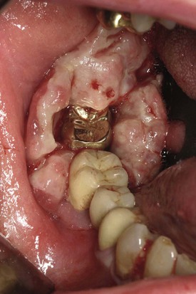
In addition to alcohol and tobacco, the use of betel quid, or paan, which is popular in India and southeast Asia, has been associated with an increased risk for developing SCCa. The quid consists of a betel leaf wrapped around a mixture of areca nut and slaked lime, commonly in combination with tobacco. The slaked lime releases an alkaloid from the areca nut, causing a feeling of euphoria. Chronic use of the quid can lead to a debilitating condition known as submucous fibrosis, which is a premalignant condition.

Human papillomavirus (HPV) types 16 and 18 have been shown to increase the risk of SCCa. Data from recent studies show that HPV 16 and 18 increase the ratio of SCCa by approximately threefold to fivefold. Tonsillar SCCa has the highest rate of HPV infection, with approximately 50% testing positive. These HPV-positive oropharyngeal tumors are usually treated with chemoradiation.

Other known risks factors include chronic sun exposure leading to cutaneous SCCa of the lip. Several studies have suggested that oral lichen planus, particularly the erosive form, is associated with an increased risk for SCCa. Severe iron deficiency presenting as Plummer-Vinson syndrome is associated with an increased risk for pharyngeal and esophageal SCCa. Previous radiation exposure is linked to an increased risk for developing SCCa.

Premalignant conditions. There are several well-known entities pathologically that have a distinct association with SCCa: leukoplakia, erythroplakia, and lichen planus.

Leukoplakia is a white patch or plaque that cannot be characterized clinically or pathologically as any other disease. Erythroplakia is defined as a red lesion of the oral cavity that cannot be classified clinically or pathologically as any other lesion (see the section on Oral Leukoplakia in Chapter 7).

Oral lichen planus has been a subject of controversy in the literature concerning its possible role as a premalignant condition. Several recent studies have shown that the transformation rate of oral lichen planus to SCCa is approximately 0.04% to 1.74%.

Early oral SCCa usually presents as one of the premalignant conditions discussed: a white, red, or mixed red and white lesion. As the lesion matures, it can become centrally ulcerated, and the borders become less distinct. The surface can become exophytic with papillary projections or endophytic with raised, rolled borders.

The tongue is the most common site of SCCa (30%), followed closely by the floor of the mouth (28%). These sites are at the higher risk, probably due to carcinogens that pool with saliva in these areas, contributing to a greater exposure. The thin, nonkeratinized layer of epithelium in these areas may contribute to the greater susceptibility. The other areas of prevalence, in descending order, are the upper and lower alveolar ridges (including the hard palate), the retromolar trigone, buccal mucosa, and the lips.

Broders’ classification system is an index of malignancy based on the fact that less differentiation (differentiation is defined as the degree of keratinization) is proportionally related to greater malignancy of the tumor. The classification is as follows:

Histopathologic factors correlating with a poorer outcome include depth of invasion, perineural invasion, and extracapsular spread.

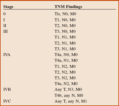
The 5-year survival rate remains about 50% and is related to the stage at diagnosis (Table 11-1). Unfortunately, there has been only a modest improvement in survival over the past several decades.

Table 11-1

Oral Squamous Cell Carcinoma Survival Rates by Stage

image

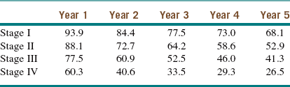
Modified from the National Cancer Institute, US National Institutes of Health: Surveillance epidemiology and end results (SEER). Available at http://seer.cancer.gov/publicdata/access.html. Accessed December 2012.

Bibliography

Braakhuis, BJM, Tabor, MP, Kummer, JA, et al. A genetic explanation of Slaughter’s concept of field cancerization: evidence and clinical implications. Cancer Res. 2003; 63:1727–1730.

Broders, AC. Carcinomas of the mouth: types and degrees of malignancy. Am J Roentgenol Radium Ther Nucl Med. 1927; 17:90–93.

Funk, FF, Karnell, LH, Robinson, RA, et al. Presentation, treatment, and outcome of oral cavity cancer: a national cancer data base report. Head Neck. 2002; 24:165–180.

Hillbertz, NS, Hirsch, JM, Jalouli, J, et al. Viral and molecular aspects of oral cancer. Anticancer Res. 2012; 32:4201–4212.

Kademani, D, Bell, RB, Bagheri, SC, et al. Prognostic factors for intraoral squamous cell carcinoma: the influence of histologic grade. J Oral Maxillofac Surg. 2005; 63:1599–1605.

Marur, S, D’Souza, G, Westra, WH, et al. HPV-associated head and neck cancer: a virus-related cancer epidemic. Lancet Oncol. 2010; 11:781–789.

McClure, SA, Mohaved, R, Salama, A, et al. Maxillofacial metastases: a retrospective review of one institution’s 15-year experience. J Oral Maxillofac Surg. 2012; 16(2):181–188.

Neville, BW, Day, TA. Oral cancer and precancerous lesions. CA Cancer J Clin. 2002; 52:195–215.

Oliver, AJ, Helfrick, JF, Gard, D. Primary oral squamous cell carcinoma. J Oral Maxillofac Surg. 1996; 54:949–954.

Reichart, PA, Philipsen, HP. Oral erythroplakia: a review. Oral Oncol. 2005; 41:551–561.

Schmidt, BL, Dierks, EJ, Homer, L, et al. Tobacco smoking history and presentation of oral squamous cell carcinoma. J Oral Maxillofac Surg. 2004; 62:1005–1058.

Schwartz, LH, Ozsahin, M, Zhang, GN, et al. Synchronous and metachronous head and neck carcinomas. Cancer. 1994; 74:1933–1938.

Shafer, WG, Waldron, CA. Erythroplakia of the oral cavity. Cancer. 1975; 36:1021–1028.

Smith, GI, O’Brien, CJ, Clark, J, et al. Management of the neck in patients with T1 and T2 cancer in the mouth. Br J Oral Maxillofac Surg. 2004; 42:494–500.

Syrjanen, S. Human papillomavirus (HPV) in head and neck cancer. J Clin Virol. 2005; 32S:S59–S66.

Todd, R, Donoff, RB, Wong, DTW. The molecular biology of oral carcinogenesis: toward a tumor progression model. J Oral Maxillofac Surg. 1997; 55:613–623.

Van der Meij, EHDDS, Schepman, KP, Van der Waal, I. The possible premalignant character of oral lichen planus and oral lichenoid lesions: a prospective study. Oral Surg Oral Med Oral Pathol Oral Radiol Endod. 2003; 96:164–171.

Waldron, CA, Shafer, WG. Leukoplakia revisited: a clinicopathological study of 3,256 oral leukoplakias. Cancer. 1975; 36:1386–1392.

Wooglar, JA. Histological distribution of cervical lymph node metastases from intraoral/oropharyngeal squamous cell carcinomas. Br J Oral Maxillofac Surg. 1999; 37:175–180.

Verrucous Carcinoma*

Stay updated, free dental videos. Join our Telegram channel

Jan 12, 2015 | Posted by in Oral and Maxillofacial Surgery | Comments Off on 11: Oral Cancer

VIDEdental - Online dental courses

Get VIDEdental app for watching clinical videos