3: Anesthesia

Anesthesia

Administration of anesthesia remains an important part of office-based oral and maxillofacial surgery. The most important step in delivering safe and effective anesthesia is preparation. Preparation begins with a thorough knowledge and understanding of the anatomy, physiology, and pharmacology relevant to anesthesia. From this point, safe anesthetic techniques are developed and used based on the preoperative patient evaluation, practitioner’s preference, and individual clinical situations. Preparation also includes measures to prevent and manage emergencies. Despite a thorough preoperative patient evaluation, use of safe and proven anesthetic techniques, and vigilant monitoring, emergency situations may arise. For these reasons, it is essential that all practitioners continually hone their skills in the recognition and management of life-threatening emergencies in the office or operating room. Our excellent safety record is evidence of oral and maxillofacial surgeons’ training and preparation in the delivery of safe anesthesia.

Each of the following teaching cases deals with the management of specific clinical scenarios. We also include a new case discussing trigeminal neuralgia/facial pain. The sections are structured to emphasize the key points in the preoperative evaluation and recognition of impending emergencies. Strategies for reducing the risk of emergent situations—and for their management when they do arise—are discussed. The highlighted clinical pearls in the preoperative patient evaluation should be incorporated into all practitioners’ routine preoperative assessment.

The intent of this section is to familiarize readers with the risk factors and clinical signs associated with disastrous outcomes involving anesthesia (local, sedation, or general).

Laryngospasm

HPI

Preoperative evaluation of the patient revealed no recent respiratory tract infections. The lungs were clear to auscultation. After ECG, blood pressure, pulse oximeter, and capnography monitors were applied, the patient was administered 4 L of oxygen and 2 L of nitrous oxide via nasal hood. Sedation was achieved using 4 mg of midazolam and 50 µg of fentanyl titrated to effect. Prior to administration of local anesthesia, 40 mg of propofol was infused. During the first extraction, respiratory stridor (a high-pitched, inspiratory “crowing” sound) was noted. A noisy, harsh sound was heard on inspiration through the precordial stethoscope, and the patient’s oxygen saturation decreased from 99% to 65%. Capnography indicated no ventilation. At this point, the respiratory noises ceased. Tracheal tug and paradoxical chest wall motion were observed (signs of upper airway obstruction), and the patient began to appear cyanotic.

Examination

General. A harsh inspiratory noise, or crowing, is audible on inspiration, which is best heard through the precordial stethoscope. The patient’s skin color is assessed for signs of cyanosis, which is seen with severe hypoxemia. In pediatric patients, hypoxemia is often a late finding of decreased ventilation or apnea. End tidal CO2 monitoring and use of the precordial stethoscope indicate hypoventilation or apnea prior to changes in pulse oximetry.

Oropharynx. The throat pack is removed, and there is no evidence of foreign bodies. Copious amounts of mucous secretions are observed. (Blood and mucus are common stimuli for airway irritation.)

Neck and chest. There is evidence of tracheal tug and paradoxical chest wall motion (despite chin-lift and jaw-thrust maneuvers). This phenomenon is the result of forced inspiration against a closed glottis.

Vital signs. The patient’s heart rate is 160 bpm, blood pressure 145/78 mm Hg, Etco2 0, and respirations 0 breaths per minute.

Oxygen saturation. Oxygen saturation decreased from 99% to 65% with the onset of laryngospasm. (Continued decline in the oxygen saturation can result in respiratory acidosis.)

ECG. The patient is in sinus tachycardia. (This is a common finding, but hypoxia can trigger more life-threatening cardiac arrhythmias. Hypoxemia in children may result in bradycardia.)

Treatment

Prompt recognition and treatment of laryngospasm usually result in a good outcome. Upon diagnosis, the airway should be suctioned clear of noxious stimuli and the surgical site should be packed. Any foreign bodies are removed from the oral cavity and 100% oxygen is administered. Positive pressure ventilation should be attempted, ideally with a two-person technique and jaw-thrust maneuver; this often “breaks” the laryngospasm (jaw thrust and pressure at the angle of the mandible may also assist in breaking laryngospasms).

A technique described by Dr. N.P. Guadagni also has been found to effective at “breaking” laryngospasm. This involves placing the middle finger of each hand anterior to the mastoid and posterior to the condyle. The fingers then press inward while at the same time positioning the mandible forward. If the patient cannot be ventilated, the plane of anesthesia may be deepened with a short-acting intravenous general anesthetic; this often obviates the need for a skeletal muscle relaxant.

In rare situations these methods are unsuccessful, and it is necessary to administer succinylcholine, a short- and fast-acting depolarizing neuromuscular blocking agent. If intravenous access is not available, succinylcholine may be administered intramuscularly at a dose of 4 mg/kg. A dose of 20 mg intravenously is usually sufficient to break the spasm (pediatric dose, 0.25 mg/kg). However, up to 60 mg can be administered if laryngospasm persists. Rapacurium, rocuronium, and mivacurium can be used for patients in whom succinylcholine is contraindicated. The longer half-life of these nondepolarizing muscle relaxants may require continuous bag-mask ventilation until spontaneous respiration resumes.

Bradycardia is not uncommon after administration of succinylcholine. This usually occurs in children and in adults after repeated doses. Atropine may be administered in an effort to prevent this. Intravenous lidocaine 2 mg/kg administered before extubation was found to be effective in preventing postextubation laryngospasm in patients undergoing tonsillectomy. Other studies have found the prophylactic use of intravenous lidocaine to be ineffective.

Complications

Laryngospasm may produce partial or complete respiratory obstruction. Fortunately, early recognition and management allow for rapid resolution and minimal morbidity. However, with prolonged hypoxemia, the complications can be devastating. Laryngospasm may result in an acid-base disturbance, such as respiratory acidosis. Rare complications of laryngospasm include cardiac arrhythmias, anoxic brain injury, negative pressure pulmonary edema, and death.

If succinylcholine is administered, the patient may complain of general postoperative myalgia secondary to the rapid muscle depolarization. Other potential complications of succinylcholine include masticator muscle rigidity, malignant hyperthermia, and hyperkalemic cardiac arrest (secondary to the transient hyperkalemia), which can be seen in patients with undiagnosed myopathies (e.g., Duchenne’s and Becker’s muscular dystrophies).

Discussion

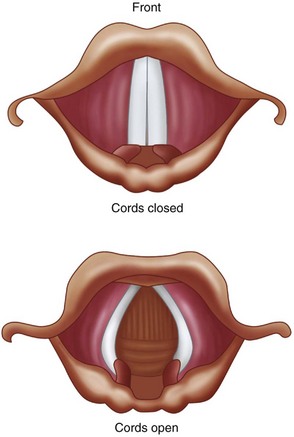
Laryngospasm results in tight approximation of the true vocal cords (Figure 3-1). It is a protective reflex that is most commonly caused by a noxious stimulus to the airway during a light plane of anesthesia. The structural and functional bases of the laryngospasm reflex were described by Rex. Secretions, vomitus, blood, pungent volatile anesthetics, painful stimuli, and oral and nasal airways may elicit this protective reflex. Mediated by the vagus nerve, this reflex is designed to prevent foreign materials from entering the tracheobronchial tree. During laryngospasm, the false vocal cords and supraglottic tissues act as a ball valve and obstruct the laryngeal inlet during inspiration. Laryngospasm has a reported occurrence of 8.7 per 1,000 patients receiving general anesthesia. It is 19 times more frequent than bronchospasm.

Laryngospasm accounts for more than 50% of the cases of negative pressure/post obstructive pulmonary edema. With the use of general endotracheal intubation, laryngospasm classically occurs during extubation in a light plane of anesthesia (stage II). Children and patients who have had a recent upper respiratory tract infection are predisposed to developing laryngospasm during anesthesia.

Efforts to prevent laryngospasm include postponing surgery in patients who have had recent upper respiratory infections, maintaining a dry surgical field, and using anticholinergics and avoiding extubation during stage II of anesthesia. Laryngospasm is not uncommon in outpatient and inpatient oral and maxillofacial surgery. Recognition and early intervention are essential in preventing morbidity and mortality.

Bibliography

Baraka, A. Intravenous lidocaine controls extubation laryngospasm in children. Anesth Analg. 1978; 57:506–507.

Ciavarro, C, Kelly, JP. Postobstructive pulmonary edema in an obese child after an oral surgery procedure under general anesthesia: a case report. J Oral Maxillofac Surg. 2002; 60(12):1503–1505.

Hartley, M, Vaughan, RS. Problems associated with tracheal extubation. Br J Anaesth. 1993; 71:561–568.

Hurford, WE, Bailin, MT, Davison, JK, et al. Clinical procedures of the Massachusetts General Hospital, ed 5. Philadelphia: Lippincott-Raven; 1998.

Larson, CP. Laryngospasm–the best treatment. Journal of the American Society of Anesthesiologists. 1998; 89(5):1293–1294.

Leicht, P, Wisborg, T, Chraemmer-Joorgensen, B. Does intravenous lidocaine prevent laryngospasm after extubation in children? Anesth Analg. 1985; 64:1193–1196.

Louis, PJ, Fernandes, R. Negative pressure pulmonary edema. Oral Surg Oral Med Oral Pathol Oral Radiol Endod. 2002; 93(1):4–6.

Rex, MAE. A review of the structural and functional basis of laryngospasm and a discussion of the nerve pathways involved in the reflex and its clinical significance in man and animals. Br J Anaesth. 1970; 42:891–898.

Stoelting, RK, Miller, RD. Basics of anesthesia, ed 3. New York: Churchill Livingstone; 1994.

Perioperative Considerations for the Pregnant Patient

HPI

The patient presents complaining of a 2-week history of dental pain in the right mandible and a 2-day history of progressive swelling of her right face. She is being followed by her obstetrician, and she states that her pregnancy is progressing without complications. Recently she has noted good fetal movements. She denies having any vaginal bleeding or leakage of vaginal fluid (a sign that amniotic fluid may be leaking from ruptured membranes). She started having pelvic cramping just recently (cramping described by pregnant patients may actually be contractions). She has been taking acetaminophen (the analgesic of choice during pregnancy) for pain and has not been able to eat adequately for the past 3 days (pregnant patients have a higher nutritional and fluid requirement). There is no history of dysphagia (difficulty swallowing), odynophagia (painful swallowing), dyspnea (difficulty breathing), or subjective fevers.

Examination

Vital signs. The patient’s blood pressure was 110/55 mm Hg, heart rate 110 to 140 bpm (tachycardic), respirations 20 per minute, and temperature 39°C (febrile).

General. She is well developed and well nourished and in no apparent distress.

Maxillofacial. There is a large right-sided facial swelling. The swelling is above the inferior border of the mandible (rules out submandibular space involvement) and below the zygomatic arch (buccal space abscess). It extends from the masseter (submasseteric space involvement) to the oral commissure. The swelling is tense, erythematous, warm, and tender to palpation. The submandibular and submental regions are normal. She has limited opening (trismus due to involvement of the lateral masticator space) secondary to pain, which limits the intraoral examination. The right mandibular first molar is grossly decayed, with adjacent vestibular swelling and purulence extravasating from the gingival sulcus. The oropharynx is not completely visible due to limited mouth opening. The floor of the mouth is nonelevated (rules out sublingual space involvement). She is in no respiratory distress and is tolerating her secretions well.

Cardiovascular. The patient is tachycardic with an II/VI systolic ejection murmur. (Early systolic ejection murmur is very common in pregnant women due to the high volume of flow; it is accentuated by acute tachycardia secondary to the elevated temperature.)

Abdominal. Examination reveals a gravid uterus (pregnant uterus) appearing larger than the stated gestational age (due to twins), nontender abdomen, and fetal heart rates of 190s for twin A (the lower presenting fetus in the uterus) and 180s for twin B (the heart rates are elevated above normal due to maternal temperature).

Extremities. The extremities are nontender with 1+ pitting edema at the ankles bilaterally (common during pregnancy), no cords, and 2+ equal pulses. She has a negative Homans sign (calf pain upon dorsiflexion of the foot, suggestive of deep vein thrombosis [DVT]).

Imaging

A panoramic radiograph is the initial diagnostic study of choice. When the oropharynx cannot be adequately visualized due to trismus or when a deep neck space abscess is suspected, computed tomography (CT) scanning of the head and neck is necessary to rule out involvement of the parapharyngeal spaces (lateral pharyngeal and retropharyngeal spaces). The use of CT with intravenous contrast material is generally considered safe during pregnancy; however, special attention must be paid to the gestational age of the fetus and the amount of ionizing radiation that would be absorbed. In this particular case, the approximate exposure rate is less than 0.05 rad per examination, which is significantly less than the 5 rad cumulative upper limit for exposure during pregnancy (Dollard). The use of iodinated IV contrast does not produce any radiation exposure. The benefit of using IV contrast outweighs any theoretical risk of transient neonatal thyroid suppression; therefore, it can be used as needed, as long as there is no other contraindication to using the contrast material.

Despite the potential theoretical effects of radiation exposure to the fetus, all necessary plain film and CT studies for diagnosing and managing head and neck infections can be safely performed as needed. The radiation exposure to the developing fetus is minimal, especially when imaging the head and neck, and is further reduced by using shielding devices. Furthermore, the benefits outweigh the risks of exposure when dealing with acute head and neck infections. Nonionization techniques, such as ultrasound scans and magnetic resonance imaging (MRI) of the head and neck, are also considered safe during pregnancy and can aid in imaging soft tissue pathology. The use of gadolinium contrast with an MRI is currently not recommended during pregnancy, although there have not been any reported adverse fetal outcomes. Gadolinium does cross the placenta and can accumulate in the amniotic fluid of the fetus. The risk of nephrogenic systemic fibrosis with gadolinium is rare but warrants special attention in patients with renal insufficiency.

In this particular patient, the panoramic radiograph showed a grossly decayed right mandibular first molar with a large periradicular radiolucent lesion. CT was not indicated, because the clinical suspicion of parapharyngeal space involvement was low.

Treatment

The ideal time to perform elective or semielective oral and maxillofacial surgical procedures is postpartum; otherwise, the second trimester is considered the safest period for performing nonelective surgery. However, urgent or emergent surgery should not be delayed at any gestation of pregnancy, especially if procedures can be carried out under local anesthesia. Local anesthesia is the preferred method for simple procedures that can be performed in an office setting (there are no contraindications to vasoconstrictors, but aspiration to avoid intravascular injection is important). If the need arises, intravenous sedation and general anesthesia (in a hospital setting, when appropriate) can be safely performed without significant risk to the mother or fetus in an uncomplicated pregnancy (this is discussed later in the chapter).

There should be a lower threshold for hospital admission in the pregnant patient with a maxillofacial infection. Fever, dehydration, inability to tolerate oral intake, and potential airway compromise are all indications for hospital admission and initiation of supportive measures. (The risk of airway and pharyngeal edema is higher during pregnancy, especially when parapharyngeal spaces are involved.) Other obstetric concerns include risk factors for preterm contractions and preterm labor (onset of labor before 37 weeks of gestation): twins, dehydration, infection, and potential early sepsis.

When a pregnant woman is hospitalized, an obstetric consultation should be obtained. Fluid resuscitation, intravenous antibiotics, fetal monitoring, and nutritional support are extremely important. Caution should be exercised to avoid excessive fluid overload that can lead to pulmonary edema, because pregnancy and sepsis both can lead to third spacing due to increased capillary permeability. Usually, a 500-ml crystalloid bolus, followed by a maintenance level of 100 to 150 ml/hr until the patient is tolerating liquids, is appropriate for the average patient. Intravenous antibiotics should also be initiated (the penicillin and cephalosporin families are considered safe first-line antibiotics during pregnancy). Oral and maxillofacial infections should be aggressively treated, because untreated infections and abscesses have been associated with preterm labor and maternal sepsis.

If a pregnant woman is admitted to the hospital, she and the fetus should be monitored for contractions and fetal well-being. Pain management with a patient-controlled analgesia pump until the patient is tolerating oral medications is appropriate. Intravenous morphine, meperidine (Demerol), and fentanyl or orally administered hydrocodone, oxycodone, or codeine with acetaminophen combinations are all considered safe during pregnancy for necessary pain control. Routine use of nonsteroidal antiinflammatory drugs (NSAIDs), such as ibuprofen and aspirin, for postoperative pain control during pregnancy is generally not recommended; however, these drugs can be used short term in the second trimester only.

The current patient was admitted to the hospital, and an obstetric consultation was obtained. Intravenous fluids and piperacillin/tazobactam 3.375 g every 6 hours were administered. The twins were evaluated for heart tones and contractions. No significant abnormalities were noted. The fetal heart rates were slightly higher than normal due to the maternal temperature but were otherwise reassuring. There were no contractions noted. The pelvic examination was unremarkable, with no evidence of cervical effacement (thinning and shortening of the cervix associated with labor) or of cervical dilatation (also a sign of labor).

The patient was taken to the operating room for incision and drainage of the submasseteric and buccal space abscess and extraction of the right mandibular first molar. Ideally, pregnant patients should be NPO (nothing to eat or drink, including chewing gum) for at least 8 hours before surgery. In addition to the slower gastric emptying time and the enlarged uterus (which takes up more abdominal space, thus leaving less room for the gastrointestinal organs), pregnancy causes relaxation of the esophageal sphincter. The combination of these conditions increases the risk of aspiration associated with general anesthesia. Therefore, preoperatively, this patient received an oral antacid (to increase the pH of gastric contents), an H2 antagonist (to decrease gastric acid production), and metoclopramide (to accelerate gastric emptying). Due to the gestational age of the patient, the uterus was enlarged (especially with twins). Therefore, a roll was placed under her right back and hip, and she was slightly tilted to the left. (A left lateral tilt of 15 to 30 degrees displaces the uterus off the aorta and inferior vena cava and prevents supine hypotensive syndrome, which is due to prolonged compression of the great vessels, leading to decreased venous return and cardiac output.)

After the patient had been positioned, she was intubated via an awake nasal fiberoptic intubation, due to her trismus (pregnant patients have edematous nasal mucosa and a potential for epistaxis, especially during a traumatic nasal intubation). The procedure was completed without complications. A thorough intraoperative examination of the oropharynx revealed no parapharyngeal involvement or edema. Her mouth opening increased to normal range after decompression of the lateral masticator (submasseteric) space. Her airway was deemed stable, without risk for postoperative upper airway obstruction, and she was successfully extubated. She remained in the hospital for a total of 4 days. She received daily antenatal testing to check for fetal well-being and contractions.

The patient was discharged home on postoperative day 4 after she had been afebrile for longer than 48 hours and was tolerating full liquids and a regular diet. She received intravenous antibiotics postoperatively until she was able to tolerate medications by mouth; she was then switched to oral antibiotics. Upon discharge, the patient’s facial swelling had decreased significantly, there was no oral purulent discharge, the WBC count had normalized and there was no bandemia, and pain control was adequate with opioid analgesics.

Complications

Complications associated with surgery under general anesthesia during pregnancy include the risk of DVT, pulmonary embolism (PE), aspiration (decreased esophageal sphincter tone, decreased gastric emptying, increased gastric pressures, and hyperemesis increase the risk of regurgitation and aspiration), pulmonary edema, acute respiratory distress syndrome (ARDS), spontaneous abortion during the first trimester, and preterm labor. These are weighed against the risks of an untreated oral or maxillofacial infection, which pose a greater direct danger to both the fetus and the mother; these risks include preterm labor and delivery with complications of a premature neonate, fetal death, maternal sepsis, and septic shock. All elective surgical procedures should be avoided during pregnancy, but necessary surgical interventions should not be delayed.

For the pregnant female undergoing general anesthesia, surgery should be performed in a setting where an obstetrician is available for consultation and where anesthesiologists are familiar with the physiologic changes associated with pregnancy. Teratogenic and abortive agents should be avoided (this is most important during the first trimester). Specific modifications may be needed as the gestational age of the fetus advances (discussed previously). A collaborative, multidisciplinary approach involving obstetricians, anesthesiologists, and oral and maxillofacial surgeons provides the most appropriate management and treatment plan for the pregnant patient.

A potential complication risk that is increased in pregnancy is the risk of deep vein thrombosis and pulmonary embolism due to the prothrombotic state of a pregnant woman. Physiologic changes that occur during pregnancy, such as increased clotting factors, increased plasma volume, increased venous stasis, decreased blood flow velocity, and decreased fibrinolytic activity, increase the risk of DVT (signs include leg pain, tenderness, edema, discoloration, palpable cord, and positive Homans sign) and subsequent PE (signs include shortness of breath, tachypnea, hypoxemia, and respiratory distress) by twofold to fourfold during pregnancy and early postpartum (Cromwell). Other exogenous factors, such as tobacco use, obesity, and immobility, add even further risks. Venous thromboembolic events are still a leading cause of maternal morbidity and mortality. Early detection of DVT (using duplex Doppler ultrasound) and initiation of heparin therapy have significantly reduced maternal mortality. Chest CT or a V/Q scan are options for evaluation if a pulmonary embolus is suspected. DVT/PE prophylaxis should be initiated upon the patient’s admission, with the use of support stockings, compression devices, and/or subcutaneous heparin.

Discussion

Several other anesthetic considerations in pregnancy are noteworthy. Propofol, thiopental, etomidate, and ketamine are examples of generally safe induction agents for the pregnant patient. The use of a 50 : 50 mixture of nitrous oxide and oxygen, and of halogenated agents (desflurane, isoflurane, sevoflurane) in low concentrations, is also considered safe. Careful examination of the patient’s oropharyngeal and neck structures, noting range of movement and any obstructing lesions, is critical. Preoxygenation, intravenous fluid resuscitation, and left lateral tilt are all important steps in preventing hypoperfusion in both the mother and the fetus. Opiates, including fentanyl and morphine, are also considered safe to administer.

The use of local anesthesia is safe during pregnancy and is well tolerated by most pregnant patients undergoing minor oral surgical procedures. Although there is a theoretical concern that epinephrine-induced vasoconstriction could lead to decreased placental blood flow, epinephrine in local anesthetics is generally considered safe. Local anesthesia without epinephrine is an alternative. Despite concerns about potential teratogenic effects of benzodiazepines in the first trimester, they can be safely administered when the usual and appropriate doses are used.

For nursing mothers, it has been historically recommended that the patient “pump and dump” after receiving a general anesthetic. With current medications, nursing mothers can pump and discard breast milk for 8 to 24 hours after receiving an intravenous sedative or general anesthetic, to err on the side of caution. However, most agents have a very short half-life and very minimal crossover into breast milk that would affect the baby. Postoperative analgesics (hydrocodone, oxycodone, morphine, ketorolac [Toradol], NSAIDs) are safe to use without pumping and discarding breast milk. Perioperative intravenous steroids to help reduce postoperative swelling can be used in pregnancy and in breast-feeding mothers if necessary. Antinausea medications, as needed, also are generally safe to use.

Bibliography

Briggs, GG, Freeman, RK, Yaffe, SJ. Drugs in pregnancy and lactation, ed 5. Philadelphia: Williams & Wilkins; 2005.

Cromwell, C. Hematologic changes in pregnancy. In Hoffman R, Benz EJ, Jr., Silberstein L, et al, eds. : Hoffman hematology: basic principles and practice, ed 6, St Louis: Saunders, 2012.

Cumminham, FG, Leveno, KJ, Bloom, SL, et al. Williams obstetrics, ed 21. New York: McGraw-Hill Professional; 2005.

Dollard, DF. Radiation in pregnancy and clinical issues of radiocontrast agents. In Roberts JR, Hedges JR, eds. : Clinical procedures in emergency medicine, ed 5, St Louis: Saunders, 2009.

Hawkins, JL, Bucklin, BA. Obstetrical anesthesia. In Gabbe SG, Niebyl JR, Galan HL, et al, eds. : Obstetrics: Normal and problem pregnancies, ed 6, St Louis: Saunders, 2012.

Lawrenz, DR, Whitley, BD, Helfrick, JF. Considerations in the management of maxillofacial infections in the pregnant patient. J Oral Maxillofac Surg. 1996; 54:474–485.

Mozurkewich, EL, Pearlman, MD. Trauma and related surgery in pregnancy. In Gabbe SG, Niebyl JR, Galan HL, et al, eds. : Obstetrics: Normal and problem pregnancies, ed 6, St Louis: Saunders, 2012.

Schwartz, N, Adamczak, J, Ludmir, J. Surgery during pregnancy. In Gabbe SG, Niebyl JR, Galan HL, et al, eds. : Obstetrics: Normal and problem pregnancies, ed 6, St Louis: Saunders, 2012.

Turner, M, Aziz, SR. Management of the pregnant oral and maxillofacial surgery patient. J Oral Maxillofac Surg. 2002; 60:1470–1488.

Respiratory Depression Secondary to Oversedation

HPI

The patient is an otherwise healthy woman for whom treatment was planned for bilateral upper and lower eyelid blepharoplasties with intravenous sedation. After the incision lines had been marked in the usual manner, ECG, blood pressure, pulse oximeter and a sidestream capnograph monitors were applied. The patient was administered 4 L of oxygen and 2 L of nitrous oxide via nasal hood (nitrous oxide decreases the amount of intravenous sedatives needed). Sedation was achieved using 5 mg of midazolam, 100 µg of fentanyl, and a propofol drip titrated to effect. Verrill’s sign (50% upper eyelid ptosis, indicating adequate sedation) was observed. Prior to administration of local anesthesia, 40 mg of propofol was administered as a bolus (propofol may cause a 20% to 25% drop in systolic blood pressure when given as a bolus). Upon administration of local anesthesia, loss of the capnogram, with no chest wall movement, was observed. (This indicates the presence of central apnea. Capnography is considered to be more sensitive than clinical assessment of ventilation in the detection of apnea. In a study by Soto and colleagues (2004), 10 of 39 patients (26%) experienced 20-second periods of apnea during procedural sedation and analgesia. All 10 episodes of apnea were detected by capnography but not by the anesthesia providers.) The apnea was attributed to the propofol bolus (combined with the respiratory depressant effects of fentanyl), which was anticipated to resolve shortly. However, the patient continued to be apneic, and her oxygen saturation decreased from 99% to 80% (pulse oximeter readings are about 30 seconds behind the real-time oxygen saturation). Tracheal tug and paradoxical chest wall motion were not observed (these would be signs of upper airway obstruction and inspiratory efforts). The patient began to appear cyanotic (bluish hue to facial skin and lips due to prolonged hypoxemia).

PMHX/PDHX/Medications/Allergies/SH/FH

A thorough medical history is important during the preoperative evaluation of any patient undergoing intravenous sedation or general anesthesia to identify potential risk factors of intraoperative or postoperative anesthetic complications.

The past medical and surgical histories are noncontributory. This patient is categorized as American Society of Anesthesiologist (ASA) Class I (Table 3-1). She does not use any medications and has no known drug allergies. She denies previous problems with local anesthetics (e.g., methemoglobinemia), intravenous sedation, or general anesthetics (problems with previous anesthesia or adverse drug reactions should alert clinicians to possible complications that may require modification of anesthetic techniques). There is no family history of complications with general anesthetics (e.g., malignant hyperthermia). She denies a history of drug or alcohol use (patients with a previous drug history or alcohol abuse may require higher doses of sedative-hypnotic drugs), and she does not smoke (smoking decreases oxyhemoglobin concentrations and increases pulmonary secretions).

Table 3-1

American Society of Anesthesiologists (ASA) Classification System for Stratifying Patients Preoperatively by Risk

image

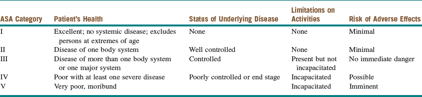
Modified from the American Society of Anesthesiologists: Relative value guide, 2003, Park Ridge, Ill, the American Society of Anesthesiologists.

Examination

Preoperative. A thorough preoperative evaluation is important to identify potential risk factors for negative anesthetic outcomes, with an emphasis on airway anatomy.

General. The patient is a well-developed and well-nourished woman in no apparent distress who weighs 60 kg.

Airway. Maximal interincisal opening (MIO) is within normal limits (difficult intubation occurs with decreased MIO). Her oropharynx is Mallampati class I (soft palate, tonsillar pillars, and uvula completely visualized), and the thyromental distance (TMD) is three finger-breadths (intubation is more difficult with retrognathia, a short TMD, and/or a higher Mallampati classification). The cervical spine has a full range of motion.

Cardiovascular. Heart is regular rate and rhythm without murmurs, rubs, or gallops.

Pulmonary. Lung fields are clear to auscultation bilaterally (preoperative wheezing may increase the risk of intraoperative bronchospasm).

Intraoperative. During the course of intravenous sedation (conscious sedation, deep sedation, or general anesthesia), it is important to continuously monitor the patient’s level of sedation and anesthesia (to prevent oversedation and respiratory depression) and to survey the ABCs (airway, breathing, and circulation [Box 3-1]).

General. The patient is sedated/unconscious and unresponsive to painful stimulus (a state of general anesthesia).

Stay updated, free dental videos. Join our Telegram channel

Jan 12, 2015 | Posted by in Oral and Maxillofacial Surgery | Comments Off on 3: Anesthesia

VIDEdental - Online dental courses

Get VIDEdental app for watching clinical videos