CHAPTER 4
Extraction Site (Socket) Preservation
Christopher Choi,1 Ray Lim,2 and Dale J. Misek2
1Private Practice, Inland Empire Oral and Maxillofacial Surgeons, Rancho Cucamonga, California, USA
2Private Practice, Carolinas Center for Oral and Facial Surgery, Charlotte, North Carolina, USA; and
Department of Oral and Maxillofacial Surgery, Louisiana State University Health Science Center, New Orleans, Louisiana, USA
A method of preserving and augmenting bone height and width immediately after tooth extraction for future dental implant placement.
Indications
- Prevention of alveolar ridge atrophy after tooth extraction
- Restoration of bony defects caused by infection, trauma, and/or traumatic extractions
Contraindications
- Sites where future implant placement is unachievable
- Medical conditions that preclude implant placement
Technique
- Preoperative antibiotics and chlorhexidine rinses are administered immediately prior to extraction.
- Atraumatic exodontia is performed to maintain the buccal cortical plate and septal bone (see Figure 4.3 in Case Report 4.1).
- All periapical and granulation tissue is curetted from the extraction site.
- The extraction site is copiously irrigated, and doxycycline paste is applied to the extraction socket walls for 5 minutes.
- A particulate grafting medium is placed and compacted within the defect.
- A collagen membrane is placed (see Figure 4.4 in Case Report 4.1) to stabilize the graft and to guide tissue regeneration. A resorbable figure-of-eight suture is used to secure the collagen membrane and to close tissue elevations.

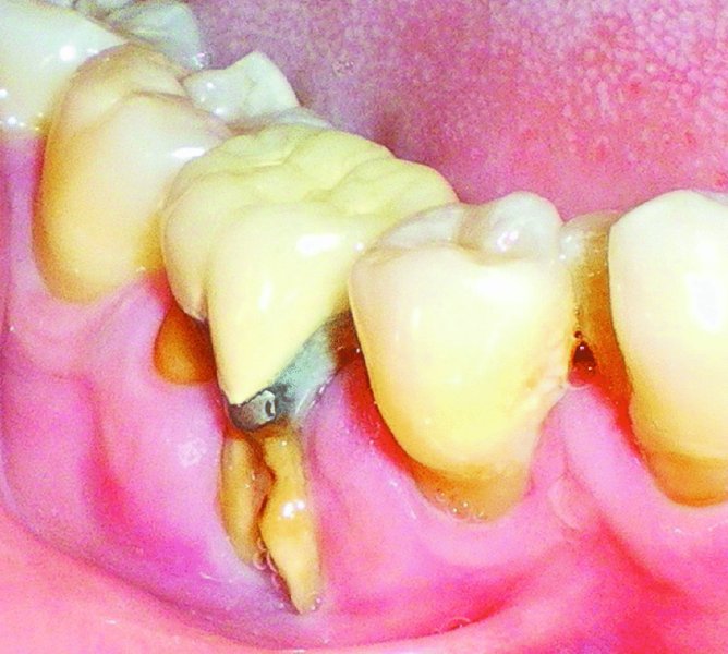
Figure 4.1. Buccal cortical defect and gingival recession associated with tooth #30.

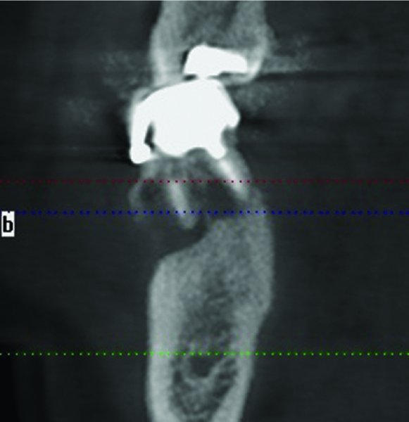
Figure 4.2. Cone beam computed t/>
Stay updated, free dental videos. Join our Telegram channel

VIDEdental - Online dental courses


