CHAPTER 4 Development and Morphology of the Primary Teeth
A complete review is available in the reference texts on oral histology, dental anatomy, and developmental anatomy listed at the end of the chapter. Furthermore, contemporary scientists are rapidly gaining knowledge of tooth development at the molecular level. We suggest that readers with a special interest in the molecular events of tooth development study the listed references by Smith1 and by Miletich and Sharpe.2
LIFE CYCLE OF THE TOOTH
INITIATION (BUD STAGE)
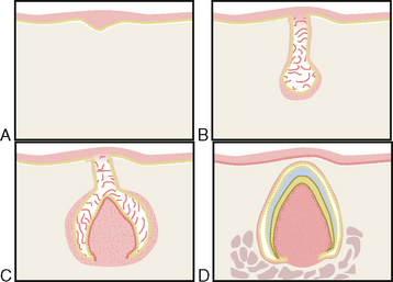
Certain cells of the basal layer begin to proliferate at a more rapid rate than do the adjacent cells (Fig. 4-1A). These proliferating cells contain the entire growth potential of the teeth. The permanent molars, like the primary teeth, arise from the dental lamina. The permanent incisors, canines, and premolars develop from the buds of their primary predecessors. The congenital absence of a tooth is the result of a lack of initiation or an arrest in the proliferation of cells. The presence of supernumerary teeth is the result of a continued budding of the enamel organ.
PROLIFERATION (CAP STAGE)
Proliferation of the cells continues during the cap stage. As a result of unequal growth in the different parts of the bud, a cap is formed (see Fig. 4-1B). A shallow invagination appears on the deep surface of the bud. The peripheral cells of the cap later form the outer and inner enamel epithelium.
As with a deficiency in initiation, a deficiency in proliferation results in failure of the tooth germ to develop and in less than the normal number of teeth. Excessive proliferation of cells may result in epithelial rests. These rests may remain inactive or become activated as a result of an irritation or stimulus. If the cells become partially differentiated or detached from the enamel organ in their partially differentiated state, they assume the secretory functions common to all epithelial cells, and a cyst develops. If the cells become more fully differentiated or detached from the enamel organ, they produce enamel and dentin, which results in an odontoma (see Fig. 7-5) or a supernumerary tooth. The degree of differentiation of the cells determines whether a cyst, an odontoma, or a supernumerary tooth develops (see Fig. 27-56).
HISTODIFFERENTIATION AND MORPHODIFFERENTIATION (BELL STAGE)
The epithelium continues to invaginate and deepen until the enamel organ takes on the shape of a bell (see Fig. 4-1C). It is during this stage that there is a differentiation of the cells of the dental papilla into odontoblasts and of the cells of the inner enamel epithelium into ameloblasts.
Histodifferentiation marks the end of the proliferative stage as the cells lose their capacity to multiply. This stage is the forerunner of appositional activity. Disturbances in the differentiation of the formative cells of the tooth germ result in abnormal structure of the dentin or enamel. One clinical example of the failure of ameloblasts to differentiate properly is amelogenesis imperfecta (see Figs. 7-32 and 7-33). The failure of the odontoblasts to differentiate properly, with the resultant abnormal dentin structure, results in the clinical entity dentinogenesis imperfecta (see Fig. 7-31).
APPOSITION
Appositional growth is the result of a layer-like deposition of a nonvital extracellular secretion in the form of a tissue matrix. This matrix is deposited by the formative cells, ameloblasts, and odontoblasts, which line up along the future dentinoenamel and dentinocemental junction at the stage of morphodifferentiation. These cells deposit the enamel and dentin matrix according to a definite pattern and at a definite rate. The formative cells begin their work at specific sites that are referred to as growth centers as soon as the blueprint, the dentinoenamel junction, is completed (see Fig. 4-1D).
Any systemic disturbance or local trauma that injures the ameloblasts during enamel formation can cause an interruption or an arrest in matrix apposition, which results in enamel hypoplasia (see Fig. 7-16). Hypoplasia of the dentin is less common than enamel hypoplasia and occurs only after severe systemic disturbances (see Fig. 7-15).
Stay updated, free dental videos. Join our Telegram channel

VIDEdental - Online dental courses


