Nervous System
• To name the basic components of the nervous system
• To describe the general makeup of a spinal nerve
• To describe how a sensory impulse causes a motor response
• To name the 12 cranial nerves and their general function
• To describe the components and general function of the autonomic nervous system
• To name the specific branches of the trigeminal nerve and which areas of the face, teeth, and oral cavity each branch supplies
• To describe the nerves and areas involved in general and special sensation of the tongue
• To discuss the nerves and pathways involved in parasympathetic innervation to major salivary glands
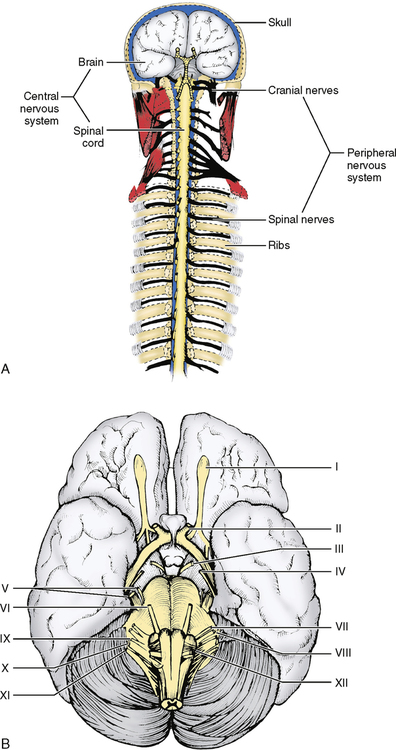
The nervous system is divided into two major categories: the central nervous system, consisting of the brain and spinal cord, and the peripheral nervous system, which comprises all nerves that extend outward from the brain and spinal cord (Fig. 34-1).
Remember from Chapter 17 that a neuron or group of neurons transmits a message or impulse in only one direction. Therefore it is necessary to have both sensory and motor nerves, or more accurately, sensory and motor neurons. A nerve is actually a bundle of neurons, some of which are sensory and some of which are motor. These neurons are what enable messages to travel to and from the brain. Most nerves have both motor and sensory neurons, although some nerves may be either purely motor or purely sensory.
CENTRAL NERVOUS SYSTEM
The brain and spinal cord are made up of neurons that are either motor or sensory, but they are only part of the chain. Imagine that you have caught your finger in a desk drawer. You feel pain and quickly pull your finger out of the drawer. Neurologically, a pain receptor or free nerve ending in the finger has picked up the message and carried it to the spinal cord. A second neuron has carried the message up the spinal cord to the lower parts of the brain, and a third neuron has taken it to the surface of the brain, where it was recognized as pain. The pain was interpreted by the brain, and past experience told the brain to remove the finger from the drawer so that it would no longer be crushed. A motor message then left the brain and traveled down the spinal cord, and a second motor neuron carried the message out of the spinal cord and along a peripheral nerve to muscles that pulled the finger out of the drawer (Fig. 34-2).
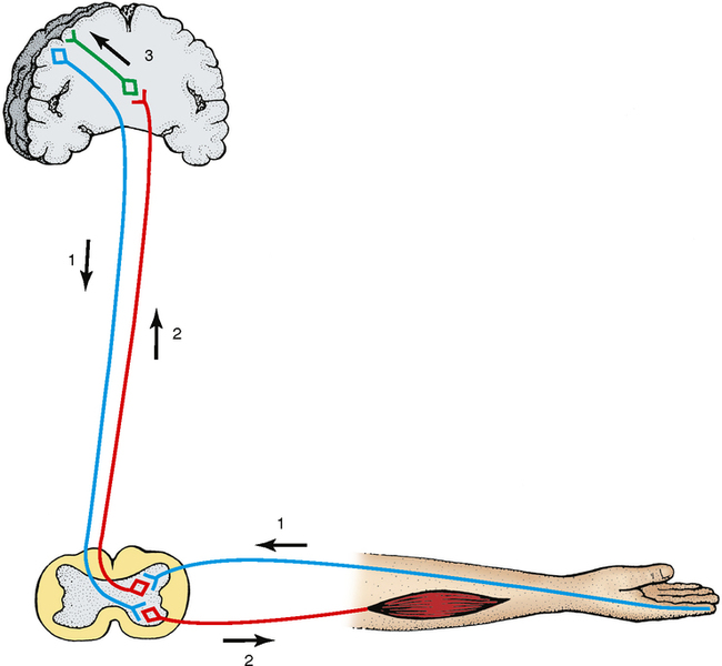
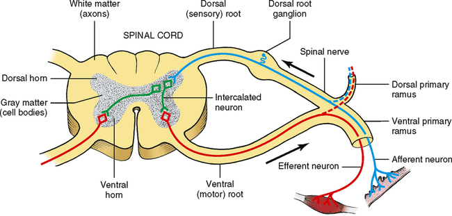
The nervous system also builds in a shortcut to this system known as a reflex arc. This shortcut takes place in the spinal cord between the sensory neuron, as it enters the spinal cord, and the motor neuron, as it leaves the spinal cord. A shorter neuron, called the intercalated neuron, runs between the two neurons and carries the message from the sensory side to the motor side without it having to go to the brain. The action is actually accomplished before the person thinks about it. Touch a hot stove, and what happens? You usually pull your hand away before you actually realize it is hot or before you consciously tell yourself to remove your hand from the stove (Fig. 34-3).
PERIPHERAL NERVOUS SYSTEM
Spinal Nerves
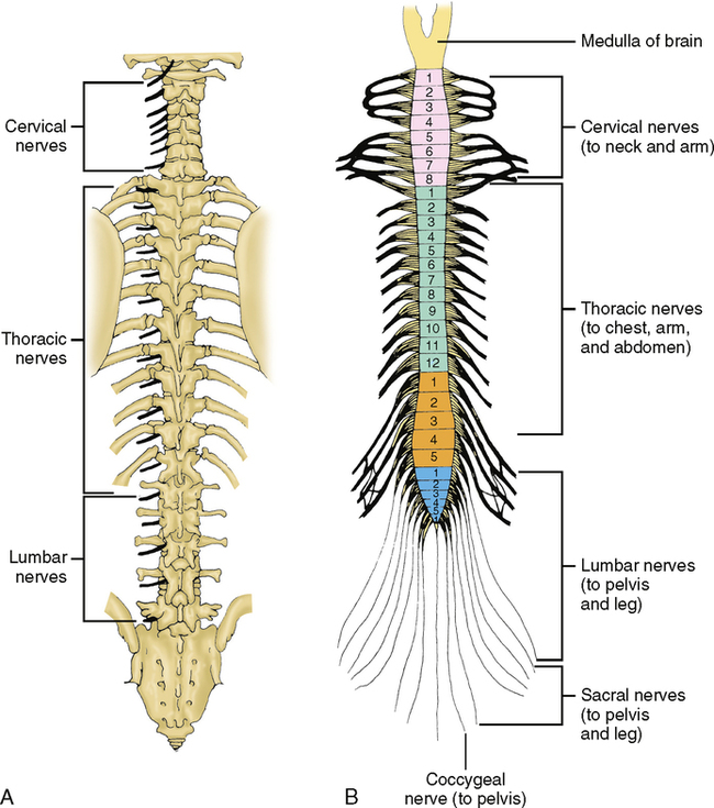
The spinal nerves extend from the spinal cord to distant parts of the body. These nerves generally have both motor and sensory neurons in them. There are 31 pairs of spinal nerves: 8 in the cervical or neck region; 12 in the thorax or chest region; 5 in the lumbar or lower back region; 5 in the sacral or hip region; and 1 in the coccygeal or tailbone region (Fig. 34-4). These nerves are distributed by region from the neck to the toes. Several of the cervical nerves innervate some of the hyoid muscles that depress the mandible and raise the larynx.
Typical spinal nerves look like Fig. 34-3. The central speckled area of the spinal cord is known as gray matter. This gray matter is made up of the cell bodies of both motor and sensory neurons, which are arranged in the shape of a butterfly in the central area of the spinal cord. The area around the gray matter is referred to as white matter and is made up of myelinated axons of both motor and sensory neurons. The spinal nerve has a dorsal root, which is the sensory root. The dorsal root has a dorsal root ganglion, which is a collection of cell bodies of the sensory neurons entering the spinal cord at that level. There is also a ventral root, which is the motor root. The point where the dorsal and ventral roots meet is the beginning of the spinal nerve, and at that point the spinal nerve contains both motor and sensory neurons. The spinal nerve then divides into a small dorsal primary ramus and a larger ventral primary ramus. Both of these primary rami are mixed motor and sensory nerves. The dorsal primary ramus carries sensation from the skin in a narrow band at the midline of the back, which is probably only a few inches wide, and also supplies the back muscles that lie immediately next to the vertebrae in the back. There are only a few dorsal primary rami that we can easily identify as nerves, and those are purely the sensory parts of the dorsal rami. The ventral primary rami are what we normally see as nerves in the body.
Cranial Nerves
I, olfactory nerve—Sensory; provides special sense of smell from the nose to the brain.
II, optic nerve—Sensory; provides special sense of sight from the eye to the brain.
III, oculomotor nerve—Motor; supplies most of the muscles that move the eye in different directions and one that raises the upper eyelid; parasympathetic innervation to the eye to cause the pupil to contract and the lens to change shape and become more rounded for close vision.
IV, trochlear nerve—Motor; supplies the superior oblique muscle, the muscle that moves the eye downward and laterally.
V, trigeminal nerve—Motor and sensory; sensory innervation from all the teeth, the oral cavity, the anterior two thirds of the tongue, the maxillary sinus, the nasal cavity, and most of the skin of the front part of the face and head; motor innervation to the muscles of mastication, a soft palate muscle (tensor veli palatini) as well as the tensor tympani muscle of the middle ear. This is the most important nerve for our consideration and will be covered in greater detail later.
VI, abducens nerve—Motor; supplies the lateral rectus muscle, the muscle that moves the eye laterally.
VII, facial nerve—Motor and sensory; motor innervation to the muscles of facial expression, the stylohyoid and posterior belly of the digastric muscles, as well the stapedius muscle of the middle ear; parasympathetic innervation to the submandibular and sublingual salivary glands, and the lacrimal gland of the eye (autonomic); sensory innervation from some areas behind the ear, and taste from the anterior two thirds of the tongue.
VIII, statoacoustic nerve—Sensory; for hearing and balance (equilibrium).
IX, glossopharyngeal nerve—Motor and sensory; motor innervation to one of the muscles of the pharynx, the stylopharyngeus; parasympathetic innervation to the parotid salivary gland; sensory innervation to the posterior one third of the tongue for taste, as well as general sensation such as pain, pressure, heat, and cold; also provides sensation to most of the mucosa of the pharynx and to a small area of the skin of the ear.
X, vagus nerve—Motor and sensory; motor innervation to the muscles of the pharynx and larynx and most of the muscles of the soft palate; parasympathetic innervation controls most of the smooth muscle of the body, cardiac muscle, and many of the glands of the body; sensory innervation from the skin around the ear and taste and general sensation from the root of the tongue and epiglottis.
XI, accessory (spinal accessory) nerve—Motor; supplies the trapezius muscle and the sternomastoid muscle in the neck. Some parts of this nerve run with the vagus nerve and supply most of the soft palate, larynx, and pharynx muscles. Many of the muscles attributed to the vagus supply actually come from XI.
XII, hypoglossal nerve—Motor; supplies most of the intrinsic and extrinsic muscles of the tongue except the palatoglossus, which is supplied by X(XI) nerve.
Stay updated, free dental videos. Join our Telegram channel
VIDEdental - Online dental courses


