Soft Palate and Pharynx
SOFT PALATE
Palatoglossal Muscle
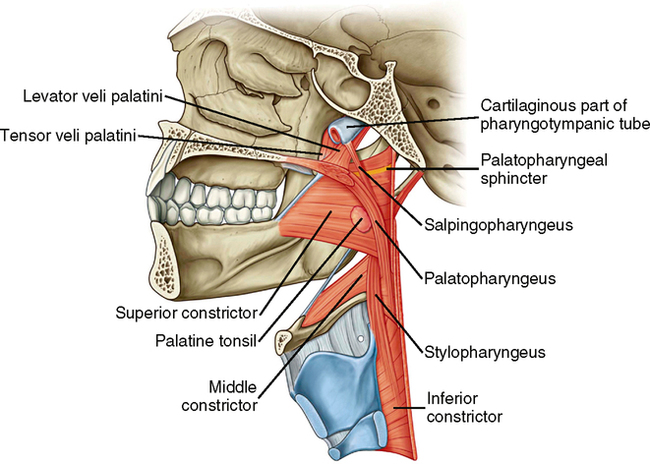
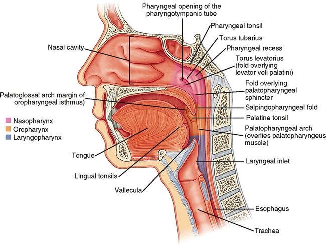
If you open the mouth and look at the tonsils on the side of the throat wall, you will see that there is a vertical fold of tissue in front of and behind each tonsil. These are called the anterior and posterior faucial pillars, or the palatoglossal and palatopharyngeal folds, respectively. Beneath the palatoglossal fold is the palatoglossal muscle. It originates from the posterior end of the hard palate and the anterior end of the soft palate. The fibers run downward, laterally, and forward to insert into the posterior and lateral part of the tongue. When the palatoglossal muscle contracts, it pulls the sides of the tongue up and back, pulls the soft palate down on the lateral edges, and narrows the space between the left and right anterior faucial pillars. The nerve that supplies this muscle is a part of the eleventh (XI) cranial nerve running with branches of the tenth (X) cranial nerve (Figs. 31-1 and 31-2).
Palatopharyngeal Muscle
The posterior faucial pillar is formed by the palatopharyngeal muscle. It originates from the posterolateral part of the soft palate and runs downward and laterally to insert into the pharyngeal constrictor muscle and the thyroid cartilage of the larynx. When it contracts, it narrows the posterior faucial pillar and elevates the pharynx and larynx. The nerve supply is also the 10th and 11th cranial nerves (see Figs. 31-1 to 31-3).
Muscles of Uvula
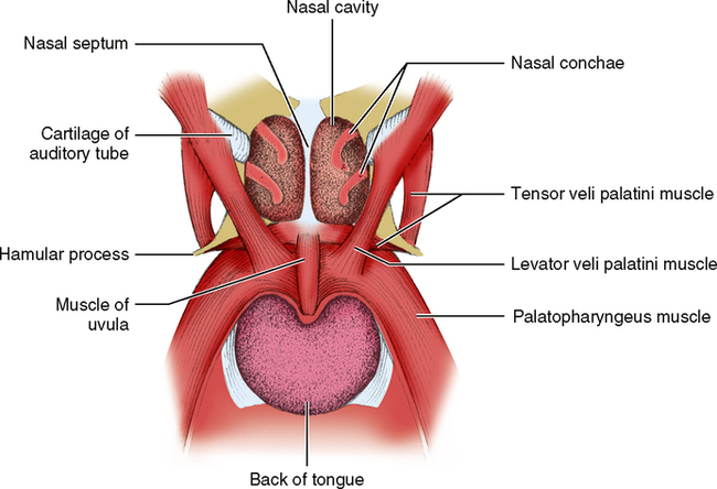
The uvula is the small fold of tissue that hangs down in the throat from the posterior part of the soft palate. It is formed by two small bands of muscle that originate from the posterior end of the hard palate and run back and down in the soft palate to form that structure. When the muscle of the uvula contracts, it shortens and broadens the uvula and changes the contour of the posterior end of the soft palate so that it adapts to the posterior throat wall when it is moved up against it. This muscle is also innervated by the eleventh and tenth cranial nerves (see Fig. 31-3).
Stay updated, free dental videos. Join our Telegram channel
VIDEdental - Online dental courses


