31: Procoagulant, Anticoagulant, and Thrombolytic Drugs

CHAPTER 31 Procoagulant, Anticoagulant, and Thrombolytic Drugs

The practice of dentistry frequently involves procedures that cause bleeding, and the dentist is often confronted with the need to achieve and maintain hemostasis. The dental practitioner must be familiar with the physiologic processes of hemostasis and the myriad conditions that cause abnormalities of these processes. Complicating matters, modern medicine has developed several therapies for systemic disease that use medications that purposefully alter normal hemostasis. When appropriate, the dentist needs to eliminate or make alterations in the dosage of these compounds before surgery. Only with a clear understanding of the complex process of hemostasis and the various drugs that affect it can the clinician manage patients with inherited or acquired bleeding disabilities safely.

HEMOSTASIS

Large or intermediate arteries and veins are generally not severed intentionally without prior ligation, but it is common during the extraction of teeth and other oral surgical procedures to sever small arteriolar, venous, and capillary vessels. Extensive blood loss may occur if hemostasis is delayed. The formation of a patent clot requires four distinct yet interdependent steps: (1) vessel constriction; (2) platelet adhesion, activation, and aggregation; (3) cross-linking of fibrin by the coagulation cascade; and (4) limitation of the blood clot to the area of damage only. Later, a fifth step becomes necessary: the controlled breakdown of the clot so that repair and remodeling can occur.

Platelet Adhesion, Activation, and Aggregation

Adhesion

The next major event is the adhesion of platelets at the severed edges of the vessel. In normal untraumatized blood vessels, platelets show little tendency to adhere to the endothelium, partly because prostacyclin, again elaborated by the endothelial cells, induces cyclic adenosine 3′,5′-monophosphate (cAMP) synthesis in platelets and inhibits platelet adhesion. Endothelium-derived relaxing factor—now believed by most investigators to be largely or entirely nitric oxide—also normally secreted by the endothelial cells, is another natural inhibitor of platelet adhesion. Injury to the intima, even if the vessel wall remains intact, leads, however, to exposure of subendothelial extracellular matrix proteins such as collagen, fibronectin, von Willebrand factor (vWF), thrombospondin, and laminin.

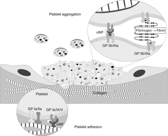
The presence of these proteins, particularly vWF, stimulates a “catch and grab” response in the platelets, causing them to leave the laminar flow of the blood and adhere to the injured area. Platelets have a high density of surface receptors that respond to these proteins, and they undergo an extremely rapid localization to the site of injury, beginning the formation of a thrombus. Two main receptors are involved in adhesion: the glycoprotein (GP) Ia/IIa heterodimer, which binds to collagen directly but weakly, and the GP Ib/IX/V heterotrimer, which binds with high shear strength to connective tissue vWF associated with the collagen surface (Figure 31-1).1 The GP Ib/IX/V–vWF linkage is more of a “tethering” of the platelet to the substrate; later, the adhesion is firmed up by GP IIb/IIIa activation. If vessels without a muscular sheath are severed, the immediate hemostatic action of platelet aggregation is especially important. The true significance of platelets in hemostasis is most evident in the management of patients with thrombocytopenia.

Activation

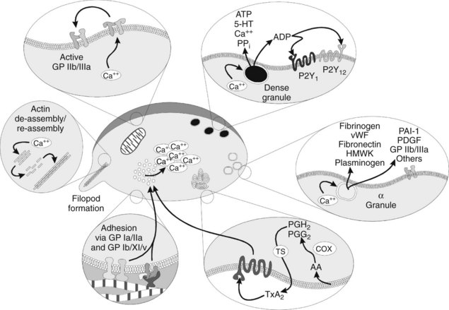
Activation of platelets is a crucial step in forming a proper thrombus. Activation can occur from various agonists, some of which are strong and some of which are weak. Examples include thrombin, adenosine diphosphate (ADP), thromboxane A2 (TXA2), 5-hydroxytryptamine (serotonin), epinephrine, vasopressin, fibrinogen, immune complexes, plasmin, and platelet-activating factor. Most plasma-derived agonists exert their effect by numerous G protein–linked membrane receptors. The strongest agonist for platelet activation is binding of vWF to the GP Ib/IX/V heterotrimeric receptors.14 When one of these receptors is bound by its specific agonist, an intraplatelet protein cascade begins that ultimately causes activation of Ca++ transporters and movement of Ca++ from stores in the platelet’s dense tubular system to the general intracellular matrix.26 The intracellular increase in Ca++ causes several other changes.

Platelets in the resting state have internal cytoskeletal actin that provides them with a smooth shape; as Ca++ increases, the actin is initially fragmented into smaller subunits, transforming the normal discoid shape of the platelet to a spherical conformation. These smaller actin subunits are rapidly reassembled into very-long-chain actin monomers, which cause the platelet to sprout filopods. The filopods are important in ultimate clot retraction. Meanwhile, as the filopods are developing, the increasing intracellular Ca++ concentrations act on cytoplasmic vesicles known as α and dense (or δ) granules (Figure 31-2), prompting them to rise to the cell surface and degranulate. The dense granules release ADP, adenosine triphosphate (ATP), the vasoconstrictor 5-hydroxytryptamine, Ca++, and inorganic pyrophosphate.18 The α granules contain numerous proteins involved in coagulation, adhesion, cellular mitogenicity, protease inhibition, and other functions (Box 31-1). Major proteins released include fibrinogen, coagulation factors, vWF, fibronectin, high-molecular-weight kininogen, plasminogen, plasminogen activator inhibitor-1 (PAI-1), platelet-derived growth factor, additional GP IIb/IIIa, and thrombospondin.18

Release of the dense granule ADP into the extracellular milieu has an autocatalytic effect on the platelet from which it came and also stimulates nearby platelets. The ADP binds to its own purinergic receptors, most notably P2Y1 and P2Y12. Activation of both of these receptors is required for maximal aggregation of the platelets to one another. P2Y1 stimulation acts to mobilize Ca++ further (an autocatalytic effect), which leads to further shape change and transient aggregation. P2Y12 activation causes inhibition of adenylyl cyclase (blocking conversion of ATP to cAMP), potentiation of secretion by the α and dense granules, and sustained aggregation. ADP also binds the transmembrane protein P2X1, an ion channel receptor linked to influx of extracellular Ca++ into the platelet.

Aggregation

As the activated platelets interact with one another, they begin to aggregate. Aggregation is initiated by the Ca++-mediated conformational activation of GP IIb/IIIa, a heterodimeric transmembrane protein. GP IIb/IIIa is a protein receptor complex unique to platelets and is expressed at extraordinarily high density on the surface of the platelets—some 80,000 to 100,000 per platelet—at an average distance of only 20 nm from one another. Another 20,000 to 40,000 units are stored in the α granules and are released onto the surface or within the local plasma milieu during degranulation. In the circulating, platelet, the resting GP IIb/IIIa receptor has little affinity for its ligands (primarily fibrinogen), so intravascular thrombus formation is minimized. On activation the GP undergoes a conformational change, however, which imparts high affinity for its ligands. Several proteins have the specific amino acid sequence necessary for binding to the GP IIb/IIIa receptor, including fibrinogen, fibronectin, vitronectin, and vWF.

As the α and dense granule contents are released extracellularly, nearby platelets become activated. The ligand proteins bind to the surface-associated GP IIb/IIIa of these adjacent platelets, forming bridges. At low shear rates, fibronectin and fibrinogen (stabilized by thrombospondin, another GP from the α granules) serve as the main adhesive proteins, whereas vWF is necessary for proper adhesion in areas of high shear. Microvascular video imaging studies show that thrombus formation initially is inefficient. Platelets bind quickly, but a significant percentage of them break free and float away. As a result, thrombus formation is much slower than would be the case if all the platelets that physically aggregate remained bound.1

Several other events occur simultaneously with activation and aggregation, but the two most important are generation of TXA2 and platelet-assisted generation of thrombin. Both of these agents accelerate the platelet-activation response. TXA2 is generated when platelet phospholipases are activated during platelet aggregation, which release arachidonic acid from glycerophospholipids of the platelet membrane. Arachidonic acid is a substrate for cyclooxygenase (COX), yielding the prostaglandin endoperoxides PGH2 and PGG2. These prostaglandins are modified by thromboxane synthase to produce TXA2, which acts at its own protein-linked receptor.

Perhaps the most remarkable effect of platelet activation is the procoagulant activity the platelets impart. In the normally resting platelet, the plasma membrane has negatively charged phospholipids, including phosphatidylserine, sequestered almost exclusively on the inner surface by processes that are not fully understood. When activating ligands bind to the platelet, the resultant increase in intracellular Ca++ causes a membrane enzyme termed scramblase to evert the phosphatidylserine to the outer surface, while simultaneously prompting the membrane to form small evaginated microvesicles. Factors Va and VIIIa (discussed subsequently) bind to the phosphatidylserine moieties and recruit factors Xa and IXa. The interaction of these complexes in toto accelerates the conversion of prothrombin to thrombin by a factor of 2.4 × 10⁶. In addition, the binding of activated coagulation factors to the platelets seems to protect the factors from plasma inhibitors, while directing the bulk of the coagulation cascade to the site of injury. The α granules contain factors V and IX; factor V is apparently complexed with multimerin, a carrier protein (see Box 31-1).

As the thrombin is generated, it activates other platelets by stimulating G protein–linked receptors. The thrombin receptors seem to be unique “suicide” receptors, requiring proteolytic cleavage to transmit an activating signal. Thrombin is a serine protease, and it acts on the receptors by cleaving the protein at a serine residue near the amino terminus. The new amino terminus acts as a “tethered ligand” to double back and stimulate the transmembrane protein to activate—hence this receptor has been named a protease-activated receptor (PAR). There are four such thrombin receptors, PAR-1 through PAR-4; only PAR-1 and PAR-4 are expressed by human platelets.23 Thrombin-induced activation seems to upregulate GP IIb/IIIa activation while downregulating GP Ib/IX/V activity. The platelets apparently are converted from a mainly adhesive role to an aggregate role when thrombin is present.

Two other important activities of platelets warrant mention. First, the α granules contain P-selectin, a membrane protein that helps recruit and tether neutrophils and monocytes into the local area. This activity is believed to be crucial for generating a local inflammatory response at the site of injury, while promoting yet limiting thrombosis.35 Second, platelets are also essential in clot retraction, an event that facilitates wound healing by bringing the severed ends of small blood vessels into closer apposition. Clot retraction, or syneresis, occurs when the filopodia expressed by platelets during activation attach to fibrin strands and contract. A number of actin-binding proteins are present in platelets.4 On activation, phosphorylated myosin monomers polymerize into filaments next to the long-chain actin filaments, which slide past one another to generate a contractile force in the presence of ATP.

Coagulation Cascade

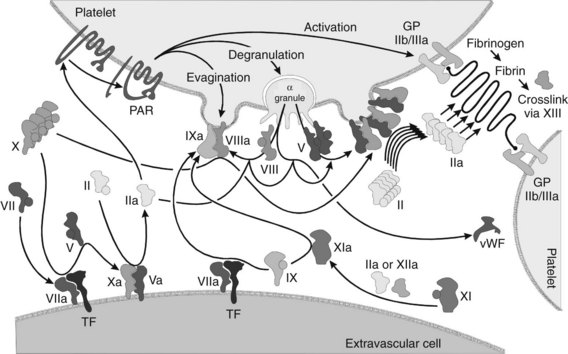
Although it is possible to separate the numerous events of hemostasis (e.g., platelet aggregation, formation of fibrin, retraction of the blood clot), the whole process occurs synergistically. Many of the factors involved are enzymatic cofactors, and most of the reaction occurs on cell and platelet membranes (Figure 31-3). Many refinements in the understanding of blood coagulation have come about through study of “experiments of nature,” in which discrete defects of the clotting process have been identified in patients with bleeding diatheses, as illustrated by the factors and deficiency states listed in Table 31-1.

TABLE 31-1 Blood Clotting Factors

INTERNATIONAL NUMBER OR TERM* PLASMA FACTOR AND ALTERNATIVE NAMES CAUSE OR DESCRIPTION OF DEFICIENCY
I Fibrinogen Liver disease
II Prothrombin Liver disease or vitamin K deficiency
III TF, thromboplastin Deficiency of TF probably does not occur
IV Ca++ Never deficient without tetany
V Proaccelerin Parahemophilia, rare
VII Proconvertin Liver disease or vitamin K deficiency
VIII Antihemophilic globulin, AHF A Hemophilia A, 80% of hemophilics
IX Christmas factor, AHF B Hemophilia B (Christmas disease), depressed with vitamin K deficiency
X Stuart-Prower factor Liver disease or vitamin K deficiency
XI Plasma thromboplastin antecedent, AHF C Factor XI hemophilia (hemophilia C)
XII Hageman factor Generally no clinical symptoms but may have thromboses, rare
XIII Fibrin-stabilizing factor, Laki-Lorand factor, fibrinase Delayed bleeding, defective healing, rare
PF3 Platelet factor 3 Thrombocytopenia
Protein C Liver disease or vitamin K deficiency
Protein S Liver disease or vitamin K deficiency
Protein M Liver disease or vitamin K deficiency
vWF von Willebrand factor vWD types I, IIa, IIb, IIc, III
Pre-K Prekallikrein, Fletcher factor  
HMWK High-molecular-weight kininogen

AHF, Antihemophilic factor; TF, tissue factor.

* Roman numerals were assigned in 1958 by the International Committee on Blood Clotting Factors. Factor VI, originally assigned to prothrombin converting principle (prothrombinase), has since been abandoned.

Most sources currently use factor numbers.

Initiation of coagulation after injury is a complex process involving an initial pathway of thrombin generation, which autocatalyzes a subsequent burst of additional thrombin generation sufficient to convert fibrinogen to fibrin (see Figure 31-3). Before the process is described, a brief review of the crucial factors and cofactors and how they function is warranted.

Vitamin K–dependent clotting factors

Synthesized in the liver, the vitamin K–dependent clotting factors comprise factors II (prothrombin), VII, IX, and X, and protein C. These five proteins are serine proteases and have similar structural elements (including a serine residue at their catalytic site). Molecular genetic evidence suggests they all are derived from a common ancestral precursor gene. They all have a preprotein leader that is cleaved away post-translationally, leaving an amino-terminal γ-carboxyglutamic acid (Gla) domain with 9 to 12 Gla residues. This sequence is followed by a hydrophobic domain and finally the serine protease domain, in which the carboxy-terminal region becomes activated by cleavage of key arginine residues.

The amino terminus Gla domain is crucial for the lipid binding of these proteases to their substrate membranes. In the presence of seven Ca++ ions intercalated within the three-dimensional structure of the Gla domain, the protein undergoes a conformational change that places its hydrophilic domain at one end of the three-dimensional protein structure, with the hydrophobic moieties facing outward. This arrangement is crucial because it allows the protein to settle into the lipid membrane and exert its effects locally rather than systemically in the vasculature. Before these events can occur, however, the Gla residues must be formed post-translationally by carboxylation of their precursor glutamate residues by a specific γ-glutamyl carboxylase that requires the uncleaved preprotein leader sequence of amino acids to bind to the protein. This carboxylase enzyme requires oxygen, carbon dioxide, and vitamin K to function (see Figure 31-8). For every glutamate residue carboxylated, one molecule of reduced vitamin K is converted to its epoxide form. A separate enzyme, vitamin K epoxide reductase, converts the vitamin K back to the reduced form. This reductase is the target of the warfarin-like anticoagulants and is discussed in greater detail later.

Each of the clotting factors mentioned is a protease with activity directed at substrate arginyl residues. Activated factors VII, IX, and X have specific cofactors associated with them—tissue factor (TF) with VIIa (a for activated), VIIIa with IXa, and Va with Xa. The cofactors bind the protease and its substrate in approximation to each other and modify the enzyme factor allosterically to have greater activity in the presence of substrate.

Enzymatic cofactors

TF is a unique protein normally constitutively expressed on the cell surfaces of many extravascular cell types. In contrast to the other coagulation cofactors, it is a transmembrane protein homologous to the receptors for interleukin-10 and interferons α, β, and γ. It seems to have procoagulant and signal transduction functions. It can be induced to become expressed on the cell surfaces of intravascular monocytes and endothelial cells in response to some bacterial products and inflammatory cytokines, perhaps as part of the body’s immunologic defense system. P-selectin, secreted by platelet α granules, can also induce TF expression in monocytes adhering to activated platelets.

When injury occurs and the vasculature gains exposure to cells with TF on their surface, circulating factor VII rapidly binds to TF and undergoes proteolytic cleavage to factor VIIa by mechanisms that are not well understood. The TF/VIIa complex serves two crucial functions: it cleaves factor X to Xa and factor IX to IXa, both of which have distinct and separate activities. Newly formed factor Xa rapidly binds to circulating factor V and activates it to Va. The factor Xa/Va complex settles into the adjacent cellular membrane (using the hydrophobic Gla domain), where it acts on circulating prothrombin to generate a very small amount of thrombin. This tiny amount of thrombin is insufficient to cleave fibrinogen significantly but instead serves four crucial functions that set up the area for a much larger burst of thrombin formation: (1) nearby platelets are activated by their PAR receptors, which causes degranulation; (2) additional factor V liberated from the platelet α granules is activated (thrombin activates factor V much more efficiently than does factor Xa); (3) factor VIII is activated and dissociated from vWF; and (4) factor XI is activated. Factor Xa exerts its effect locally; any factor Xa that escapes the TF/VIIa complex area is rapidly destroyed by tissue factor pathway inhibitor (TFPI) or antithrombin III (ATIII), both of which are discussed later.

In contrast to the factor Xa/Va complex, activation of factor IXa by TF/VIIa results in an enzyme that is not restricted to the nearby cell surface because it is not inhibited by TFPI and is only slowly affected by ATIII. As a result, factor IXa diffuses through the plasma over to nearby activated platelets. As previously discussed, activated platelets rapidly place factors Va and VIIIa on their cell surfaces. The diffusing factor IXa binds tightly to the factor VIIIa cofactor, and this IXa/VIIIa complex efficiently activates additional factor X to Xa. As before, factor Xa then binds to adjacent factor Va, and this time a much larger burst of prothrombin conversion to thrombin occurs. This much larger amount of thrombin formation is sufficient to begin cleaving fibrinogen and start clot formation and continue to perform the activating functions listed previously.

Fibrinogen and factor XIII

The final phase of blood clotting consists of the thrombin-mediated proteolytic cleavage of fibrinogen to fibrin. Fibrinogen consists of a mirror image dimer in which each monomer is composed of three intertwined and disulfide bond–linked polypeptide chains. In the dimer, the amino terminus of all six polypeptides meet in the middle of the linear molecule to form the N-terminal disulfide knot, or E domain. The carboxy termini of the three polypeptides at each opposite end form a globular protein cluster known as the D domain. Between the E and D domains, the polypeptide chains form a helical structure.

Thrombin binds to the central E domain and cleaves off peptides from the knot to expose binding sites in the E domain that match the corresponding D domains of two neighboring fibrinogen molecules. The monomers begin to form a staggered “ladder” protofibril. As the monomers continue to associate, branch points occur that allow the fibrin meshwork to become more like a net and thicken. The initial clot is unstable, being held together primarily by hydrogen bonds. With time, however, the fibrin strands become cross-linked with covalent bonds by the action of a transglutaminase, fibrin-stabilizing factor XIII. This factor cross-links proteins between the γ-carbon of glutamine in one fibrin strand and the ε-amino group of lysine in the other.

Entrapped in this coagulum are red and white blood cells and intact platelets; the latter promote clot retraction as previously described. These events are followed by the inflammatory processes of organization and wound healing, which require, among other things, an effective proteolytic (fibrinolytic) mechanism described later in this chapter.

Regulation of Coagulation

When discussing hemostatic mechanisms, consideration should be given to the natural inhibitors of blood clotting. As important as the procoagulant process is, it is equally important to ensure that inappropriate clotting does not occur. The intent of the clotting system is to seal a site of vascular compromise; powerful antithrombotic mechanisms must come into play to ensure that clotting remains limited to the injured area. Several mechanisms of antithrombosis have been elucidated; they are discussed in detail subsequently and summarized in Figure 31-4. At the heart of the matter is how to control the extremely efficient clotting cascade after it is initiated.

Strict control of the coagulation cascade is mediated by several proteins that act as natural anticoagulants, all of which rely on the first traces of thrombin from the nearby wound site to activate them. In general, the theory is simple: bind or degrade any activated procoagulant proteins if they escape the site of injury. At the same time, the site of injury must be protected from invasion or inclusion of these same inhibitory proteins.

Because thrombin is the major procoagulant protein, it makes sense that inactivation of it is a high priority. An elegant mechanism exists that, instead of destroying thrombin, uses thrombin to catalyze an important set of anticoagulant proteins, the protein C/protein S system. In the microcirculation, where there is a high cell surface-to-volume ratio, the protein C/protein S system predominates. Vascular endothelial cells normally express thrombomodulin on their membranes. Thrombomodulin is a transmembrane cofactor protein with no known enzymatic activity. It binds the thrombin that escapes from the surface of nearby platelets but is not carried off in the vascular flow.

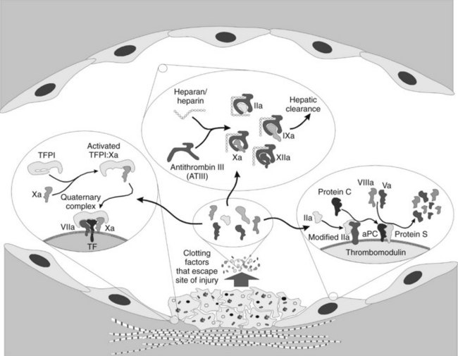
Thrombomodulin, as the name implies, alters the conformation of the thrombin and effectively removes its ability to cleave fibrinogen, activate platelets, and activate factors V and VIII. Instead, the new conformation of thrombin imparts a 2000 times greater affinity for activation of the vitamin K–dependent protein C.12 Activated protein C (aPC) has considerable homologous characteristics with the other vitamin K–dependent factors, complete with a Gla domain, hydrophobic domain, and active serine protease domain. The cofactor for aPC is protein S, a membrane-bound protein that has no inherent activity. When aPC is bound, the complex efficiently cleaves and destroys any factors Va and VIIIa that might have been liberated from the platelet surfaces, however, slowing coagulation and protecting the normal individual against random intravascular coagulation.

Another protein, ATIII, is a serine protease inhibitor (“serpin”) found in the plasma. It inhibits clotting by covalently binding 1 : 1 to the active sites of thrombin and certain other serine proteases (factors IXa, Xa, and XIIa). This reaction is normally slow but is accelerated 1000-fold in the presence of heparan sulfate, a proteoglycan synthesized on the surfaces by endothelial cells. (A similar effect is achieved therapeutically by administration of the closely related agent heparin sulfate.) Although ATIII binds to these factors only without destroying them, reactivation by unbinding probably does not occur physiologically. The ATIII-protease complexes are cleared in the liver. It is believed that ATIII is responsible for complexing with proteases that escape into the circulation.

Finally, as mentioned earlier, TFPI is a protease inhibitor found in low concentrations in the plasma, mostly bound to circulating lipoproteins or to endothelial cell membrane heparans. It is capable of inactivating factor Xa and the TF/VIIa complex; it must first bind factor Xa before it can bind to the TF/VIIa complex. TFPI seems to be the major inhibitor of free-floating factor Xa, and it may be responsible for shifting the activation of factor IX from the TF/VIIa complex to thrombin-activated factor XI. The inhibitor is found in high concentrations in patients with hemophilia A and B, presumably because fewer substrates are available for TFPI binding. This finding offers one explanation for why hemophilics bleed despite normal concentrations of TF, factor VIIa, and factor Xa at the site of injury. TFPI is synthesized in liver and endothelial cells.

PROCOAGULANT AGENTS

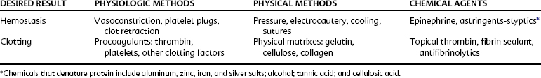
In medical and dental practice it is essential to take appropriate precautions to avoid serious hemorrhage. This admonition is particularly true for patients with hemophilia, patients with hematopoietic disease, and patients receiving therapies known to affect hemostasis. Precautions, which may include the administration of clotting factors or hospitalization or both, are prudent in these cases. In contrast, normal patients usually require no more than temporary hemostatic assistance (e.g., pressure packs, hemostatic forceps, ligation, or other locally active measures) to facilitate normal hemostasis and allow clotting to occur. Table 31-2 outlines various methods for controlling bleeding.

Intrasocket Preparations

Bleeding from bony structures, especially from extraction sockets, can be controlled by various means. If initial attempts to achieve hemostasis with sterile cotton gauze and pressure do not succeed, a gelatin sponge, denatured cellulose sponge, or collagen plug may be inserted within the bony crypt.

Gelatin sponges are intended to be a matrix in which platelets and red blood cells can be trapped. In so doing, the sponges facilitate platelet disruption and can absorb 40 to 50 times their own weight in blood, both of which aid in coagulation. They typically resorb in 4 to 6 weeks. Because they are made of gelatin, they must be applied dry; when moistened, they become difficult to handle. For this reason, many practitioners prefer to use either denatured cellulose preparations or collagen sponge.

Denatured cellulose sponge or gauze serves as a physical plug and a chemical hemostatic. The apparent coagulation-promoting action stems from the release of cellulosic acid, which denatures hemoglobin, and these breakdown products help plug the site of injury. Cellulosic acid, similar to tannic acid, inactivates thrombin; the use of cellulose sponge in conjunction with this procoagulant is ineffective. Two forms of cellulose sponge, oxidized cellulose and oxidized regenerated cellulose, are available. Both these materials cause delayed healing, particularly oxidized cellulose, which notably interferes with bone regeneration and epithelialization. Although regenerated cellulose is said to have less inhibitory action, neither dressing should be left permanently in the wound if it can be removed.

The collagen plug, similar to microfibrillar collagen, serves to accelerate the aggregation of platelets and form a physical barrier. Because it also is usually made from bovine collagen sources, occasional foreign body responses can occur. Overall, the collagen plug generally activates platelets more completely and is the preferred intrasocket product.

Topically Applied Clotting Factors

The most physiologic hemostatic aids are the blood clotting factors themselves. Assuming an otherwise normal clotting system, topical thrombin is often used clinically. It must remain topically applied; if given intravenously, thrombin causes extensive thrombosis and possibly death. Topically applied thrombin (particularly in conjunction with a compatible matrix such as gelatin sponge) operates as a hemostatic, particularly if the patient has a coagulation deficiency or is receiving oral anticoagulants, because all that is required for clotting is a normal supply of platelets, fibrinogen, and factor XIII in the plasma. If blood flows too freely, temporary physical hemostasis must be attained before topical thrombin can be of practical value. The use of thrombin is not without problems. Currently available thrombin, especially the bovine products, may be relatively crude preparations that still contain plasmin, a fibrinolytic agent (discussed subsequently). Antibodies may also be generated to the bovine thrombin or bovine factor V; the latter can cross-react with human factor V and lead to an acquired inhibition and bleeding.

Fibrin sealant, also sometimes referred to as fibrin glue, is one of the more promising hemostatic aids to appear in recent years.5 With this agent, the concept of the application of topical thrombin is taken one step further. Bovine or human thrombin and calcium chloride are mixed in one of two syringes; purified human fibrinogen with factor XIII, aprotinin, and other plasma proteins (fibronectin and plasminogen) are in the second syringe. The two solutions are mixed in a single delivery barrel, where the thrombin cleaves the fibrinogen to fibrin monomers. Initially, they are gelled by hydrogen bond formation, but in 3 to 5 minutes the factor XIII in the presence of Ca++ initiates cross-linking and increases the tensile strength of the clot.5 As the clot solidifies, the sealant becomes milky white.

The rate of fibrin clot formation depends on the concentration of the thrombin; 4 IU/mL produces a clot in approximately 1 minute, whereas 500 IU/mL requires only a few seconds. The strength of the clot depends on the concentration of the fibrinogen. If used in an area where the clot is likely to break down too soon, or in patients with compromised hemostasis, a protease inhibitor such as aprotinin can be added to delay fibrinolysis. Aprotinin functions by inhibiting plasmin, which is generally carried along with the thrombin. The term glue arises from the fact that in many medical applications this material has been literally used to adhere tissues together naturally.

Fibrin sealant is commercially available in the United States. The protein fractions are lyophilized and require careful reconstitution at 37° C under sterile conditions; proper mixing of the materials requires approximately 30 minutes to perform. As a result, the emergent use of this material is difficult; typically, it is used more in planned surgeries in patients with known bleeding disorders. It is also an expensive medication; 1 mL of the material costs several hundred dollars. Fibrin sealant works well, however, in stopping the microbleeding and oozing that often accompany dental procedures.27

Astringents and Styptics

The terms astringents and styptics are interchangeable, referring to different concentrations of the same drugs. Many chemicals have vasoconstrictive or protein-denaturing ability, but relatively few are appropriate for dentistry. The suitable preparations are primarily salts of several metals, particularly zinc, silver, iron, and aluminum. Aluminum and iron salts are quite acidic (pH 1.3 to 3.1) and irritating.36 Iron causes annoying, although temporary, surface staining of the enamel, whereas silver stains may be permanent.

Currently, astringents are generally used in dentistry only to aid hemostasis while retracting gingival tissue. Other applications, such as controlling bleeding after surgery, are not looked on as favorably as in the past, when 20% ferric subsulfate (Monsel’s solution) and 8% zinc chloride were among the most popular agents used. Aluminum and iron salts function by denaturing blood and tissue proteins, which agglutinate and form plugs that occlude the capillary orifices. In a rabbit mandible model, when ferric sulfate salts were left in an osseous wound, there was an intense foreign body reaction and delayed healing in many of the experimental sites compared with the control sites.24 When the salts were irrigated, and the coagulum was curetted away, this response was markedly diminished with no persistent inflammation or delay of osseous repair.

It is imperative that if these compounds are used in dentistry, they are used briefly and with copious irrigation and debridement to remove the breakdown products. They should not be applied to areas of exposed osseous material so as to avoid inflammation or complications of retarded healing such as the distressful dry socket. Tannic acid (0.5% to 1%) is an effective astringent; it also precipitates proteins, including thrombin, but is often incompatible with other drugs and metal salts used therapeutically. Finally, the use of an astringent in a patient with even a mild bleeding tendency may provide temporary hemostasis, but subsequently lead to a larger area of delayed oozing after the chemically affected tissue sloughs.

Systemic Measures

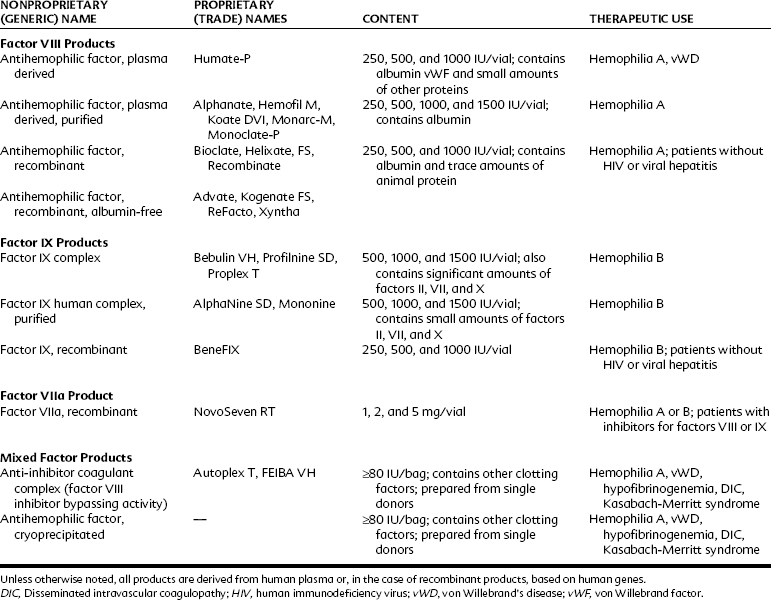
Patients with acquired or genetic bleeding disorders usually have deficiencies in platelet number, platelet function, or faulty or missing clotting factors. Bleeding may develop several hours after trauma or surgery. Uncontrolled bleeding does not generally appear with superficial abrasions, but hemarthrosis and hemorrhage are common with deeper injuries. Thrombocytopenia is frequently drug-induced or associated with other myelogenous diseases; hemophilia disorders are generally inherited. With proper evaluation and supportive therapy (Table 31-3), extensive surgery can usually be accomplished without serious incident.

Stay updated, free dental videos. Join our Telegram channel

Jan 5, 2015 | Posted by in General Dentistry | Comments Off on 31: Procoagulant, Anticoagulant, and Thrombolytic Drugs

VIDEdental - Online dental courses

Get VIDEdental app for watching clinical videos