30: Antianemic and Hematopoietic Stimulating Drugs

CHAPTER 30 Antianemic and Hematopoietic Stimulating Drugs

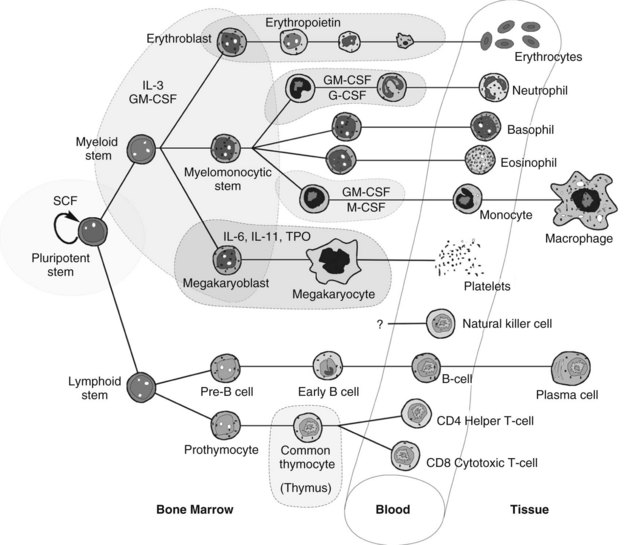
Hematopoiesis is the intricate system of growth and differentiation of immature pluripotent/multipotent stem cells into all the formed elements of the blood (Figure 30-1). These stem cells, derived embryologically in the liver and later from bone marrow,33 divide early in development into either myeloid or lymphoid precursors. The myeloid precursors differentiate into the erythrocytes, megakaryocytes (which give rise to thrombocytes [platelets]), neutrophils, and monocytes. The lymphoid precursors give rise to the T-cell and B-cell lymphocytes, natural killer cells, and all their respective subtypes. The derivation of eosinophils and basophils lies in the myeloid stem line, but appears to be downstream of the common myeloid precursors.26 Hematopoietically active bone marrow retains essentially the same mass throughout life, and although cell-producing bone marrow is found in practically all bones through adolescence, it becomes restricted to the vertebrae, sternum, ribs, pelvis, scapulae, parts of the skull, and epiphyseal ends of the long bones after approximately age 20 years.

Hematopoiesis is a dynamic, continuous process because mature cells of the blood have a limited life span in periods of sickness and health. Because of its complexity, ubiquity, and high rate of activity, the hematopoietic system is often the first organ system to show evidence of underlying systemic disease. This chapter discusses the pharmacologic interventions currently available to correct perturbations in marrow function. Conditions such as anemia, thrombocytopenia, neutropenia, and volume depletion and novel approaches to medical care that involve the hematopoietic system are discussed in detail.

ANEMIA

Anemia comprises a multifactorial group of illnesses with a wide range of underlying causes. As a result, anemia is a generic term indicating only that the concentration of hemoglobin in whole blood is less than normal. Anemia is not a disease, but a sign of underlying disease. When discussing anemia, it is important to diagnose the nature and the cause of the anemia. There are three general categories of diseases that cause anemia: (1) diseases that cause blood loss, (2) diseases that disturb red blood cell production, and (3) diseases that increase endogenous destruction of red blood cells.

Blood loss can occur either acutely, as in hemorrhage from trauma or surgery, or chronically, as with excessive menstrual bleeding or the occult bleeding of esophageal varices or gastric/duodenal ulcers. Disturbed red blood cell production is associated with nutritional deficiencies, disorders that suppress erythrocyte production (such as in aplastic anemia and with some antiretroviral therapy), and myelophthisic (marrow-displacing) diseases. Finally, anemia can be caused by increased destruction of the red blood cells, such as in sickle cell disease, thalassemia, hemolytic immune reactions, and genetic disorders such as glucose-6-phosphate dehydrogenase deficiency.

When a patient is suspected to have a type of anemia, the first tests to consider are a simple hematocrit and erythrocyte count. These two tests tell whether the production-to-loss ratio of red blood cells is normal. The hematocrit is defined as the ratio of red blood cells to the total blood volume. It is expressed as a percentage and is determined by comparing the packed cell volume (which is composed largely of erythrocytes) to the total volume of centrifuged whole blood. Normal values for women are 36% to 45%, and normal values for men are 38% to 50%. In an anemic patient, the hematocrit level is reduced, often into the 20s and in severe cases into the teens or lower. Conversely, the hematocrit increases (referred to as polycythemia vera if moderate and erythroleukemia if severe) in patients with poor pulmonary function, in patients with some chronic cardiac conditions, in patients with certain marrow tumors, and in patients living at high altitudes. In acute hemorrhage, because plasma and red blood cells are lost together, the hematocrit does not initially reflect the loss until the body or exogenous medical intervention has had the opportunity to replenish the lost plasma volume. In these cases, there may be no indication of a problem until several hours later.

The erythrocyte (reticulocyte) count is a simple determination of the absolute number of cells (in millions) per microliter. Normal values for women are 3.8 to 5 million/µL, and normal values for men are 4.4 to 5.6 million/µL.

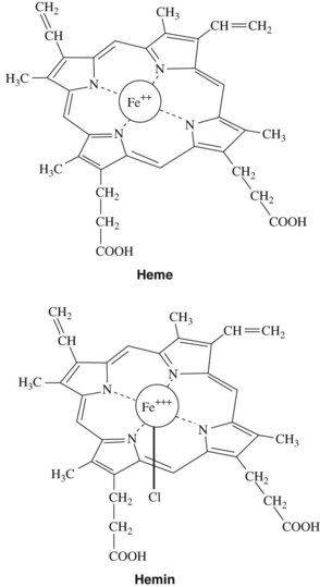
After anemia has been detected, it can be characterized by evaluating the hemoglobin in the erythrocytes. Normal amounts of hemoglobin per unit volume of blood (assayed on peripheral blood draw) are 15.2 ± 2.2 g/dL for men and 13.7 ± 2.1 g/dL for women. Hemoglobin is the oxygen-carrying component of red blood cells. It comprises three components: iron, porphyrin rings, and globin chains. Alterations in any one of these three components can be a cause for a clinical anemia. In normal hemoglobin, iron in the ferrous form (Fe++) is chelated into the middle of the porphyrin chemical ring to yield heme, the nonprotein component of hemoglobin (Figure 30-2).

The globin chains constitute the main protein constituents of hemoglobin. There are four forms of globin chains: α (141 amino acids), β (146 amino acids), δ, and γ (δ and γ are variants of β). Approximately 97% of normal hemoglobin (hemoglobin A) consists of two α and two β chains (α2β2); 1% to 2% consists of the α2δ2 combination (hemoglobin A2). The α2γ2 tetramer forms hemoglobin F, or fetal hemoglobin. Hemoglobin F is the major form during gestation and until approximately 6 months of age. In adults, hemoglobin F makes up less than 1% of normal hemoglobin. One heme ring is accommodated within each of the four structural folds of the tetramer, allowing each molecule of hemoglobin to bind four oxygen molecules.

Hemoglobin accounts for approximately 95% of the dry weight of mature erythrocytes. Any significant changes in hemoglobin are often directly reflected in the way the erythrocytes look or behave grossly. Classically, laboratory analyses for anemia have reviewed erythrocyte size, shape, and color intensity. The size is determined by the mean corpuscular volume (MCV). The normocytic, or normal size, range is 80 to 100 fL/cell. Cells that are too small are termed microcytic, whereas cells that are too large are termed macrocytic. The shape of the red blood cells is also important in diagnosing the cause of anemia. Box 30-1 lists terms that describe various shapes found on a peripheral blood smear. The color intensity of the cell is reflected in the mean corpuscular hemoglobin (normally 26 to 34 pg/cell) and the mean corpuscular hemoglobin concentration (normally 31 to 36 g/dL). These two parameters, along with MCV, collectively referred to as the red blood cell indices, are extremely helpful in delineating the causes of a particular anemia.

BOX 30-1 Descriptive Terms of Red Blood Cell Morphologic Characteristics

TERM DESCRIPTION
Poikilocytosis Irregular erythrocyte shape
Anisocytosis Irregular erythrocyte size
Polychromasia Change in amount of hemoglobin
Sickling Sickle cell disease and trait
Targeting “Bull’s-eye” look to the erythrocytes caused by hemoglobin C and liver disease
Leptocytes Hemoglobin in the border with pigmentation in the center; found in thalassemia, obstructive jaundice, any hypochromic anemia, hemoglobinopathy, and after splenectomy
Spherocytes Round erythrocytes (not biconcave), caused by hereditary spherocytosis or by immune or microangiopathic hemolysis
Schistocytes Fragments of erythrocytes; found in hemolytic transfusion reactions, microangiopathic hemolysis, and other severe anemias
Acanthocytes Distorted (“thorny”) erythrocytes with protoplasmic projections; seen in severe liver disease and with high titers of bile, fats, or toxins
Howell-Jolly bodies Smooth, round remnants of nuclear chromatin; seen in megaloblastic and hemolytic anemias and after splenectomy
Nucleated erythrocytes Found in severe bone marrow stress (e.g., hemorrhage, hemolysis), marrow replacement by tumor, extramedullary hematopoiesis

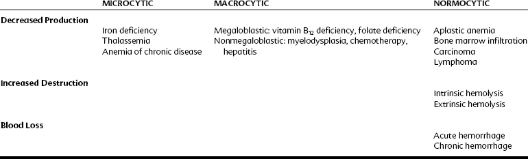
The various kinds of anemia are classified by their typical effect on the erythrocytes (Table 30-1). When anemia results from a loss of blood (intrinsically from hemolysis or extrinsically from hemorrhage) or because of a decrease in production of normal erythrocytes, the cells are still normal, just fewer in quantity. These anemias are normocytic and normochromic. When anemia is caused by a decrease in the production of properly formed hemoglobin, the cells tend to be smaller (because hemoglobin comprises such a high percentage of erythrocyte content) and paler in color. These forms of anemia are known as microcytic and hypochromic and are usually the result of defective or inadequate iron absorption. Forms of anemia that cause the red blood cells to mature incompletely and retain some DNA content result in larger cells; these are known as macrocytic or megaloblastic anemia. They generally occur as a result of a deficiency in vitamin B12, folic acid, or both nutrients. In these forms of anemia, the cells may also have a darker or hyperchromic color.

Iron and Iron Deficiency Anemia

Nutrition and physiologic characteristics

Iron deficiency anemia is the most common cause of anemia worldwide and may occur for many reasons: inadequate nutrition in relation to rate of growth (qualitative or quantitative); defective absorption, transport, or storage (e.g., congenital atransferrinemia or inability to release iron from transferrin to the red blood cells and their precursors); or blood loss from hemorrhage (most commonly gastrointestinal), menstruation, or blood donation. In the United States, iron deficiency anemia is found in 7% of infants, 4% to 5% of children, and 9% to 16% of menstruating women.5 Only 2% to 3% of men have iron deficiency anemia, and women taking oral contraceptives tend to have lower rates because progestins reduce menstrual blood loss. Children 6 months to 2 years old are particularly vulnerable because of their high growth rate coupled with weaning off breast milk and onto cow’s milk. Cow’s milk is low in absorbable iron and may irritate the intestines. Pregnancy may precipitate iron deficiency anemia by rapidly increasing the blood volume, sometimes requiring two to five times the normal intake of iron. According to a World Health Organization technical report,17 women who have sufficient iron reserves to support the increase in hemoglobin production during pregnancy and who breastfeed their infants are generally capable of meeting their iron needs by diet alone, although supplementation is still recommended. In a nonpregnant, normal, healthy individual, iron reserves and recycling are so effective that even extreme reduction of iron intake may be insufficient to cause severe anemia.12

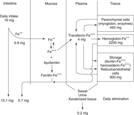
Men average 3.8 g total iron (50 mg/kg) and women average 2.3 g (35 to 42 mg/kg). Approximately 60% to 80% of the iron in the body is incorporated into hemoglobin (Figure 30-3). Anemia is the primary presenting sign of iron deficiency. Approximately 10% to 25% is sequestered in reticuloendothelial cells in the storage forms ferritin and hemosiderin (described later), and another 10% to 15% is associated in parenchymal cells with myoglobin. Less than 1% is used in various enzymes, most notably the cytochromes, and trace amounts are linked to the plasma transport protein transferrin. The amount of stored iron varies with intake and demand, averaging 400 mg in women and 1000 mg in men.

The average American ingests 10 to 20 mg of iron per day. Iron is obtained through the diet, most commonly by heme or iron complexed to various organic compounds. Foods considered high in iron (>0.5% by weight) are liver, heart, oysters, egg yolks, and yeast. Other meats and green vegetables have less iron. Absorption of iron from dietary sources is ordinarily 10% efficient or less, but it increases when iron stores are depleted. Therapeutic iron, generally in the form of inorganic salts or complexes, has an even poorer absorption profile than dietary iron because the Fe++ must be liberated from the salt before it can be absorbed across the intestinal mucosa.

Iron absorption occurs along the entire length of the intestine, but maximum absorption occurs in the duodenum and proximal jejunum because iron is absorbed primarily as Fe++, and an acid medium favors the breakdown of salts to the ionic form. In the lower portions of the gastrointestinal tract there is a trend toward increasing alkalinity, which favors the formation of less soluble iron salts and complexes. Iron ingested as heme iron is absorbed five to seven times more efficiently than Fe++ salts. Iron absorption is hindered by coffee, tea, phosphates, and antacids, particularly calcium carbonate and aluminum or magnesium hydroxide. Absorption of nonheme iron is facilitated by vitamin C. How ethanol interacts with iron is not well elucidated, but approximately 50% of alcoholics exhibit some iron depletion or anemia.

Iron is absorbed by active transport across the intestinal mucosa, where it is converted intracellularly to ferric iron (Fe+++). Depending on the body’s acute need for iron, Fe+++ is either bound to transferrin or converted to ferritin or hemosiderin for storage in the intestinal mucosa. Transferrin is a transport glycoprotein electrophoretically migrating with the β globulins; it specifically binds two molecules of Fe+++. It enters the plasma and carries Fe+++ to the bone marrow and developing erythroblasts. The erythroblasts present membrane transferrin receptors that bind diferric transferrin and then internalize the complex by endocytosis. Inside the cell, the transferrin receptor, transferrin, and Fe+++ are broken apart, with the iron being used in hemoglobin synthesis and the transferrin and transferrin receptor being carried back intact to the surface for recycling. The typical developing erythroblast can process 25,000 to 50,000 transferrin molecules per minute.

A test for transferrin is total iron-binding capacity (TIBC). In a normal adult, approximately 20% to 50% of transferrin is replete with Fe+++. In an iron-deficient individual, transferrin saturation may decrease to 15% or less. The capacity to bind iron is considerably greater, and the TIBC value increases. Normal values are 250 to 450 µg/dL.

If the body is not in acute need of iron, most of the ingested iron is stored as ferritin. Twenty-four apoferritin monomers bind together to form a hollow spherical shell 130Å in diameter and fenestrated with small pores through which 4000 Fe++ atoms can enter. When inside, the Fe++ is oxidized to Fe+++ and stored in the form of hydrous ferric oxide phosphate. Ferritin, the resulting apoferritin-iron complex, is a very effective storage mechanism, allowing the binding and release of iron to occur rapidly and efficiently. Mature ferritin is found in virtually all cells of the body and in plasma. Although the amount in plasma is small, it reflects the total ferritin stores in the body and is measured to diagnose iron deficiency anemia. Normal values for serum ferritin are 16 to 300 mg/mL in men and 4 to 160 mg/mL in women.

The other minor storage component of iron is hemosiderin. It is found in the monocyte/macrophage system of the marrow and in the Kupffer cells of the liver. Hemosiderin is an insoluble compound that seems to be aggregated ferritin cores partially or completely stripped of the apoferritin protein shell. In pathologic conditions (hemosiderosis), it can be found in large quantities in most tissues of the body.

The concentration of iron in the plasma at any one time represents a balance between the absorption rate, storage capacity, rate of hemoglobin formation, and rate of iron excretion. Iron is remarkably well conserved in the body; less than 0.1% is excreted on a daily basis, or approximately 0.5 to 1 mg/day. The major pathway of iron excretion is through the feces by exfoliation of gastrointestinal cells and their intracellular stores of ferritin when the mucosal cells are replaced by new epithelium. Iron is also lost in considerably smaller amounts by excretion through urine, exfoliation of dermal cells, and perspiration. Menstruation causes the amount of lost iron roughly to double to 2 mg/day. Uncommon sources of iron loss include excessive blood loss or excessive destruction of erythrocytes. Hemorrhage depletes heme iron, whereas excessive turnover of erythrocytes releases it back into the circulation, where it can be recycled. A normal individual can lose a quarter to a third of the erythrocyte mass through hemorrhage without need for iron therapy. Because iron is so well conserved in the body and most people have large reserves, chronically insufficient intake of iron is almost always the cause of iron deficiency anemia.

Iron therapy

The intuitive treatment of any disease state that is accompanied by extreme fatigue, weakness, and loss of color includes increased dietary intake, and the ancient Greeks, Hindus, and other early peoples turned to iron in many forms simply because it represented “strength.” Although Sydenham is generally credited with the first rational use of iron (iron filings in wine) for treating anemia in 1681, it was not known that iron was actually present in blood until 30 years later, when Lemery and Geoffry demonstrated its presence. Shortly thereafter, Menghini, an Italian physician, showed that foods with iron actually increase blood iron, but it was not until approximately 1830 that a pill containing iron (ferrous sulfate and potassium carbonate, 1 : 1) was introduced into medicine by Blaud,3 an event that marked the beginning of modern treatment of iron deficiency anemia.

Iron therapy is indicated in iron deficiency anemia; it is contraindicated in anemia of any other cause. Iron is available in the form of Fe++ salts (sulfate, gluconate, and fumarate), which are reasonably well absorbed, and a Fe+++-containing compound (iron polysaccharide), which is not as well absorbed. The most commonly used Fe++ preparation, and the agent of choice for uncomplicated iron deficiency anemia, is ferrous sulfate. It is normally given in doses (325 mg three times a day) much larger than should theoretically be needed because of its limited absorption (≤15%). The response to oral iron preparations is usually evident in 5 to 10 days and is first manifested by an increase in reticulocytes. Adverse effects associated with orally administered iron are gastrointestinal symptoms, chiefly nausea and vomiting, because of direct irritation of the stomach. The patient has black stools as a result of therapy, which may obscure the diagnosis of melena. The drug is unquestionably best absorbed when taken between meals, but gastrointestinal distress is reduced if the medication is taken with meals and if the dose is started at a lower level and slowly increased with time. In general, the hematocrit returns halfway to normal in approximately 3 weeks and is fully corrected in roughly 8 weeks. To replenish iron stores, a course of therapy of 3 to 6 months is generally required.

Although parenteral iron preparations are available, they are not generally used because of the simplicity of oral medication and the much greater risk of serious side effects and higher expense. Iron should be administered parenterally only if the oral preparations are inadequately absorbed or poorly tolerated, such as in patients with enteritis or colitis, or if it is absolutely necessary to replace a serious iron deficit quickly. The classic parenteral form is iron dextran, a sterile colloidal solution of ferric hydroxide and low-molecular-weight dextran, which is administered by intramuscular or intravenous injection. Adverse reactions include pain and straining at the site of injection (intramuscular), urticaria, fever, arthralgia, lymphadenopathy, nausea, and vomiting. Rarely, severe or fatal anaphylactic reactions have occurred after the use of this preparation. Another parenteral form, iron sucrose, is a polynuclear ferric hydroxide sucrose complex. It is believed that the antigenic potential of iron dextran lies in the Fe++ and dextran polysaccharides and that this preparation is less allergenic. Iron sucrose is commonly used in patients undergoing renal dialysis who are receiving erythropoietin (EPO) therapy.

Acute iron poisoning is uncommon, but can occur, particularly because many iron formulations are brightly colored and attractive to children. Ingestion of large doses of iron causes the transferrin to become saturated, and free iron enters the blood in excess. Unbound iron is toxic and has caused severe gastrointestinal disturbances and may lead to circulatory collapse. Chelating agents have been used in the treatment of acute iron toxicity; deferoxamine, a potent and specific iron-chelating compound, is capable of removing iron from ferritin and transferrin but not from hemoglobin. It is, however, no substitute for more immediate measures, such as inducing vomiting, gastric lavage, and fluid administration, that should be carried out in the event of iron poisoning.

Perhaps the most important consideration for the dental professional is the finding that people who are taking oral iron supplements may have altered absorption profiles of other drugs. The quinolone class of antibiotics, tetracyclines, and thyroid replacement hormones all form complexes with iron and result in significantly poorer (36% less) absorption profiles. Simply having the patient stagger the iron and the interacting competing medication by 2 or more hours is usually sufficient to avoid this difficulty.

PORPHYRIA

Although iron deficiency anemia is the most commonly encountered form of anemia, it is not the only disorder in which insufficient functional heme is produced. The porphyrias, a cluster of disorders that involve decreased or disordered production of the porphyrin ring, can be associated with anemia depending on the variety and severity of the presentation of the diseases. Heme is a major component of hemoglobin, but it is also crucial to several enzyme systems, most notably the large family of cytochrome P450 enzymes involved in steroid synthesis and drug metabolism.

Porphyrin is produced in an eight-step process that occurs in the mitochondria and in the cytosol. The two principal cell types involved are the developing erythroblasts and reticulocytes of the bone marrow (mature erythrocytes lack mitochondria and are unable to synthesize porphyrin) and the liver hepatocytes. As a result, two general classifications of porphyria exist—erythropoietic and hepatic—that are divided further into nine varieties (Table 30-2), each corresponding to a particular enzyme deficiency in the synthetic pathway of porphyrin. These deficiencies may be genetic in nature or caused by medications.

TABLE 30-2 Classification of Porphyrias

PORPHYRIA SITE OF EXPRESSION PRINCIPAL CLINICAL FEATURE
Acute intermittent porphyria Liver Neurologic
δ-Aminolevulinic acid dehydratase deficiency porphyria (rare) Liver Neurologic
Hereditary coproporphyria Liver Neurologic, photosensitivity
Porphyria cutanea tarda Liver Photosensitivity
Variegate porphyria Liver Neurologic, photosensitivity
Hepatoerythropoietic porphyria Liver, bone marrow Photosensitivity
Congenital erythropoietic porphyria Bone marrow Photosensitivity
Erythropoietic protoporphyria Bone marrow Photosensitivity
X-linked sideroblastic anemia Bone marrow Hemolytic anemia

Acute exacerbations usually occur when there is a significant demand for heme synthesis that cannot be met by the limited enzyme function. This deficiency in heme inhibits the negative feedback cycle on δ-aminolevulinic acid synthase, causing induction of this rate-limiting enzyme. Because the heme synthesis pathway is damaged, induction instead leads to the excessive production in the liver of the porphyrin precursors δ-aminolevulinic acid and porphobilinogen, which build up and cause the acute symptoms. This accumulation of protoporphyrin precursors results in neurologic disorders, photocutaneous disturbances, or both.

Probably the most common genetic form of porphyria is acute intermittent porphyria. Its mode of transmission is autosomally dominant and results from a partial enzymatic deficiency (<50% of normal) in the third step of porphyrin synthesis. Because synthetic activity is diminished but not lost, most patients remain asymptomatic throughout normal life. Acute exacerbations, which give rise to the name, have highly variable symptoms that last from days to months. The most common presentation is neurologic, including mental changes; seizures; and acute sensory neuropathies such as abdominal pain, chest and back pain, and limb pain. The severity of the pain can be great enough to mimic other acute disorders and result in unnecessary surgical intervention such as laparotomy. Motor neuropathies, especially in the cranial nerves, are often seen. Occasionally, motor paralysis of the respiratory diaphragm has resulted in death. Gastrointestinal disturbances—primarily nausea, vomiting, and diarrhea—are common.

Several events can precipitate an attack. Physiologic stressors, such as surgery, excessive alcohol intake, illnesses, and infections, may induce hepatic heme oxygenase, which breaks down heme. Endocrine changes, such as may occur around a woman’s menses, or synthetic estrogens and progestins may also induce an attack. More than 1000 medications have been categorized with regard to their porphyrinogenicity,32 of which a few reactions are well documented and many are still anecdotal. What is accepted is that endocrine properties of the drug, affinity for cytochrome P450, hepatic load, and capacity to modulate nuclear receptors affecting gene transcription (particularly 5-aminolevulinate synthase [ALAS1]) all play a role in how porphyrinogenic a drug might be.

Several medications commonly used in dentistry and medicine (Box 30-2) are steroid-based or metabolized by, and induce the synthesis of, the cytochrome P450 enzyme system, which leads to increased accumulation of porphyrin precursors.22 The response of any individual to any of these medications can be highly variable; the proposed unsafe medications should be discussed with the physician on a case-by-case basis. In susceptible porphyric patients, which also includes individuals with hereditary coproporphyria and variegate porphyria, dose reductions or avoidance of specific medications may be necessary. Thunell and colleagues32 have proposed a standardized method to determine a drug’s probable porphyrinogenicity, and an Internet database is available at http://www.drugs-porphyria.org. Poor nutritional intake has also been associated with acute attacks.

BOX 30-2 Drugs Considered Safe or Unsafe for Use in Patients with Acute Intermittent Porphyria, Variegate Porphyria, and Hereditary Coproporphyria

SAFE POSSIBLY UNSAFE
Acetaminophen Alcohol
Amitriptyline Alkylating agents
Aspirin Barbiturates (severe)
Atropine Carbamazepine
Chloral hydrate Chlordiazepoxide
Clorazepate Chlorpropamide
Diazepam Chloroquine
Digoxin Clonidine
Diphenhydramine Dapsone
Glucocorticoids Ergots
Guanethidine Erythromycin
Hyoscine Estrogens, synthetic
Ibuprofen Food additives
Imipramine Glutethimide
Insulin Griseofulvin
Labetalol Hydralazine
Lithium Ketamine
Naproxen Meprobamate
Nitrofurantoin Methyldopa
Opioid analgesics Metoclopramide
Penicillamine Nortriptyline
Penicillin and derivatives Pentazocine
Phenothiazines Phenytoin
Procaine Progestins
Propranolol Pyrazinamide
Selective serotonin reuptake inhibitors

Streptomycin Succinimides Succinylcholine Sulfonamides (severe) Tetracycline Theophylline Thiouracil Tolazamide Vitamins B and C Tolbutamide   Valproic acid

Porphyria cutanea tarda is the most common porphyria and is representative of the erythropoietic porphyrias. Symptoms commonly include photosensitivity, which results from sequestration of protoporphyrins in the skin and subsequent deposition of iron in the integument. Porphyrin and its precursors undergo photoactivation at 400 nm in the presence of oxygen, causing cellular destruction by release of oxygen free radicals. In skin exposed to light, the porphyrins become photoexcited, and clinically evident cellular damage occurs. Porphyrin-laden erythrocytes also undergo phototoxicity when circulating through light-penetrated tissues. The damage may be sufficient to result in hemolytic anemia.

Management of acute intermittent porphyria has been primarily aimed at avoiding exacerbating conditions. Adequate caloric intake, prompt diagnosis and treatment of infections (including odontogenic and other orofacial infections), and care in not taking medications known to trigger attacks are strategies the patient can use to minimize the risk of developing a crisis. In patients who have photoreactive porphyria, avoidance of sunlight, wearing clothing to cover the skin, and generous use of sunscreen lotion are helpful. If an acute attack occurs that is not amenable to glucose infusion, a medication of choice is lyophilized hemin with sodium carbonate. Hemin is ferric heme that has a Cl on one of the two available coordination sites for Fe+++ (see Figure 30-2). On mixing with sterile water, hemin is converted in the resulting alkaline solution to hematin by replacement of the Cl with an OH group. Hematin serves as an enzymatic inhibitor of porphyrin synthesis by decreasing the concentration of the precursors porphobilinogen and δ-aminolevulinic acid. The reconstituted drug is unstable, however, and has been frequently associated with thrombophlebitis and increased coagulopathy. Palliative use of opioid analgesics is also often indicated during porphyric exac/>

Only gold members can continue reading. Log In or Register to continue

Stay updated, free dental videos. Join our Telegram channel

Jan 5, 2015 | Posted by in General Dentistry | Comments Off on 30: Antianemic and Hematopoietic Stimulating Drugs

VIDEdental - Online dental courses

Get VIDEdental app for watching clinical videos