Development of the oral facial region
Learning objectives
Key terms
Alveolar process
Angular process
Aortic arch vessel
Auditory capsules
Basioccipital cartilages
Basion
Body
Common carotid arteries
Condylar
Coronoid process
Cranial base
Cranial base cartilages
Ethmoid
Ethmosphenoid and sphenoccipital articulations
Eustachian tube
External auditory canal
External carotid
Facial bones: zygomatic, maxillary, frontal, and temporal
Facial suture types: simple, serrated/interdigitating, squamosal
Facial sutures: zygomaticomaxillary, frontomaxillary, and zygomaticotemporal
Frontal, parietal, and squamous portions
Hyoid: superior hyoid, inferior hyoid
Incus
Inferior parathyroids
Internal carotid
Laryngeal cartilages
Malleus
Mandibular arch
Masseter, medial, and lateral pterygoid and temporalis muscles
Maxillary tissues
Meckel’s cartilage bar, Meckel’s cartilages
Middle ear
Nasion
Oropharyngeal membrane
Oropharynx
Palatine tonsils
Pharyngeal arch
Pharyngeal pouches
Premaxillary, maxillary, zygomatic, and petrous portions
Pulmonary circulation
Sella turcica
Sphenoid
Stapes
Superior parathyroids
Sutures
Synchondrosis
Syndesmosis
Synostosis
T cells
Temporal and intraoccipital bones
Thymus
Tympanic membrane
Ultimobranchial body
Overview
This chapter concerns development and orientation of the tissues that form the human face and neck. During the fourth week of development, the human embryo consists of a flat disk that bends down at its anterior extremity as the overlying brain expands and enlarges. This action pushes the heart beneath the brain. A pit develops in the midline between the brain and the heart and becomes the oral cavity, or stomodeum (see Fig. 3-2). Beneath this pit the first pharyngeal arch, termed the mandibular arch, forms. The maxillary tissues that form the cheeks grow from this first arch. Below the mandibular arch four other pharyngeal arches or bars appear during the fourth to seventh prenatal week. The second arch is called the hyoid (see Fig. 3-3). These parallel arches are important in the development of the face and neck, and each contains blood vessels, muscles, nerves, and skeletal elements. Aortic arch blood vessels, which course through each pharyngeal arch from the heart below to the brain above, are important to craniofacial development.
The first, second, and fifth of these vessels soon disappear. The third arch vessel quickly assumes the role of supplying nutrients to the tissues of the first and second arches. This third arch vessel also shifts the blood supply to the face from the internal carotid vessels to the external carotid. Muscles arise in each of the pharyngeal arches: the mandibular arch muscles become the masticatory muscles; the second arch muscles become the facial expression muscles; and the muscles of the third and fourth arches become the constrictor muscles of the throat. Cranial nerves enter each of these muscle masses as they arise. The fifth nerve enters the mandibular arch to innervate the muscles of mastication. The seventh innervates the second arch muscle mass, and other cranial nerves innervate the muscles of the neck. Cartilage also appears in each arch: Meckel’s cartilage bar in the first, the superior hyoid in the second, the inferior hyoid in the third, and the laryngeal cartilages in the fourth. The cranial base cartilages arise to support the brain, and from them come the auditory and olfactory sense capsules. All the creative events described in this chapter take place from the fourth to seventh prenatal weeks, the short time required for facial organization.
Development of the oropharynx
The oropharynx is composed of the primitive oral cavity and the area of foregut called the pharynx. The oral pit first appears in the fourth week of development, when the neural plate bends ventrally as the neural folds develop to form the forebrain. This cephalocaudal bend pushes the heart ventrally, and the yolk sac becomes enclosed to form an elongating tube known as the foregut (Fig. 3-1).
The deepening oral pit then appears between the forebrain and the heart and eventually becomes the oral cavity (Fig. 3-2). At its deepest extent is the oropharyngeal membrane, which ruptures in the fifth week, opens the oral cavity to the tubular foregut, and soon becomes the oropharynx (Figs. 3-3 and 3-4). The mandibular arch will grow laterally to the oral pit, developing the maxillary process, which forms the cheeks.
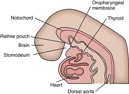
The oropharyngeal membrane separated the oral pit (stomodeum) and the pharyngeal cavities. The membranes will then rupture, allowing the two cavities to join.
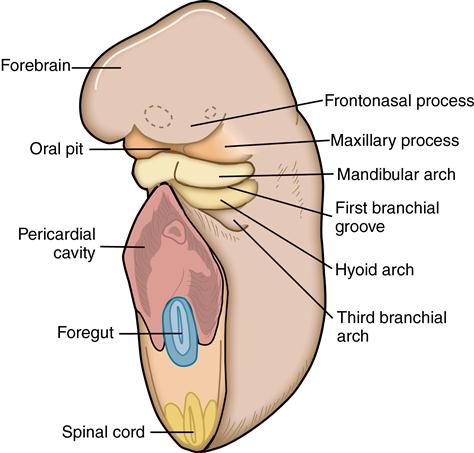
The oral pit is surrounded by the facial primordia, which are the frontonasal processes, the maxillary processes, and the mandibular arch. The pharyngeal arches are defined by grooves between each arch. The heart develops in the thorax, which is in the pericardial cavity.
The pharyngeal arches are seen in the wall of the pharynx. The aortic arch vasculature leads from the heart through these arches to the face.
The enlarging heart now becomes positioned below the mandibular arch in the thorax and begins beating at the end of the fourth week (see Fig. 3-2). Blood is forced through the vessels in the pharyngeal arches supplying the face, neck, and brain. The forming face now grows away from the forebrain and presses against the chest and heart.
Development of the pharyngeal arches
 CLINICAL COMMENT
From the initial development, each pharyngeal arch has a specific cranial nerve associated with it. The nerves and the musculature of each arch emerge together and follow defined pathways to their functional positions. These events are closely regulated genetically during development, and few errors occur.
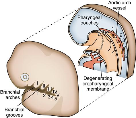
The pharyngeal arches are so termed because they bend around the sides of the pharynx as bars of tissue. Each arch is separated by vertical grooves on the lateral sides of the neck at the fifth week. Within the pharynx, grooves called pharyngeal pouches separate each arch. These pouches match the pharyngeal clefts on the external aspects of the neck (see Fig. 3-4).
 CONSIDER THE PATIENT
A patient appears with a swelling in the lateral area of the neck and states that the swelling subsides from time to time but then resumes. He asks you what you think the cause may be.
The five arches with their clefts resemble the embryonic gill slits of fish and amphibians. This is one of many similarities between human embryos and other embryos during early development. The first arch is termed the mandibular arch because it will later form the bony mandible and the associated muscles of mastication, nerves, and blood supply. The second, or hyoid, arch forms the facial muscles, vessels, and hyoid bone. The third, fourth, and fifth arches consist of paired right and left bars that are divided before they reach the midline by the presence of the bulging heart (see Fig. 3-2). The arches become progressively smaller anterior to posterior. The outer surface of each arch is covered with ectoderm as are the inner surface of the first arch and the covering of the anterior surface of the second. This ectoderm will become the epithelial lining of the oral cavity. The pharyngeal surface of the remaining four arches is, however, lined by endoderm, which is the same as the lining of the gastrointestinal tract (see Fig. 3-4). The cores of the arches—the blood vessels, muscles, nerves, cartilages, and bones—will differentiate and are important in the development of the adult human face.
 CLINICAL COMMENT
The face develops during the short span from the fourth to seventh prenatal weeks. Environmental factors can cause a facial or pharyngeal arch defect, which would probably affect these tissues before the fourth week. This is the time to be especially careful of irradiation and chemical, hormonal, dietary, or stress-related factors.
Pharyngeal grooves and pharyngeal pouches
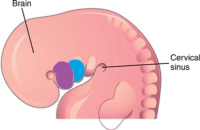
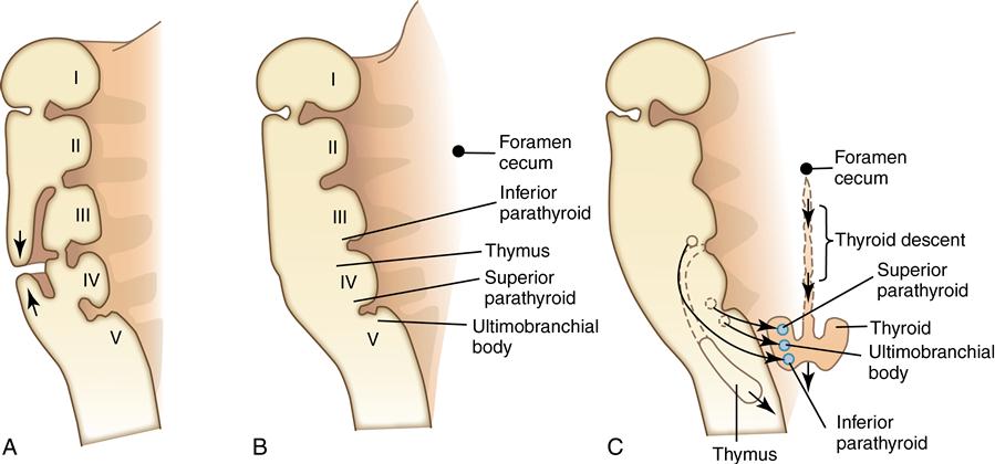
The first pharyngeal groove deepens to become the external auditory canal leading to the middle ear. The membrane at the depth of this tube becomes the tympanic membrane. The middle ear and eustachian tube develop from the corresponding first pharyngeal pouch. After the fifth week, no other pharyngeal grooves are seen externally as the tissues of the second and fifth arch grow over the other arches and grooves and make contact with each other (Fig. 3-5, A). This overgrowth obscures the tissue of both the arches and the grooves externally, although their internal structures are unaffected and provide an important role in facial and body development (see Fig. 3-5, A and B).
Stay updated, free dental videos. Join our Telegram channel
VIDEdental - Online dental courses


