Root Formation and Attachment Apparatus
• To discuss the role of the epithelial root sheath in root formation and dentin formation
• To describe the fate of the epithelial root sheath
• To describe the beginning of cementum formation, the two varieties, and where they are found
• To define and diagram alveolar bone and its components
• To define periodontal ligament and list its various groups and subgroups of fibers
• To briefly describe bone’s reaction to pressure and tension and how this affects tooth movement
ROOT FORMATION
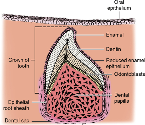
Root formation begins after the outline of the crown has been established but before the full crown is calcified. If you refer to Figs. 19-4 and 20-3 (bell stage), you will see that the point where the outer enamel epithelium (OEE) meets the inner enamel epithelium (IEE) is located at the deepest part of the enamel organ and is known as the cervical loop. There are no interposing layers of stellate reticulum or stratum intermedium here, which one would see more coronally in the crown. These layers of OEE and IEE, which make up the epithelial root sheath (Hertwig’s epithelial root sheath), begin to undergo rapid mitotic division and grow deep into the underlying connective tissue—the beginning of root formation. It is important to keep in mind the relationship of the dental papilla and the dental sac to this growing epithelial root sheath. The dental papilla is on the inside, and the dental sac is on the outside (Fig. 21-1). As the apical growth continues, the tip of the epithelial root sheath turns horizontally inward; this turned-in portion is known as the epithelial diaphragm of the root sheath. The epithelial root sheath and epithelial diaphragm guide the shape and the number of roots.
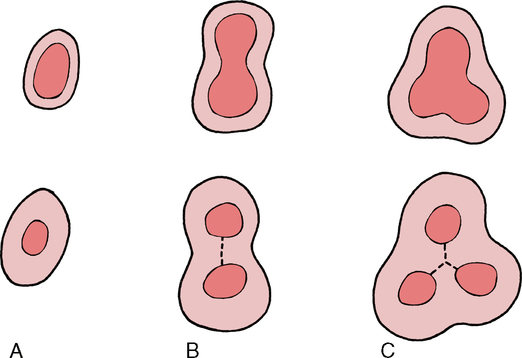
Figs. 21-1 and 21-2 are two-dimensional representations of a three-dimensional object. To help you better understand this three-dimensionally, visualize a paper cup. The rim of the cup represents the cervical line of the tooth, and the side of the cup is the epithelial root sheath. If you cut a round hole in the bottom of the cup about two thirds the diameter of the cup and made a vertical cut through the middle, the horizontal bottom of the cup that remained would represent the epithelial diaphragm. The way in which this epithelial diaphragm continues to grow inward determines whether the tooth will have one, two, or three roots.
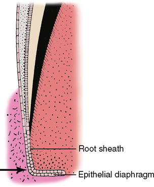
Changes continue to occur at the deep surface of the epithelial diaphragm. As the vertical epithelial root sheath grows longer, forming root length, the horizontal epithelial diaphragm continues to grow in toward the middle of the tooth. If the entire circumference of the diaphragm grows evenly, it will eventually form a single-rooted tooth. If two areas opposite one another grow inward more rapidly and meet, it will then separate into two columns of root formation to form a bi-rooted tooth. If three areas grow inward to meet, a tri-rooted tooth will be formed (Fig. 21-3). In multi-rooted teeth, the point where the epithelial diaphragm meets is the bifurcation or trifurcation of the tooth.
ATTACHMENT APPARATUS
Dentinocemental Junction
Fig. 21-1 shows the relationship of the epithelial root sheath to the dental papilla and dental sac. As the root sheath grows from the cervical line deeper into the connective tissue, it influences the peripheral cells of the dental papilla to change into odontoblasts, similar to what happens in the crown of the tooth. The odontoblasts begin to secrete matrix and calcify. Once dentin begins to form next to the epithelial root sheath, cellular influence causes the root sheath to begin to break up. There is still some debate as to which cells cause this breakup. It may be the inside odontoblasts, or it may be the cells of the dental sac on the outside.
To envision this deterioration of the epithelial root sheath, imagine that the sheath is originally a solid wall of cells surrounding the developing tooth root. Later it becomes riddled with holes, like a piece of Swiss cheese. With the appearance of these holes, there is no longer any barrier separating the odontoblasts and dentin on the inside from the cells of the dental sac on the outside. Some of the dental sac cells begin to change into cementoblasts, move through the holes, and begin to form cementum. This cementum is laid down against the previously formed dentin and establishes the dentinocemental junction (Fig. 21-4). Remember that the epithelial root sheath is perforated; therefore the cementoblasts that contact dentin are able to accomplish the transformation only in the areas where the sheath has broken up. While this occurs, the remaining root sheath cells pull away from the dentin, and the cementoblasts contact all of the dentin and establish the rest of the dentinocemental junction. Occasionally, some epithelial root sheath cells do not pull away and may become ameloblasts, forming small globs of enamel on the surface of the dentin. These are referred to as enamel pearls and are usually found in bifurcations and trifurcations of roots. Other defects occurring at this time may lead to the formation of accessory root canals, which make it difficult for a dentist to remove all pulpal tissue completely if the tooth has to be treated endodontically.
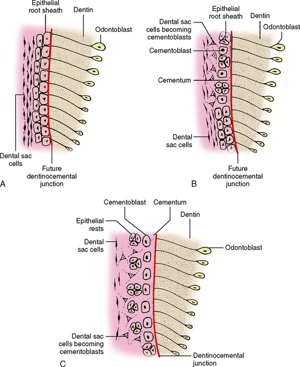
After the cells of the epithelial root sheath have broken up and moved away from the dentin, the remaining root sheath cells are found in the periodontal space next to the tooth and are called epithelial rests of Malassez or simply epithelial rest cells. If these cells later begin dividing, they may lead to the formation of periodontal cysts in the jaws, similar in origin to the palatal cysts mentioned in Chapter 18.
Stay updated, free dental videos. Join our Telegram channel
VIDEdental - Online dental courses


