20: Problem Solving the Challenges Faced in the Restoration of Endodontically Treated Teeth

Chapter 20

Problem Solving the Challenges Faced in the Restoration of Endodontically Treated Teeth

Problem-Solving List

Problem-solving issues and challenges in the restoration of endodontically treated teeth addressed in this chapter are:

Postendodontic and Intertreatment Temporary Restorations
Restoring Access Openings
Problem-Solving Key Factors That Influence Restorative Choices in Anterior and Posterior Teeth

    Restoration of anterior teeth
    Restoration of posterior teeth
    Intraradicular posts
Exposed and Contaminated Root Fillings Due to Coronal Leakage

“I have made a diligent search to find whether or not teeth from which the pulp had been removed, and the canals and pulp chamber filled without admitting the fluids of the mouth, were subject to a similar deterioration of strength. It is now well known that teeth treated in this way retain their color almost perfectly; and from my observation of the relation of color to strength of the teeth I am prepared to entertain the suppositions that they retain their strength also. Only one tooth came into my hands with a root filling. It was a central incisor, and had not sufficient tissue for me to obtain a block; but it showed no undue percentage of water, and the color was good. Up to the present time I have been unable to obtain the history of the filling of this root.”< ?xml:namespace prefix = "mbp" />21

G.V. Black, 1885

The concept of restoring endodontically treated teeth usually evokes challenging images of teeth with significant coronal tooth loss. In reality it may be the simple closure of an access cavity in a satisfactory coronal restoration to the total reconstruction of a tooth with no clinical crown due to the ravages of caries, faulty restorations or traumatic episodes, both short and long term (e.g., accidents, occlusal wear, abnormal function, etc.). Regardless of the clinical situation, the two primary objectives are to preserve the integrity of the root canal filling and rebuild the tooth into a symptom-free, functional, stable contributor in the patient’s dentition.29,47,51,90

In the last 25 to 30 years, a plethora of scientific studies, clinical-technique articles, and the introduction of new materials have addressed the enigmatic issue of restoring an endodontically treated tooth.49,93 During this same period, however, the significance of coronal leakage relative to endodontic failure has been highlighted, although this problem was identified over 90 years ago.33 The application of the principles of problem solving must now include, through careful and deliberate diagnostic assessments, the elements of identification and prevention as well as the need to manage these issues using either evidence-based or the best evidence directives. Although scientific insights and technologic improvements have provided a clearer understanding of this issue, sound clinical judgment and experience must still enter into the decision-making process in the restoration of these teeth. This includes the choice and placement of interim restorations before and during root canal treatment as well.

In Chapter 8, the importance of removing questionable restorative materials and all caries, along with proper tooth isolation, was stressed in conjunction with (1) the assessment of the restorability of the tooth, (2) identification of defects in the tooth structure, and (3) the development of a functional endodontic access opening in the tooth to be treated. To enhance this important phase of treatment, the use of magnification and a caries detector is usually beneficial, but the latter may also stain sound dentin that has decreased mineral content. Recent advances in the development of caries-detecting agents may have eliminated this problem.59 In many cases requiring root canal treatment, two or more treatment visits may be indicated, and this requires that a proper interim temporary restoration be placed. The same dictate applies to each tooth upon completion of the root canal treatment if a permanent restoration is not going to be placed immediately. However, the effectiveness of temporary cements can vary,10,99 so the importance of these temporary restorations cannot be overemphasized relative to the prognosis for positive treatment outomes.56,74

Postendodontic and Intertreatment Temporary Restorations

With the initial access opening preparation, all caries, undermined tooth structure, and areas that are questionable relative to their integrity are to be removed or investigated (cracks, stained lines, etc.) so the tooth can be assessed for restorability prior to the actual root canal procedures. Some clinicians choose not to do these procedures, claiming there might be leakage if a wall were removed or if all the caries under a crown were eliminated. The claim is also made that the tooth cannot be isolated properly if specific tooth structure is removed. In essence, these approaches destroy the problem-solving concept and actually create environments conducive for problems. For example, salivary and bacterial leakage through carious dentin or poorly placed temporary materials can cause interappointment flare-ups and the opportunity for bacteria and their biofilms and byproducts to gain a foothold into an open canal or even one that has been obturated (see Chapter 5).*

As a basic principle in restorative dentistry, restorations usually leak in time, and no materials are perfect. When considering temporary restorative materials for endodontic access openings, everything will leak sooner rather than later.68,116 Even in well-done root canal procedures, coronal leakage can result in reinfection of the apical tissues in as little as 19 days,69,135 with relative certainty within 3 months.76 Therefore prevention is essential in these cases by first considering immediate, permanent restoration following root canal treatment. If not possible, preventive techniques are necessary to minimize the possibility of this devastating occurrence.

Many premixed preparations have enjoyed popularity as interim or post root-treatment temporary filling materials, such as Cavit (3M ESPE, St. Paul, MN, USA) or IRM (Dentsply Caulk, Milford, DE, USA). Laboratory studies indicate, however, that their ability to prevent the ingress of bacteria over time is poor.121471 A second complication arises in that cotton, which is commonly used to maintain space in the pulp chamber prior to permanent restoration, may become entrapped in the materials along the cavity wall and provide an avenue for leakage.95,145 A well-placed temporary restoration can be expected to protect against bacterial leakage no longer than 1 month.14

While all materials leak to some extent,7,17,105,132,154 zinc oxide eugenol cements have long been regarded as possessing the best sealing properties of all cement materials because of their antimicrobial properties.28,146 However, zinc oxide/calcium sulfate materials such as Cavit tend to be more resistant to leakage than zinc oxide eugenol materials.146 The combined use of both materials may minimize leakage,12 but in either case, a minimum of 3 mm of thickness is essential.16,68,99,146 Resin-based materials can be used, but they have a tendency to shrink and result in leakage.26

The placement of a temporary post and core creates a different set of circumstances, especially as it relates to an impervious seal.34,43 In these circumstances, a barrier40,82 using a self-curing material may need to be placed over the coronal, intracanal termination of the root canal filling material.

A better consideration for a temporary filling, if the tooth is not to be restored permanently in the immediate future, would be placement of a temporary material to the pulpal chamber floor without the use of a cotton pellet.116 The seal may be enhanced by thickness (usually greater than 3 mm). If a cotton pellet is to be used, however, orifice barriers are recommended. First, countersink the orifice with a small round bur, followed by a thorough cleaning with alcohol or a detergent to remove excess cement and debris. Second, if possible use air abrasion to free the dentin from films and debris. Third, place a temporary or permanent restoration in the orifices and over the floor of the chamber. In this regard, bonded materials or glass ionomers are favored.19,20,45,148,149 Mineral trioxide aggregate (MTA) may also be considered,4,77 but a clear material would assist the restoring clinician in identifying the orifice(s).

If temporary materials are used to fill the entire chamber in lieu of an orifice barrier, they may create a problem for the restoring clinician for two reasons. Firstly, careful removal of the material at the time of restoration is essential to prevent gouging the pulp chamber floor or walls or removing excess tooth structure in the cervical area. Secondly, weakening of the tooth in this area is a potential problem relative to future tooth fracture. Retention of sound dentin, especially in the cervical portion of the tooth, is very important. Thirdly, if eugenol-based materials have been used, future bonding in the chamber may be affected.

None of the laboratory studies that have investigated the efficacy of many of the temporary filling materials has included perhaps one of the most devastating of complications, namely the forces of occlusion on a temporary filling over time. Under clinical conditions and involving access cavities in the occlusal surfaces of teeth in function, the ability of any temporary restoration to maintain a seal is questionable. This is especially true in occlusional relationships with deep plunging cusps that can destroy the temporary, resulting in coronal leakage or in tooth fracture. These potential problems have been identified by some as a rationale for one-visit root canal treatment for all teeth; however, it does not consider those patients who delay the ultimate restorative process.

For interappointment temporary fillings, the most reasonable approach to prevent bacterial ingress would be to use calcium hydroxide as an intracanal medicament or, as some studies have pointed out, the combination of chlorhexidine and calcium hydroxide (see Chapter 11). This material or combination is not only antibacterial but also provides a physical barrier at the orifice. Secondly, if desiccated, it will not compress when the temporary filling material is placed. This would also be the method of choice in the case of a tooth with prepared post space, in which the post could not be permanently cemented immediately. The choice of temporary materials could still include a zinc oxide eugenol cement if the interval between appointments will be short. If the interval will be extended, or if the tooth is under heavy occlusal loading, composite resins or amalgam would provide better sealing under function. In the experience of the authors, glass ionomer cements have not been durable under occlusion, especially when placed in the access opening that has been made in a metalloceramic crown.

A second complication that may arise with the use of temporary filling materials over long periods of time is the presence of unrestored post space.11,34,91 If coronal leakage occurs, the significantly shorter gutta-percha fillings have been shown to leak much more rapidly than fully filled canals.1,2,86,87 In these situations, the use of a calcium hydroxide/2% chlorhexidine dressing in the post space would seem to be advantageous.100 At the time of restoration, removal of the dressing and rinsing of the post space with additional antimicrobial solutions such as 2% chlorhexidine would be of benefit.117

Regarding the use of intermediate restorative material (IRM, Dentsply Caulk, York, PA, USA) as a temporary material, the issue of a eugenol-based material is controversial relative to the ultimate use of a bonded restoration, composite, or core material for the final restoration.42,150 Since dentinal tubules account for only ±15% of the material retention,53 the dentinal surface, in particular the collagen matrix in the intertubular dentin, will influence the bulk of the ultimate retention.101,129,130 Eugenol-contaminated dentin will lessen the adhesiveness of the bonded restorations, whether by stopping the polymerization reaction or interfering with the actual bonding mechanism,96 and therefore the surfaces must be etched and rinsed to regain full bonding capability.102,148 Various approaches have been advocated to achieve this goal, such as air abrasion techniques (EtchMaster [Gorman Dental, Ann Arbor, MI, USA]) and etch-and-rinse management.

There are other problem-solving concepts that must be addressed when using composites to restore access openings in root-treated teeth. First, while there are multiple generation bonding systems available for this purpose, the three-step adhesives (fourth generation) seem to work the best and are the most durable.116 They might be the best materials to use with eugenol-contaminated surfaces.100,102,148 Secondly, dentin bonding agents will lose bond strength as early as 3 months, followed by the potential for leakage over time.23 Finally, self-etching materials may not fare well with intact enamel rods around the margins; therefore, the margins must be beveled.

Postoperatively, the very best solution is to restore the tooth immediately and avoid temporization completely. If this approach is not possible, the treatment plan should seek to accomplish as much permanent closure of the root canal system as possible. The pulp chamber should be permanently restored immediately if a post is not required. If the tooth requires a post and core, it would be best to proceed to these procedures immediately on completion of the root canal treatment. The crown could be completed at a later time. An alternative would be temporary closure of the pulp chamber and immediate crown preparation so that the integrity of the root canal procedures and the internal anatomy could be further protected with a temporary crown. The important goal is the preservation of all the efforts made to clean, disinfect, and obturate the root canal system.

image

BOX 20-1 Case Study Highlighting the Problems Encountered With Failure to Restore Endodontically Treated Teeth in a Timely and Appropriate Manner

Case Study

A 49-year-old female was referred for evaluation of the mandibular right posterior teeth. She had a recent history of spontaneous pain episodes in the area and was now experiencing prolonged pain to heat. Clinical examination revealed a large carious lesion on the distal surface below the margin of the full gold crown of the mandibular right first molar (Fig. 20-1, A). Pulp sensibility testing indicated tooth number 30 was abnormally responsive to heat stimulation. The crown was removed, the caries was excavated, and complete root canal treatment was performed. The distal root was prepared for an intraradicular post, and the tooth was temporized with IRM. The patient was dismissed with the expectation that the tooth would be restored as soon as possible.

image

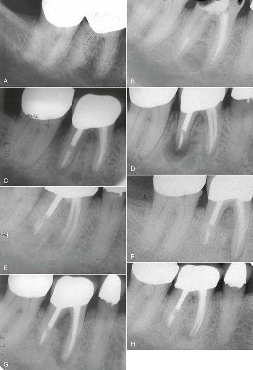
FIGURE 20-1 A, Preoperative radiographic image of mandibular molars. Note carious defect on distal margin of crown on the first molar. B, Two months posttreatment with degraded temporary restoration. C, A 17-month reexamination radiograph indicating lesion on distal apex. D, Immediate postsurgical radiograph. E, Eight-month postsurgical radiograph indicating healing of the surgical site. F, Nine years after the surgery, a reexamination radiograph indicates failure of the root canal treatment in the mesial canals. G, Radiograph following nonsurgical revision of mesial canals. H, Nine-month reevaluation indicating healing of the lesion on the mesial apex.

Two months later, the patient called with the complaint that the temporary filling had come out. Upon examination, the IRM was found to be intact over the mesial orifices, but the distal canal was exposed to saliva (see Fig. 20-1, B). The distal canal was immediately recleaned and reobturated, again leaving space for a post. The patient was urged to have the tooth restored immediately.

Seventeen months later, the patient was referred for evaluation of a draining sinus tract lateral to the mandibular right first molar. The tooth was now restored, and a radiograph indicated that a post had been placed in the distal canal. A large periapical lesion was identified on the same root (see Fig. 20-1, C), but there was no apparent pathosis associated with the mesial root apices. The patient recalled that again there had been a delay of several weeks before she was able to get an appointment for the restoration. Periapical surgery was performed on the distal root only (see Fig. 20-1, D), which upon 8-month reexamination appeared to have healed completely (see Fig. 20-1, E).

Nearly 9 years after the original treatment, the patient was referred again for reevaluation of the mandibular right first molar, as the referring dentist had discovered another draining tract in the buccal vestibule. The radiograph indicated that there was a periapical lesion at the apex of the mesial root which extended coronally to the furcation (see Fig. 20-1, F). Clinically, the restoration was in excellent condition without evidence of caries or marginal leakage. The mesial canals were revised nonsurgically through the existing restoration (see Fig. 20-1, G) and on reexamination 9 months later, the lesion on the mesial root appeared to be healing normally (see Fig. 20-1, H).

Problem-Solving Analysis

In retrospect, the most obvious etiology for the multiple postoperative problems was coronal leakage that occurred before the final restoration of the tooth initially. This could be attributed to an inadequate temporary restoration and prolonged delays before permanent placement of the post and crown. Leakage into the prepared post space most likely accelerated the failure of the root canal treatment in the distal root. The most reasonable explanation for the eventual failure of the root treatment of the mesial canals was residual bacteria in the pulp chamber from the original leakage problem. If the tooth had been restored immediately and periods of temporization had been avoided, none of the subsequent problems would likely have occurred. These problems are identified daily in private practice and can be prevented with a cognizant approach to the protection of all root-treated teeth with immediate attention to proper restoration.

image

Restoring Access Openings

Root canal procedures are commonly required for teeth that have satisfactory existing restorations, primarily crowns that have good margins and no evidence or deterioration (if metalloceramic no obvious craze lines or fractures). In doing so, potential weakening of the restoration or the tooth structure supporting the restoration is a valid concern.9 A number of studies have addressed the clinical scenario.54,92,136139,151 For this reason, conservative access cavities are always preferable to generous and often excessive preparations. Keep in mind, however, that when crowns have been placed on rotated teeth or roots and canals exit the coronal part of the tooth at unusual angles, access openings may have to be enlarged or altered in shape significantly in order to find the entire canal system (see Chapter 8).

The crowns of many teeth are frequently reconstructed entirely of amalgam or composite resin filling materials. Following root canal treatment, it is common to repair the access cavities with restorations of the same material. Nevertheless, research has demonstrated that routine access through large, multiple surface amalgam restorations significantly compromises the fracture strength of the repaired restoration postoperatively.54 For this reason, the placement of a full crown is worthy of consideration because the prognosis of this type of restoration will enhance tooth retention.5 The effect of access preparations through coronal reconstructions with composite resin is an unknown entity, and good restorative principles must be applied based on individual case demands. If any area of exposed enamel exists during the access opening preparation, beveling of the enamel margin followed by a self-etching adhesive system would be favored38 to protect the weaker dentin bonding beneath the surface.36

For onlays and full crown restorations, the structural integrity of the underlying tooth structure and restorative materials is more of a concern than the structure of the crown itself. Therefore, the clinician should use a caries detector upon entry to ensure that there are no hidden carious pathways that have undermined the restoration.84,116 This examination is also enhanced with magnification (i.e., loupes or microscope). Access openings made through an existing restoration result in loss of retention92,151 and strength.54 When the access opening is restored, loss of retention is reversed,92,151 and if a post is added, additional retention can be gained.151

The most significant structural consideration in making access though full crowns is the presence of ceramic material. As discussed in Chapter 8, access cavities can be safely made through metalloceramic and full ceramic crowns without serious risk of fracture, although craze lines have been noted.139 Once the root canal procedures have been completed, the restoration of the access cavity must be completed. Bonding to dentin deeper in the access opening is not as effective as bonding to enamel.36 In these cases, however, there is a metalloceramic or ceramic margin. Etched ceramic materials can form a strong and durable bond with resin.67 A micromechanical bond can be obtained by roughening the porcelain with a bur, air abrasion, or etching with hydrofluoric acid,128 but acid etching appears to be the most effective method.3,89,124,133 This acid is provided in a 10% concentration in a syringe, and adherence to the manufacturer’s instructions is essential to prevent problems.8 Adhesion between a resin and porcelain may be enhanced by the use of a silane coupling agent.120 Some systems can bond well to both composites and amalgams, thereby reducing leakage with either material.31 This could provide direction for restoring a root-treated tooth that is not in a load-bearing area or has little contact in function, such as opposing a removable denture or partially edentulous space.

The metallic ring in the metalloceramic crowns is usually not significant when restoring an access opening. However, if the occlusal boundaries of the access opening are all metal, then some type of mechanical adhesion is necessary.89 The use of burs or air-abrasion techniques is indicated; chemical adhesion is also possible with metals that form an oxide layer, but silane has no effect on bonding to metal.142

Traditionally it has been common in restorative dentistry to place a cement base for deeper preparations. A more contemporary evolution of this concept is the intracoronal barrier as discussed above. Research has recently shown the effectiveness of sealing the canal orifices against coronal leakage with a 3-mm layer of glass ionomer cement, flowable composite resin, compomer, or MTA.* Following placement of the intracoronal barrier, the remainder of the access cavity may be restored as discussed earlier, depending on the nature of the coronal material. The technique for composite repair of the access cavity made through a gold three-quarter crown is seen in Fig. 20-2. The intracoronal barrier would be indicated even if the final restoration of the occlusal surface were amalgam or gold.

image

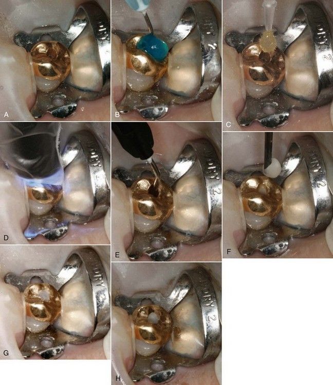
FIGURE 20-2 A, Access cavity to be restored following routine root canal treatment. Sealer has been thoroughly removed with solvent. B, Acid etch. C, Placement of the bonding system. D, Light curing of bonding system. E, Placement of compomer in 1-mm increments. Each increment is light cured, creating an intracoronal barrier. F, Remainder of cavity is restored with composite resin. G, Resin restoration cured. H, Completed restoration.

Problem-Solving Key Factors That Influence Restorative Choices in Anterior and Posterior Teeth

Multiple factors that must be considered in choosing the final restoration include:

Amount of remaining sound tooth structure
Occlusal function and the use of cuspal coverage in posterior teeth
Nature of the opposing dentition
Position of the tooth in the arch
Length, width, and curvature of the roots
Alterations in the tooth architecture caused by removal of the roof of the pulp chamber and the loss of cusps and marginal ridges
Changes that occur in the dentin of endodontically treated teeth, and the ability of the tooth to function under stress
The need for crown lengthening (see Chapter 17)
The need for a ferrule to retain and stabilize the restoration
Preservation of cervical tooth structure
Use of agents (eugenol-based) that impact on bonding posts in the root canal55,85 and on coronal tooth structure25,27

Alterations in the tooth that result in the potential for structural weakness are not attributable to changes in moisture content,51,98 but rather these changes are attributable to architectural changes in the remaining compromised dentin, both on a macroscopic and microscopic basis.51,108 Studies in the biomechanical properties of root-treated teeth and their contralateral vital pairs indicate that teeth do not become brittle after root canal treatment, as was previously thought.118 However, the strongest tooth will always be the one in which sound dentin and enamel can be retained and used to rebuild the tooth.29,62,94,117 Multiple restorative options are available to best serve the patient’s needs; these options must be chosen carefully for each particular situation.

Restoration of Anterior Teeth

The use of a composite resin in combination with dentinal bonding systems is the restorative material of choice in teeth that have intact marginal ridges, cingulum, and incisal edges.121123,136139 Additionally, prevention of coronal leakage is a primary concern. When restorations are placed without sufficient adherence to restorative protocols, failure is likely to result (Fig. 20-3). Because most anterior teeth experience fewer functional forces than posterior teeth do, the routine removal of sound tooth structure in favor of extensive post-core restorations is rarely warranted.52,58,140 In this regard, many root-treated and discolored teeth may be bleached to an esthetically acceptable color by use of a chemical or “walking bleach” technique without placement of a veneer or ceramic crown.

image

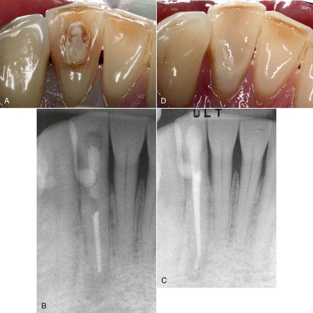
FIGURE 20-3 A, Mandibular incisor with failed access restoration. B, Periapical lesion resulting from treatment failure. C, Completed revision with satisfactory intracoronal barrier and final restoration. D, Clinical view of restoration.

Some root-treated anterior teeth require complete coronal coverage. The most common indications are loss of tooth structure in the incisal half of the crown from trauma or from fracture associated with large or multiple restorations. Such heavily restored teeth often become unaesthetic as well through staining and wear. Full coverage is both aesthetic and durable and should be considered routinely in these situations.

The retention of a sound cingulum in anterior teeth also appears to be a major consideration, especially as it relates to the “ferrule effect” to be discussed later in this chapter.117 Often, removal or undermining of the cingulum has been encouraged in anterior teeth that have the propensity to have two canals, such as mandibular anterior teeth. The lingual canal is usually located beneath the cingulum, and failure to find it has led to many failures in these teeth. Ironically, the destruction of the cingulum in an attempt to find the canals may adversely affect the ultimate retention of an artificial crown on these teeth. Of greater importance, however, may be the concerns that come with treatment planning the retention of these teeth in the presence of significant lingual decay or, more frequently seen, lingual resorption that destroys the integrity of the cingulum.

Restoration of Posterior Teeth

Primarily because of the loss of structural integrity and the amount of occlusal force during function, these teeth have a different set of restorative needs.47,115 Contemporary thought in research, clinical practice, and outcomes studies supports the placement of a restoration with full cuspal protection.5,29 Restorations of this nature are essential to prevent fracture when occlusal forces from the opposing cusps tend to wedge and separate cusp tips during function. Retrospective evaluation of endodontically treated teeth up to 8 years has shown that 85% of the teeth that are lost were not protected with full-coverage restorations.123 Furthermore, studies have shown that endodontically treated posterior teeth that do not receive full, protective coverage fail as much as six times more than those restored with full coverage.5 In cases of posterior teeth opposing a partial or complete denture, the forces of mastication and cuspal interdigitation may be reduced significantly, and tooth restoration with complete coronal coverage may or may not be warranted.

Of initial restorative concern is the seal of the pulp chamber. A separate intracoronal barrier should be placed before the construction of the final restoration begins. During core construction, care must be taken to insure that areas of deep excavation or fracture near the future crown margins are well sealed to prevent possible marginal leakage. When there is substantial remaining coronal tooth structure, any restorative material can be used for the core as the support for the crown is derived principally from dentin. However, if the coronal preparation would eliminate or reduce the remaining dentin thickness to where it is of no value, then a bonded composite or core paste-type material is indicated. If the final preparation will be in sound dentin, even a glass ionomer would be appropriate as a core buildup material, h/>

Only gold members can continue reading. Log In or Register to continue

Stay updated, free dental videos. Join our Telegram channel

Jan 2, 2015 | Posted by in Endodontics | Comments Off on 20: Problem Solving the Challenges Faced in the Restoration of Endodontically Treated Teeth

VIDEdental - Online dental courses

Get VIDEdental app for watching clinical videos