2: The Tooth: Functions and Terms

The Tooth: Functions and Terms

FUNCTION OF TEETH

Teeth are important in many functions of the body. They are essential for protecting the oral cavity, in acquiring and chewing food, and in aiding the digestive system in breaking down food. The teeth form a hard physical barrier that protects the oral cavity. This shield not only affords protection to the oral structures, but the teeth themselves are formidable weapons. One group of mammals classified to the order Carnivora demonstrates this particularly well. Lions and tigers are members of this order and have well-developed canines that they use as weapons to defend themselves and to attack and kill their prey. The teeth also function in communication. They are necessary for proper speech, phonetics and even whistling. In many cultures their appearance can be a very positive sexual attraction. In dental anatomy the teeth are studied individually and collectively, including their functions, anchorages, and relations to one another. Our study therefore begins with a discussion of the individual tooth.

CROWN AND ROOT

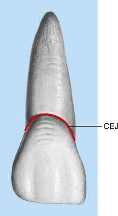
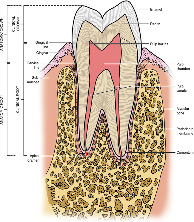
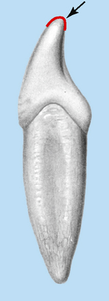
Each tooth has a crown and root portion. The crown is covered with enamel, and the root portion is covered with cementum. The crown and root are joined at the cementoenamel junction (CEJ). The line that demarcates it is called the cervical line, a line that is formed by the junction of the cementum of the root and the enamel of the crown (Fig. 2-1).

The crown portion of the tooth erupts through the bone and gum tissue. After eruption it will never again be covered with gum tissue. Only the cervical third of the crown in healthy young adults is partly covered by gingiva (gum tissue). The tooth continues to erupt from the bone and gingival tissue until all of the crown is exposed (Fig. 2-2).

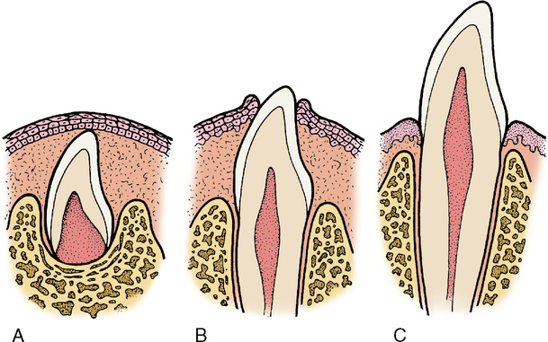
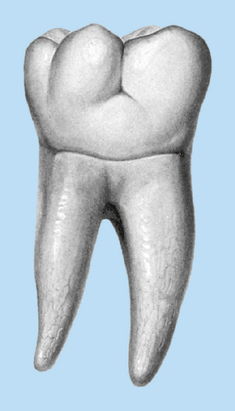
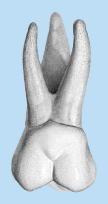
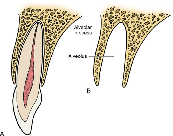
A clinical difference is evident between the amount of crown that could be erupted and the actual amount that is visible in the mouth. The anatomic crown is the whole crown of the tooth that is covered by enamel, regardless of whether it is erupted. The clinical crown is only that part seen above the gingiva. Any nonerupted area is not a part of the clinical crown of the tooth. Therefore, if all of the anatomic crown does not erupt, the part that is visible is considered the clinical crown, and the unerupted portion is part of the clinical root (Fig. 2-3). Eruption of a tooth is thus the moving of that tooth through its surrounding tissues so that the clinical crown gradually appears longer. The tooth may have a single root (see Fig. 2-1) or a multiple root with bifurcation or trifurcation—that is, division of the root portion into two or three segments (Figs. 2-4 and 2-5). Each root has one apex or terminal end. The root is held in its position relative to the other teeth in the dental arch by being firmly anchored in the bony process of the jaw. The portion of the jaw that supports the teeth is called the alveolar process. The bony socket in which the tooth fits is called the alveolus (Fig. 2-6). Teeth in the upper part of the jaw are called maxillary teeth because they are anchored in the maxillary bone. In the lower jaw they are called mandibular teeth because they are anchored in the bone called the mandible.

TOOTH TISSUES

The four tooth tissues are enamel, dentin, cementum, and dental pulp (Fig. 2-7). The first three are hard tissues; the pulp is soft tissue.

Enamel

Enamel forms the outer surface of the anatomic crown. It is thickest over the tip of the crown and becomes thinner until it ends at the cervical line. The color of enamel varies with its thickness and mineralization. The thicker the enamel, then the whiter it appears. The thinner the enamel, then the more it varies, from grayish white at the crown cusps’ edges to white in the middle of the tooth to yellow-white at the cervical line where the thin enamel covering is translucent enough to show the yellow tint of the dentin underneath. The more mineralized the enamel, then the more it lends itself to translucency. These two factors, the mineralization and thickness of enamel, coupled with skin pigmentation, determine the color of the enamel. Older individuals and people with darker skin coloration often display brownish or grayish tones of coloration in addition to or instead of the yellow tones. Individuals with red or auburn colored hair often exhibit a slight reddish or brown-red coloration. Although these darker tones are more heavily concentrated at the gingival area, they are also dispersed throughout the entire tooth.

Enamel is the most densely mineralized and hardest tissue in the human body. The chemical composition of enamel is 96% inorganic and 4% organic matter and water. This dense mineralization gives enamel the ability to resist the wear that the crown of a tooth is subjected to. The hard enamel does not wear very readily; rather, it wears down, grinds up, and crushes almost anything to which it is subjected, including nuts; seeds; ice cubes; and even particles of bone, grit, sand, and leather.

Our ancestors wore the enamel off of their teeth. Their diet consisted of hard uncooked nuts, fruits, and grains. In addition, their food was unprocessed so that pieces of rock, sand, dirt, and grit were embedded in their food. Their lifestyle required chewing leather or bone to fabricate clothing. Often the life expectancy of our ancestors was directly related to whether they still possessed functional teeth. Those who were fortunate enough to have adequate enamel (durable and wear-resistant) were able to live longer and produce more offspring.

Present-day humans rarely wear the enamel off of their teeth. Enamel is not only resistant to wear, but it is very durable and rather resistant to bacteria, mild acids, and tooth decay. The densely packed enamel is smooth, which gives the crown of the tooth a self-cleaning ability, making it difficult for food particles, bacteria, sticky carbohydrate material, and other debris to adhere to the surface of the tooth crown. This self-cleaning ability of enamel and its extreme hardness and resistance to wear make it a nearly perfect outer covering for the crown.

Dentin

Dentin forms the main portion or body of the tooth; it comprises the greatest bulk of the tooth because it forms the largest portion of the crown and root. Dentin is wrapped in an envelope of enamel, which covers the crown, and an envelope of cementum, which covers the root.

Dentin is a hard, dense, calcified tissue. It is softer than enamel but harder than cementum or bone. It is yellow in color and elastic in nature. Its chemical composition is 70% inorganic and 30% organic matter and water. Unlike enamel, dentin is capable of adding to itself. When it does this, the new dentin is called secondary dentin.

Secondary dentin is formed throughout the pulp chamber after the tooth erupts. In time, secondary dentin could completely fill the pulp chamber. Reparative dentin is the dentin that is laid down in response to caries or trauma.

Cementum

Cementum is a bonelike substance that covers the root, although the root is not covered with a perfect layer of cementum; voids expose small patches of dentin. Its main function is to provide a medium for the attachment of the tooth to the alveolar bone. It is not as dense or as hard as enamel or dentin but is more dense than bone, to which it bears a physiologic resemblance. The chemical composition of cementum is 45% to 50% inorganic and 50% to 55% organic components, making it less durable tissue than dentin or enamel. Cementum is quite thin at the cervical line but increases slightly in thickness at the apex of the root. The union of cementum and dentin is called the dentinocemental junction.

The two types of cementum are cellular cementum and acellular cementum. Acellular cementum covers the entire anatomic root. Cellular cementum is confined to the apical third of the root and can reproduce itself, thereby compensating for the attrition (wear) that occurs on the crown of the tooth. Cellular cementum derives its name from the fact that the very cells that lie down and form the cementum eventually become entrapped within newly formed cementum. The cells that produce cementum are called cementoblasts.

Cementum gives the tooth a mechanism of anchorage that protects and supports the tooth, yet it is self-adjusting and independent of the tooth’s main nourishment system. The nutrition for cementum is derived from the outside of the tooth through blood vessels that come directly from the bone.

Pulp

Dental pulp is the nourishing, sensory, and dentin-reparative system of the tooth. It is composed of blood vessels, lymph vessels, connective tissue, nerve tissue, and special dentin-formation cells called odontoblasts.

The pulp is housed in the center of the tooth, with dentin surrounding the pulp tissue. The walls of the pulp cavity are lined with odontoblasts, the chief function of which is to lay down primary and secondary dentin. The odontoblasts form secondary dentin when the tooth is subjected to trauma from chemical, mechanical, or bacterial causes. Blood vessels bring in the nourishment necessary to activate and support the formation of secondary dentin. In addition, the blood vessels also supply the white blood cells necessary to fight bacterial invasion within the pulp. The lymph tissue filters the fluids within the tooth, and the nerve tissue responds to pain and does not differentiate the cause.

Anatomically the pulp is divided into two areas: the pulp chamber and the pulp canals or root canals. The pulp chamber is housed within the coronal portion of the tooth, and the pulp canals are located within the roots of the tooth. Together the pulp chamber and pulp canals are referred to as the pulp cavity; thus the pulp cavity runs the entire length of the interior of the tooth from the tip of the pulp chamber, the pulp horns, to the apex of the root canal (see Fig. 2-7).

Stay updated, free dental videos. Join our Telegram channel

Jan 4, 2015 | Posted by in General Dentistry | Comments Off on 2: The Tooth: Functions and Terms

VIDEdental - Online dental courses

Get VIDEdental app for watching clinical videos