2: Pharmacokinetics: The Absorption, Distribution, and Fate of Drugs

CHAPTER 2 Pharmacokinetics

The Absorption, Distribution, and Fate of Drugs

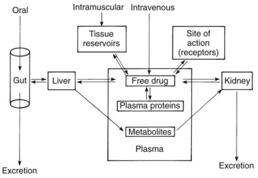
When the magnitude of a drug’s pharmacologic effect is quantified as a function of dose, the tacit assumption is that the drug concentration vicinal to the site of action is linearly related to the amount administered. Although this assumption may strictly apply to an in vitro test, it ignores the temporal factors that modify drug effects in vivo. Drug concentrations are rarely static; they increase and decrease as dictated by the processes of absorption, distribution, metabolism, and excretion. This chapter examines these processes (Figure 2-1) and how they influence the passage of drugs through the body.

PASSAGE OF DRUGS ACROSS MEMBRANES

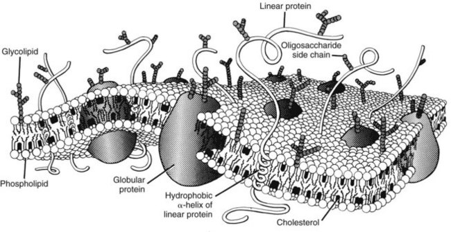
For a drug to be absorbed, reach its site of action, and eventually be eliminated, it must cross one or more biologic membrane barriers. These may consist of a single plasma membrane or constitute a layer of closely packed cells. Because such barriers to drugs behave similarly, the cell membrane can serve as a prototype for all. The plasma membrane is composed of a bimolecular sheet of lipids (primarily phospholipids and cholesterol) with proteins interspersed throughout and extending beyond the lipid phase of the membrane (Figure 2-2).8,51 The presence of protein molecules spanning the entire thickness of the membrane provides a necessary link between the extracellular environment and the cell interior, which is consistent with the concept that drug activation of a membrane-bound receptor on the external surface of a cell can be directly translated into an intracellular response. Specific transmembrane proteins also provide important pathways for the uptake and extrusion of drugs.

Passive Diffusion

The passage of drugs across biologic membranes can involve several different mechanisms. Of these, passive diffusion is the most commonly encountered. The defining characteristic of passive diffusion is that the drug moves down its electrochemical gradient when crossing the membrane.

Simple diffusion

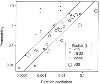
Studies by Overton and Meyer more than a century ago showed that the cell membrane acts for the most part as a lipoid barrier. As shown by Collander (Figure 2-3), the rate of transfer of nonelectrolytes across a membrane is directly proportional to the lipid/water partition coefficient. (The partition coefficient is a measure of the relative solubility of an agent in a fat solvent, such as olive oil or octanol, versus its solubility in water.) A drug with a high partition coefficient (i.e., a lipophilic drug) readily enters the lipid phase of the membrane and passes down its concentration gradient to the aqueous phase on the other side. More molecules are then free to enter the membrane and continue the transfer process. With poorly lipid-soluble compounds, however, few molecules enter the membrane per unit of time, and the rate of passage is depressed.

The absence of an ionic charge is one major factor favoring lipid solubility. Drugs with a fixed charge, such as drugs containing a quaternary nitrogen atom, permeate membranes slowly if at all. The reason for the relative solubility of nonionized molecules in lipids relates to their exclusion from polar media. Simple ions and charged molecules are stabilized in water by the hydration shells that surround them, a consequence of the tendency of charged species to orient polar molecules. This process excludes nonpolar substances, and the resulting segregation causes them to coalesce in a manner analogous to the formation of oil droplets on the surface of water. The term hydrophobic bonding, introduced in Chapter 1, refers to the tendency for water-insoluble molecules to be drawn together; this behavior is responsible for the preferential tendency of lipid-soluble drugs to penetrate cell membranes by way of the lipid components. Ionized compounds are so stabilized by their interaction with water that movement into a lipid phase is markedly restricted. Many therapeutic agents are weak electrolytes; depending on the pH of their aqueous environment, they can exist in ionized and neutral forms. Because charged molecules penetrate membranes with considerable difficulty, the rate of movement of these drugs is governed by the partition coefficient of the neutral species and the degree of ionization. As illustrated in Figure 2-4, acidic conditions favor the transport of weak acids, and the opposite holds true for basic compounds.

The same concept of water interaction used to explain the aqueous solubility of ions also applies to many nonionic molecules. Although unsubstituted aliphatic and aromatic hydrocarbons have little or no tendency to react with water, affinity for water molecules is not restricted to structures with a formal charge. Organic residues possessing electronegative atoms such as oxygen, nitrogen, and sulfur can interact with water through the formation of hydrogen bonds to provide some degree of aqueous solubility.

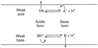
Figure 2-3 shows that lipid solubility is not the only factor influencing the simple diffusion of uncharged drugs across cell membranes; molecular size is also important. Water, glycerol, and some other small molecules permeate much more readily than would be predicted from their respective partition coefficients. Figure 2-3 also shows that some large organic molecules diffuse more slowly than expected. Nonelectrolytes containing numerous hydrophobic groups are often so insoluble in water that their transit across the lipid/water interface may be retarded despite a favorable partition coefficient.26 This finding suggests that some degree of water solubility is necessary for the passive diffusion of drugs across membranes. No matter how lipid soluble an agent is, it will never cross a membrane if it cannot first dissolve in the extracellular fluid and be carried to the structure. Benzocaine, an active local anesthetic when applied directly to nerves, is ineffective after injection because its water insolubility precludes significant diffusion away from the administration site and toward its locus of action within the neuronal membrane. When inside the membrane, a drug with an extremely high partition coefficient may be so soluble in the lipid phase that it has little tendency, despite moderate solubility in water, to diffuse out of the membrane down its concentration gradient.38 A review of human clinical data involving more than 2400 compounds suggests that simple diffusion will be poor if a drug has two or more of the following characteristics: (1) more than five H-bonding donor groups, (2) more than five H-bonding acceptor groups, (3) more than 10 N and O atoms, (4) a molecular weight greater than 500 Da, and (5) a partition coefficient greater than 10,000 : 1.28

Simple diffusion across capillary walls warrants special comment. In addition to the transcellular pathway of drug diffusion just described for lipid-soluble agents, an aqueous paracellular pathway formed by 10-nm to 15-nm clefts between the endothelial cells of most capillaries permits the aqueous diffusion of water-soluble drugs between the plasma and extracellular space. Hydrophilic molecules up to small proteins in size can use this route; fixed negative charges along the diffusion pathway tend to promote the movement of positively charged macromolecules while restricting movement of those with net negative charges.

Adding to the paracellular movement of drugs across capillaries is the bulk flow of water that moves in relation to the net balance of hydrostatic and osmotic forces between the vascular and interstitial compartments. This net transfer of fluid, termed convection, carries with it dissolved drugs and other solutes. Convective movement of most drugs is quantitatively inconsequential; however, it may play a major role in the movement of proteins and other macromolecules that avoid filtration by the endothelium, especially in inflamed tissues. The small amounts of albumin and other plasma proteins that reach the extracellular space (4% per hour for albumin) are largely returned to the circulation by lymphatic convection.

Facilitated diffusion

Water, small electrolytes, and hydrophilic molecules of biologic importance generally move across plasma membranes much more readily than would be predicted by simple diffusion. In these instances, transmembrane proteins that circumvent the lipid bilayer facilitate diffusion. The simplest mechanism involves a transmembrane pore, such as aquaporin 1. Discovered in 1991, aquaporin 1 is a 28-kDa polypeptide that forms a 3-Å channel through which water can enter or leave cells. More than 10 aquaporins have been discovered in mammalian tissues and are especially prominent in cells and organs involved with the transcellular movement of water: kidneys, capillaries, secretory glands, red blood cells, choroid plexus, brain glia, eyes, and lungs.1,24 Some aquaporins are selective for water only, increasing its membrane permeability by a factor of 10 to 100; others permit the passage of glycerol and several other molecules in addition to water.

The movement of specific ions (e.g., Na+, K+, and Ca++) across the cell membrane is facilitated by the presence of transmembrane channels, such as the nicotinic receptor described in Figure 1-2 and the Na+ channel illustrated in Figure 16-4. The opening of these gated channels (in contrast to porins, which are always open) is regulated by the electric potential across the membrane or by the presence of specific ligands, such as neurotransmitters. When a channel is open, passive diffusion of an ion capable of traversing it depends on the electric potential across the membrane and the ion’s own chemical gradient. Boosting the electrochemical gradient by manipulating the voltage across the cell membrane is an effective method of increasing ionic flow. Even in the absence of specific ion channels, the transport of fixed ions and weak electrolytes across tissue barriers can be facilitated by the appropriate use of electric current (as in iontophoresis, discussed subsequently).

Numerous lipid-insoluble substances are shuttled across plasma membranes by forming complexes with specific membrane constituents called carriers or transporters. Carriers are similar to receptors in many ways; they are proteins, often quite selective in the agents with which they combine, and subject to competitive inhibition. Because the number of transporter molecules is finite, carrier-mediated diffusion can be saturated at high drug concentrations. The GLUT family of glucose transporters is representative of carrier proteins that facilitate the movement of hydrophilic solutes across cell membranes. The initial step in the facilitated diffusion of glucose is its binding to the exposed active site of the transporter protein. This binding sequentially causes an external barrier or gate to close and interior gate to open, after which the glucose is released into the cell. The loss of glucose causes the internal gate to close and the external gate to open, exposing the active site and completing the cycle.

Active Transport

Active transport is the term given to the carrier-mediated transfer of a drug against its electrochemical gradient. In addition to exhibiting selectivity and saturability, active transport requires the expenditure of energy and may be blocked by inhibitors of cellular metabolism. Active transport permits the efficient absorption of substances vital for cellular function (and certain drugs that resemble them structurally) and the selective elimination of waste products and foreign chemicals, including many drugs. Approximately 2000 genes—7% of the total human genome—code for transporters and associated proteins. Two superfamilies of transporters are of special significance to pharmacokinetics: ATP-binding cassette (ABC) transporters, and solute carrier (SLC) transporters.

Approximately 49 ABC transporters hydrolyze adenosine triphosphate (ATP) to provide the energy directly needed for molecular transport and are referred to as primary active transporters. P-glycoprotein (P for altered permeability), also known as multidrug resistance protein-1 (MDR-1) and given the designation ABCB1 by the Human Gene Nomenclature Committee, is the most extensively researched representative. Originally identified in 1976 for its ability to expel numerous antineoplastic drugs from mutated cells that overexpress it, P-glycoprotein is a 170-kDa glycoprotein composed of two subunits in a head-to-tail arrangement (Figure 2-5).44,48 Each subunit contains a transmembrane domain of six α-helices that span the plasma membrane and help form the pump itself, and a nucleotide-binding domain (also known as the ABC cassette) that hydrolyzes ATP to power the transport. Many ABC transporters are referred to as half transporters because they consist of only a single subunit and must dimerize to create the active pump. P-glycoprotein preferentially promotes the cellular extrusion of large (300 Da to 2000 Da) hydrophobic substances and neutral or positively charged amphiphilic molecules. Transported drugs include numerous anticancer agents (e.g., doxorubicin, vinblastine, and paclitaxel), antiviral compounds (e.g., ritonavir), Ca++-channel blockers (e.g., diltiazem), digoxin, antibiotic and antifungal drugs (e.g., erythromycin and ketoconazole), hormones (e.g., testosterone), and immunosuppressants (e.g., cyclosporine).

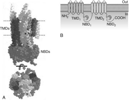
Drug binding occurs within the plasma membrane near the cytoplasmic surface, limiting transport to drugs with good lipid solubility or sufficient length to reach the active site. P-glycoprotein is expressed in various cells, but the highest concentrations are located in intestinal epithelial cells; renal proximal tubular cells; canalicular membranes of hepatocytes; the capillary endothelium of the brain, choroid plexus, testes, and placenta; placental trophoblasts; adrenocortical cells; and stem cells.30 Other ABC transporters important in pharmacokinetics include the multidrug resistance-associated protein (MRP) family. Collectively, the MRP transporters are also widespread and involved in the vectorial (one-way) movement of drugs and other xenobiotics. In contrast to P-glycoprotein, the MRP transporters pump amphiphilic molecules with at least one negative charge. These substrates include bile salts, nucleotide analogues, and conjugates of glutathione, glucuronic acid, and sulfate.

The known SLC transporters encompass 48 families encoded in 400 genes. Because the SLC transporters do not directly use ATP as an energy source for transport, they are more accurately referred to as secondary active transporters. The Na+ pump (or Na+/K+-ATPase), which uses about one fourth of the body’s ATP production, is the principal driving force for secondary active transport. By maintaining a large electrochemical gradient for Na+ across the plasma membrane, movements of molecules that are energetically coupled to Na+ (or another ion with a strong electrochemical potential difference across the membrane) can occur against their own concentration gradients. Secondary active transporters that move the coupled substances in the same direction as the linked ion are termed cotransporters or symporters. In contrast, antiporters or exchangers move the coupled substances in the opposite direction. Many SLC transporters (including the GLUT family described previously) allow the transmembrane movement of specific chemicals down their own electrochemical gradients and support facilitated diffusion. In contrast to the ABC transporters, SLC transporters can facilitate the bidirectional movement of substrates based on their existing concentrations across the cell membrane.

Organic anion transporters (OATs) and organic anion–transporting polypeptides (OATPs) are important families of SLC transporters involved in pharmacokinetics.35 As a group, they promote the cellular uptake of acidic drugs into the liver, kidney, intestine, lung, and brain, and their excretion via the bile and urine. An analogous family of organic cation transporters (OCTs) provides similar handling of positively charged drugs.

Endocytosis and Exocytosis

The processes of endocytosis and exocytosis are together the most complex methods of drug transfer across a biologic membrane. The term endocytosis refers to a series of events in which a substance is engulfed and internalized by the cell. (Phagocytosis, or “cell eating,” is a variant of endocytosis associated more with the removal of particulate matter by macrophages than with drug transport.)

Endocytosis usually begins with the binding of a compound, usually a macromolecule, to be absorbed by its receptor on the membrane surface. Two good examples are the attachment of low-density lipoprotein (LDL) and insulin to their respective receptors. With time, the bound agent-receptor complex is concentrated in an indentation of the membrane called a coated pit. (This migration also occurs spontaneously with the LDL receptor.) Clathrin, a cytoplasmic protein that attaches to the internal surface of the plasma membrane, serves to capture the receptors within the pit while excluding other surface proteins.49 Internal rearrangement of its structure deepens the pit, forming a coated bud. A second protein, termed dynamin, is believed to congregate around the collar of the invaginated bud and initiate separation from the membrane. When released, the vesicle loses its clathrin coat and fuses with an organelle called the endosome. Some of the captured contents, such as LDL receptors, are recycled back to the plasma membrane by transport vesicles; the remainder undergo lysosomal processing and release into the cytoplasm.

An alternative method of endocytosis involves indentations on the plasma membrane termed caveolae. Caveolae contain large amounts of cholesterol covalently linked to caveolin-1, the primary structural protein of these structures. The internalization process also involves vesicle formation, but clathrin and endosomes do not participate in the internalization process.

The complementary process of exocytosis occurs when vesicles, such as those produced by the Golgi apparatus, fuse with the plasma membrane and discharge their contents outside the cell. Exocytosis is the primary method by which cellular products such as regulatory hormones are secreted by the cell. The term transcytosis is descriptive of a coupled form of endocytosis and exocytosis leading to the transfer of drug from one epithelial surface of a cell to another. In this scenario, the endosomal vesicle described previously avoids lysosomal capture, is transported across the cell, and fuses with the plasma membrane to release its contents extracellularly.

Cells generally are capable of endocytosis; however, exocytosis and transcytosis are most intensive in tissues adapted for the absorption, distribution, and export of important foodstuffs, regulatory hormones, and secretory products. Endocytosis/transcytosis is probably responsible for the absorption of antigenic proteins and certain toxins from the small intestine and for the transfer of large molecules between tissue compartments. It plays a minor role in the transport of most drugs.

ABSORPTION

Absorption refers to the transfer of a drug from its site of administration into the bloodstream. The particular route of administration selected greatly influences the rate and perhaps the extent of drug absorption.

Oral Ingestion

Oral ingestion was the first and is still the most commonly used method for the administration of therapeutic agents. The major advantages of the oral route lie in three areas: convenience, economics, and safety. Patient acceptance of oral medication is good because the technique itself is painless, and trained personnel are not required for its accomplishment. The convenience and low cost with respect to other modes of therapy are especially prominent for drugs that must be given several times daily on a long-term basis. The oral route is relatively safe because drug absorption is comparatively slow. Sudden high blood concentrations are not nearly as likely to be achieved by the ingestion of drugs as they are by parenteral injection. Allergic reactions are also less likely to occur, especially serious reactions. The oral route does have some drawbacks, however. Because self-administration is the rule, patient compliance is required for optimal therapy. Drug absorption is likely to be delayed (on a clinical average of 30 to 60 minutes) and may be incomplete. Metabolic inactivation or complex formation may also occur before the drug has a chance to reach the systemic circulation. These limitations to the oral route translate into an increased variability in patient response. Finally, the spectrum of adverse reactions caused by oral medication can extend from one end of the gastrointestinal tract to the other.

Drugs taken orally may be absorbed along the entire alimentary canal, but the relative degree of contact with the mucosa determines the amount of uptake in each segment. Variables affecting absorption include the duration of exposure, the concentration of the drug, and the surface area available for absorption. Under normal circumstances, the oral and esophageal mucosa are exposed too briefly to a drug during the process of swallowing for any absorption to occur. The colon normally plays no role in the uptake of orally administered compounds because, with the exception of some sustained-release preparations, little absorbable drug usually reaches it. By exclusion, the bulk of drug absorption must occur in the stomach and small intestine.

Influence of pH

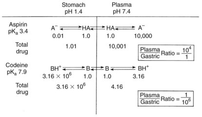
As previously discussed, absorption is favored when the drug ingested is lipid soluble. For weak electrolytes, the pH of the surrounding medium affects the degree of ionization and drug absorption. Because the H+ concentrations of the stomach and small intestine diverge widely, the two structures seem to be qualitatively dissimilar in their respective patterns of drug absorption. Figure 2-6 illustrates this difference and its effect on the previously commonly used analgesic combination of aspirin plus codeine. Aspirin is an organic acid with a pKa (negative log of the dissociation constant) of 3.49. In gastric juice (pH 1 to 3), aspirin remains largely nonionized, and its passage across the stomach mucosa and into the bloodstream is favored. The plasma has a pH of 7.4, however. On entering this environment, the aspirin becomes ionized to such an extent that return of the drug to the gastrointestinal tract is prevented by the low lipid solubility of the anionic species. When equilibrium is established, the concentration of nonionized aspirin molecules on both sides of the membrane is the same, but the total amount of drug (ionized plus neutral forms) is much greater on the plasma side. The relative concentration of drug in each compartment can be calculated with the Henderson-Hasselbalch equation, as follows:

image

This unequal distribution of drug molecules based on the pH gradient across the gastric membrane is an example of ion trapping. The biologic process that sustains this partitioning is the energy-consuming secretion of H+ by the gastric parietal cells. Because few organic acids have a pKa low enough to permit significant ionization at stomach pH, almost all acidic drugs should theoretically be effectively absorbed across the gastric mucosa.

For bases such as codeine (pKa 7.9), the opposite applies. Codeine is almost completely ionized in the acidic environment of the stomach; absorption is negligible. At equilibrium, virtually all the drug remains within the stomach. Only very weak bases are nonionized at gastric pH and available for absorption. The ion trapping of basic compounds within the gastric lumen is sometimes useful in forensic medicine. Many drugs subject to abuse are organic bases (e.g., heroin, cocaine, and amphetamine). Even when injected intravenously, they tend to accumulate in the stomach by crossing the gastric mucosa in the reverse direction. Questions of intravenous overdosage can often be answered from the analysis of stomach contents.

When the gastric fluid passes into the small intestine, it is quickly neutralized by pancreatic, biliary, and intestinal secretions. The pH of the proximal one fourth of the intestine varies from 3 to 6, but it reaches neutrality in more distal segments. Under these more alkaline conditions, aspirin converts to the anionic form, whereas a significant fraction of the codeine molecules give up their positive charge. Although basic drugs are favored for absorption over acids in the small intestine, ion trapping is not as extensive because the pH differential across the intestinal mucosa is small. Differences in intestinal absorption based on pH are more concerned with the rate of uptake than with its extent. As one might expect, neutralization of gastric contents by the administration of antacids or ingestion of food temporarily removes the qualitative disparity in electrolyte absorption normally observed between the stomach and the small intestine.

Gastric emptying

Because almost any substance that can penetrate the gastrointestinal epithelium is best absorbed in the small intestine, the rate of gastric emptying can significantly affect drug absorption, particularly for organic bases that are not absorbed at all from the stomach. Gastric emptying is accomplished by contraction of the antrum of the stomach. A cyclical pattern of activity occurs in fasting patients where periods of quiescence (about 1 hour each) are followed by contractions that increase in intensity over a 40-minute period before terminating in a short burst of intense contractions that migrate from the stomach to the distal ileum. Ingesting a tablet or small volume of liquid may result in gastric retention of the drug for 1 hour or longer. After eating a meal, sustained antral and pyloric contractions help break up the ingested food and permit the extrusion of liquid into the duodenum while retaining particles more than 1 mm in diameter within the stomach. A mixed meal of solids and liquids usually begins to enter the duodenum in about 30 minutes and requires about 4 hours to leave the stomach completely. A glass of water ingested on an empty stomach is moved into the small intestine in exponential fashion, with half of the water expelled from the stomach in 15 minutes, and essentially all of the liquid removed by 1 hour.

A major variable in delaying gastric emptying is the presence of fat. Unless drug-induced irritation of the gastric mucosa must be avoided, most oral medications should be taken in the absence of food but with a full glass of water. This procedure speeds drug entry into the small intestine and provides maximum access to the gastrointestinal mucosa. Occasionally, the presence of a fatty meal promotes the absorption of a drug that has a high lipid but low water solubility. The antifungal agent griseofulvin, the protease inhibitor saquinavir, and the fat-soluble vitamins are examples of substances that are better absorbed in the presence of lipids. In these instances, the delay in gastric emptying produced by the high fat content of the chyme is compensated for by a more complete absorption.

Additional situations in which food enhances drug uptake have been reviewed.33 Nevertheless, because gastric emptying is often a limiting factor in the rate of drug absorption, many unrelated drugs exhibit latency periods (the lag phase between oral ingestion and onset of drug effect) of a similar magnitude.

Influence of dosage form

Although the times required for gastric emptying and for diffusion across the mucosal barrier undoubtedly contribute to the delayed onset of action of drugs taken orally, situations exist in which these events are not rate limiting. Most drugs intended for oral use are marketed in the form of capsules or solid tablets. In contrast to solutions, these preparations must first dissolve in the gastrointestinal fluid before absorption can occur. If dissolution is very slow, it can become the controlling factor in drug absorption.

The first step in the dissolution process is the disintegration of the tablet (or the capsule and its granules) to yield the primary drug particles. Various excipients are usually included in solid drug preparations to promote disintegration and particle dispersion. If disintegration is impaired, drug absorption is depressed accordingly. The dissolution of drug particles occurs by a diffusion-limited mechanism. The diffusion layer of solvent surrounding each particle becomes saturated very quickly with drug molecules escaping from the solid. Because saturation of the diffusion layer occurs far more rapidly than does diffusion from it into the bulk solution, the entire process proceeds no faster than the rate of drug diffusion. Several methods can be used, however, to accelerate the dissolution rate. Because the total surface area of the particles determines the area available for diffusion, reducing the mean particle size through the process of micronization promotes solubilization. A decrease in particle size of 85% with a compensating increase in particle number doubles the rate of dissolution.27 Another useful approach is to manufacture drugs in the form of water-soluble salts. The concentration of drug in the dissolution layer is enhanced (often by many times), and the rate of diffusion is increased.

The dissolution process may be considered rate limiting whenever a drug solution produces a systemic effect faster than a solid formulation of the same agent does. Sometimes discrepancies in absorption between dosage forms are of such magnitude that clinical differences are noted. With aspirin, the concentration of drug in the plasma 30 minutes after administration can be twice as high for a solution as for a solid tablet.27 Although it is unclear whether this difference results solely from drug dissolution or from other factors, such as the more rapid gastric emptying typical of liquids, dissolution is probably at least partially responsible.

The influence of dosage form on drug absorption is often taken advantage of by drug manufacturers. Some drugs (e.g., erythromycin) are unstable at a low pH, and others (e.g., ammonium chloride) are irritating to the gastric mucosa. To avoid release of these drugs within the stomach, they are often prepared in the form of enteric-coated tablets. An enteric coat consists of a film of shellac or some polymeric substitute. The covering is insoluble under acidic conditions, but does break down to permit tablet disintegration in the more alkaline environment of the small intestine. Although these preparations are often beneficial, their usefulness nevertheless is negatively affected by an increased variability in patient response. Because drug absorption cannot begin until the tablet passes into the duodenum, the time required for gastric transit becomes an important variable. The passage of a single insoluble tablet from the stomach into the intestine is a random event that can take several minutes to more than 6 hours.15

Sustained-release preparations represent another method of capitalizing on the influence of formulation on drug absorption. These products are usually designed to release a steady amount of drug within the gastrointestinal tract for 12 to 24 hours. Some preparations also provide an initial loading dose that is readily available for absorption. Sustained release may be accomplished by using a porous matrix, with the drug located in the interior spaces and on the external surface. An alternative is to make spheres of drug that dissolve at different rates because of various coatings. An intriguing form of sustained-release tablet is the “elementary osmotic pump,” in which the drug is enclosed in a semipermeable membrane that lets water in, but restricts drug egress. Constant release through a small hole in the membrane is achieved by the osmotic pressure that builds up within the tablet as the drug is slowly dissolved. Advantages claimed for these drug products include greater patient compliance and smaller fluctuations in blood concentration between dosages. Studies with some preparations have documented a greater variability in performance, however, than is normally encountered with conventional dosage forms. Because sustained-release products contain several conventional doses of medication, a danger exists that a too-rapid release of drug from these preparations might cause unexpected toxic concentrations. Conversely, inordinately slow or incomplete release could lead to inadequate drug therapy. Uncertainty over the effects of these formulations is recognized by the U.S. Food and Drug Administration (FDA), which regards them as new drugs and requires that efficacy and safety be shown before they can be marketed.

The sensitivity of gastrointestinal absorption to variations in drug formulation is best exemplified by the concern over bioavailability. In many instances, chemically identical drugs have proved in the past to be biologically nonequivalent. In one study of tetracycline hydrochloride, nine preparations of different manufacture were compared with an aqueous solution of the same drug.29 Although seven brands produced blood concentrations ranging from 70% to 100% of the reference solution, two products exhibited relative bioavailabilities of only 20% to 30%. Differences in bioavailability are most likely to be clinically important with drugs that are poorly absorbed, have low margins of safety, and are inactivated by capacity-limited processes. Since 1977, federal law has required that bioequivalence testing be performed on all new drugs, and the FDA has mandated such testing of existing products for which a problem of nonequivalence is known to exist. Bioavailability considerations related to drug selection are considered further in Chapter 55.

Active transport

Most drugs intended for oral use are absorbed by passive diffusion. Active transport systems do exist, however, for specific dietary constituents that sometimes increase the absorption of certain drugs. The absorption of levodopa and baclofen from the intestine is enhanced because they are amino acid analogues and actively transported into intestinal cells by the large neutral amino acid transporter (LNAT, an SLC transporter). Valacyclovir is likewise much better absorbed than is its congener acyclovir because it is a substrate for PepT-1, another SLC transporter.

Active transport mechanisms can also inhibit drug absorption.30 P-glycoprotein is highly expressed along the luminal surface of intestinal epithelial cells, where it exports xenobiotics that would otherwise be absorbed. This function is in concert with the “chemoimmunity defensive” role P-glycoprotein plays in protecting cells from exposure to potentially toxic compounds.48 Although P-glycoprotein may delay the absorption of many drugs and prevent altogether the uptake of pharmaceuticals of low absorptive potential, it is probably of minor significance regarding the extent of absorption of most drugs intended for oral use, whose concentrations in the chyme are sufficient to overwhelm the capacity of P-glycoprotein to export them.47 Figure 2-7 depicts the active transport of drugs into and out of intestinal cells and at other important sites.

image

FIGURE 2-7 Transepithelial or transendothelial transport of drugs across the liver (absorption), brain capillaries (distribution), and liver and kidneys (elimination). ABC, ATP-binding cassette transporter; SLC, solute carrier transporter.

(Adapted from Giacomini KM, Sugiyama Y: Membrane transporters and drug response. In Brunton LL, Lazo JS, Parker KL, editors: Goodman & Gilman’s the pharmacological basis of therapeutics, ed 11, New York, 2006, McGraw-Hill.)

Drug inactivation

A shortcoming of oral ingestion is the inactivation of drugs before they reach the systemic circulation. The destruction of some agents (e.g., epinephrine and insulin) is sufficiently great to preclude their administration by this route. With other drugs (e.g., penicillin G), losses may be smaller, but still large enough to make oral administration inefficient. Gastric acid is one of the principal causes of drug breakdown within the gastrointestinal tract, but degradation also results from enzymatic activity. Vasopressin, insulin, calcitonin, and other polypeptides are subject to hydrolysis by pancreatic and intestinal peptidases. Intestinal cells also contain intracellular enzymes for metabolizing drugs. Of particular importance are the presence of monoamine oxidase for the inactivation of biogenic amines and the presence of CYP3A4/5 enzymes (described later) for the oxidation of numerous compounds. Enteric bacterial enzymes may also destroy certain ingested agents, such as chlorpromazine. Finally, intestinal contents can alter the effectiveness of many orally administered drugs. Binding to constituents of chyme, chelation with divalent cations, or formation of insoluble salts may decrease the amount of drug available for absorption.

A special fate exists for substances that are successfully absorbed from the gastrointestinal tract. The venous drainage of the stomach, small intestine, and colon is routed by the hepatic portal system to the liver. A first pass of high drug concentration through this enzyme-laden organ can significantly reduce the quantity of agent reaching the systemic circulation. Lidocaine is metabolized so rapidly in the liver that virtually all of an oral dose is destroyed during its first pass. Although less pronounced, disparities in opioid analgesic and antibiotic efficacies observed between the oral route and other modes of administration are of clinical importance to the practice of dentistry.

Inhalation

The alveolar membrane is an important route of entry for some drugs and many noxious substances. Although the alveolar lining is highly permeable, it is accessible only to agents that are in a gaseous state or are inhaled in sufficiently fine powders or droplets to reach the deepest endings of the respiratory tree. Gaseous agents include the therapeutic gases, carbon monoxide, the inhalation anesthetics, and numerous volatile organic solvents. The second category of alveolar membrane penetrants is collectively known as aerosols. This term refers to liquid or solid particles small enough (usually ≤10 µm in diameter) to remain suspended in air for prolonged periods. Particles of this sort include bacteria, viruses, smoke, pollens, sprays, and dusts. Any such finely divided material, when inhaled, reaches some portion of the respiratory tree and is affected by the processes of sedimentation and inertial precipitation. Most aerosols contain a mixture of particle sizes. Relatively large particles (≥5 µm) impact on the terminal bronchioles and larger branches of the respiratory tree and are removed from the lungs by a cilia-driven blanket of mucus flowing continuously toward the pharynx. Smaller particles, which do reach the alveolar sacs, can be absorbed through the lining cells into the bloodstream, taken up by the process of phagocytosis, or carried by an aqueous film covering the alveolar cells to the terminal bronchioles where they join the mucous blanket. Although two of these three possible fates involve particle uptake, the mechanism for removing solids is remarkably efficient. Only a minute portion of the inhaled dusts of a lifetime fails to be removed by ciliary transport.

Therapeutic use of aerosols is not widespread, but some emergency medications are prepared in this form. Because the onset of effect is extremely rapid after inhalation of an aerosol drug, this route can provide a means of quick self-medication for individuals in danger of acute allergic reactions to venoms or drugs. Epinephrine is one such emergency agent that is marketed as an aerosol. Many respiratory drugs are also prepared in aerosol form because they are highly effective by this route while minimizing systemic exposure. The rapidity and efficiency of alveolar membrane absorption can occasionally pose problems for therapy, however, as illustrated by the use of pressurized aerosols containing isoproterenol. Although 97% of an isoproterenol spray is swallowed under normal conditions and inactivated by various enzymes, overmedication can produce toxic effects. Data gathered over a 7-year period in the United Kingdom suggested that the undisciplined use of these preparations increased mortality in asthmatic patients. Restriction of over-the-counter sales and warnings to physicians were accompanied by a decline in mortality.21 Findings such as these reflect the hazards of aerosols when abused and provide a caveat for uncontrolled self-medication with any potentially dangerous drug. Concern over aerosols is also related to questions of toxicology, such as the absorption of heavy metal dusts by industrial workers.

Parenteral Injection

Drugs are frequently given by parenteral injection when oral ingestion is precluded by the patient’s condition, when a rapid onset of effect is necessary, or when blood concentrations greater than those obtainable with the enteral route are required. The method of injection selected varies with the particular drug and therapeutic need of the patient.

Intravenous route

The administration of drugs by infusion or injection directly into the bloodstream is particularly useful when immediate effects or exact blood concentrations are desired. Because absorption is bypassed, intravenous injection circumvents the delays and variations in drug response characteristically associated with other modes of administration. Rapid dilution in the bloodstream and the relative insensitivity of the venous endothelium to drugs often permit the successful administration of compounds or solutions too irritating for other routes (e.g., alkylating anticancer drugs and hypertonic fluids). Also, through the technique of titration, the intravenous route provides an avenue for the controlled administration of drugs that have a very narrow margin of safety between therapeutic and toxic concentrations. The infusion of lidocaine to prevent ventricular arrhythmias and the incremental injection of antianxiety drugs during intravenous sedation are two examples in which titration is used to achieve a desired effect while avoiding adverse reactions. Although many intravenous agents do not require titration and may be given in standardized doses, they should still be injected slowly. If administered too quickly, a dose may move initially through the heart, lungs, and major arteries as a bolus of high drug concentration. Nonspecific but potentially disastrous cardiopulmonary side effects may result, even from the rapid injection of simple salt solutions. Most drugs should be administered over a period of 1 minute, which approximates the circulation time of blood through the body. This procedure avoids high, transient concentrations and permits discontinuance if any untoward effect is observed during the course of injection.

A major disadvantage of the intravenous route is that, once the drug is injected, very little can be done to remove it from the bloodstream. When an adverse response is noted with another route, further absorption usually can be delayed or perhaps even prevented. Toxic reactions to drugs given intravenously are often instantaneous and severe. Life-threatening anaphylactic events are also more likely because of the possibility of a massive antigen-antibody reaction. Other complications of intravenous injection include vasculitis and embolism (from drug irritation, particulate matter in the injected solution, or needle trauma), fever (from injection of pyrogens such as bacterial lipopolysaccharides), infection, and hematoma formation. Finally, the accidental intra-arterial injection of drugs (e.g., promethazine) intended for intravenous use has led to arteriospasm, gangrene, and loss of limbs.

Intramuscular route

The intramuscular route is often selected for drugs that cannot be given orally because of slow or erratic absorption, high percentage of drug inactivation, or lack of patient cooperation. The rate of absorption from an intramuscular site is governed by the same factors influencing gastrointestinal uptake, such as lipid/water partition coefficient, degree of ionization, and molecular size. Many drugs are absorbed at approximately the same rate, however, regardless of these factors. The only barrier separating a drug deposited intramuscularly from the bloodstream is the capillary endothelium, a multicellular membrane with large intercellular gaps. Many lipid-insoluble substances can enter the vascular compartment through these gaps, and even proteins are capable of being absorbed. In these circumstances, blood flow through the tissue is often the primary determinant of the rate of drug absorption. Muscles with high blood flows (e.g., deltoid) provide faster absorption rates than muscles with lesser flows (e.g., gluteus maximus). Generally, 5 to 30 minutes is required for the onset of drug effect, but this latency period can be controlled to some extent. Exercise markedly speeds absorption by stimulating local circulation. Conversely, uptake may be minimized by the application of ice packs or (in an emergency) tourniquets.

With the exception of a few drugs that are relatively insoluble at tissue pH (e.g., diazepam, phenytoin), absorption from an intramuscular injection is usually rapid and complete. Formulations have been developed, though, to provide for prolonged and steady drug release. These depot preparations consist of drugs manufactured as insoluble salts or dispensed in oil vehicles, or both, such as procaine penicillin suspended in peanut oil. Relatively large volumes of solution may be given by this route, but soreness at the injection site is frequent, and some drugs (e.g., doxycycline) are too irritating to be administered in this manner.

Subcutaneous route

Injection of drugs into the subcutaneous connective tissue is a widely used method of administration for agents that can be given in small volumes (≤2 mL) and are not locally damaging. Subcutaneous absorption is similar to that of resting muscle, and onset times are often comparable. As with the intramuscular route, absorption can be delayed by diminishing blood flow, either through the application of pressure or by surface cooling. Pharmacologic interruption of circulation with vasoconstrictors is also a common strategy, especially in local anesthesia. Because of the ease of subcutaneous implantation, compressed pellets of drugs, sometimes mixed with insoluble matrix material, can be inserted to provide nearly constant drug release for weeks or months. Testosterone and several progestational contraceptive agents (e.g., levonorgestrel) have been successfully administered by this approach. Slow absorption also can be achieved through the use of depot forms as described for intramuscular injections.

When subcutaneous administration is chosen for a systemic effect, the hastening of drug absorption is sometimes advantageous. Toward this end, warming the tissue promotes drug uptake by improving local circulation. Massage of the injection site, in addition to stimulating blood flow, helps spread the drug and provides an increased surface area for absorption. This latter effect can also be accomplished through the coadministration of hyaluronidase, an enzyme that breaks down the mucopolysaccharide matrix of connective tissue. The lateral spread of aqueous solutions is so enhanced that hyaluronidase is sometimes used to permit the injection of large fluid volumes in situations in which continuous intravenous infusion is difficult or impossible.

Topical Application

Drugs are often applied to epithelial surfaces for local effects and less frequently for systemic absorption. Penetration of drugs across the epithelium is strongly influenced by the degree of keratinization.

Skin

The epidermis is a highly modified tissue that isolates the body from the external environment. The outer layer of skin (stratum corneum) is densely packed with the protein keratin. This layer is impervious to water and water-soluble drugs, and its relative thickness and paucity of lipids in contrast to other biologic membranes retards even the diffusion of strongly lipophilic agents. The impermeable nature of skin to water-soluble drugs often requires that agents (e.g., antibiotics, fungicides) intended for dermatologic conditions be administered by a systemic route despite the accessibility of the skin. For lipid-soluble drugs, however, the percutaneous route is often successful for local problems. Disruption of the keratinized layer markedly enhances drug absorption, especially of hydrophilic compounds. The underlying connective tissue (dermis) is quite permeable to many solutes, although it differs from most tissues in having an abundant supply of arteriovenous shunts, which may cause systemic absorption to be particularly sensitive to changes in temperature.

The general resistance of the intact skin to drugs does not invalidate the need for caution when dealing with potentially toxic chemicals. Sufficient documentation of epidermal absorption of foreign substances has established that certain compounds may readily penetrate the skin to cause systemic effects. These drugs include organic solvents, organophosphate and nicotine-based insecticides, and some nerve gases. Severe poisoning has also resulted from the excessive application of sunburn creams containing local anesthetics. Even lipid-insoluble substances such as inorganic mercury can diffuse across skin if exposure is prolonged.

The benefits of improving and sufficiently controlling percutaneous absorption to make it a reliable route of drug administration have prompted several strategies. A “transdermal therapeutic system” has been developed to provide continuous systemic uptake of nitroglycerin, scopolamine, fentanyl, and nicotine for prophylaxis of angina pectoris, prophylaxis of motion sickness, management of chronic pain, and assistance with smoking cessation. The system is a complex patch that consists of an outer impermeable backing, a reservoir containing the drug in a suspended form, a semipermeable membrane, and an inner adhesive seal.

In the early 1960s, it was discovered that the industrial solvent dimethyl sulfoxide promotes the percutaneous absorption of water-soluble drugs. The potential of simplified therapy for arthritic and other patients that this drug carrier offered generated much enthusiasm. Subsequent reports of adverse reactions in animals caused interest to wane, however, until the late 1970s, when it was promoted as an effective agent for the symptomatic relief of a wide variety of musculoskeletal and collagen disorders. Although widely available as an herbal remedy, dimethyl sulfoxide is currently approved by the FDA only for the treatment of interstitial cystitis.

Another approach to improving drug penetration through the epidermis is the use of occlusive dressings. These dressings retain moisture and break down the horny layer through the process of maceration. A final technique, iontophoresis, is discussed subsequently.

Stay updated, free dental videos. Join our Telegram channel

Jan 5, 2015 | Posted by in General Dentistry | Comments Off on 2: Pharmacokinetics: The Absorption, Distribution, and Fate of Drugs

VIDEdental - Online dental courses

Get VIDEdental app for watching clinical videos