2
Monitoring
Routine monitoring can be divided into three categories.
The anaesthetist
The anaesthetist is continuously present during the entire administration of an anaesthetic. Information obtained from clinical observation of the patient, monitoring equipment and the progress of the operation allows for the provision of a balanced anaesthetic in terms of: anaesthesia and analgesia, fluid balance, muscle relaxation and general appearance (skin colour, temperature, sweatiness etc.).
The patient
The minimum monitoring consists of: electrocardiogram (ECG), pulseoximetry, non-invasive blood pressure, capnography and other gas analysis (O2, anaesthetic vapour), airway pressure, neuromuscular blockade; see Chapter 12.
The equipment
This includes: oxygen analyser, vapour analyser, breathing system, alarms and infusion limits on infusion devices. A means of recording the patient’s temperature must be available, as well as a peripheral nerve stimulator when a muscle relaxant is used.
Other monitoring devices are used depending on the type of operation and the medical condition of the patient (e.g. to measure cardiovascular function). These include invasive blood pressure monitoring, central venous pressure, echocardiography, oesophageal Doppler and awareness monitors.
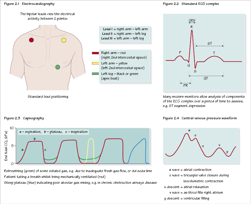
Electrocardiography (ECG)
Continuous assessment of the heart’s electrical activity can detect dysrhythmias (lead II) (Figures 2.1 and 2.2) and ischaemia (CM5 position). Most commonly, the standard lead position is used. From this the monitor can be used to measure the electrical activity between two of the leads whilst the third acts as a neutral.
It is important to remember that the heart’s electrical activity does not reflect cardiac output or perfusion, for example pulseless electrical activity (ECG complexes associated with no cardiac output) may be recorded.
Oximetry
A pulse oximeter consists of a light source that emits red and infrared light (650 nm and 805 nm) and a photodetector. The absorption of light at these wavelengths differs in oxygenated and deoxygenated haemoglobin. Thus the relative amount of light detected after passing through a patient’s body can be used to estimate the percentage oxygen saturation. The detecting probe is typically/>
Stay updated, free dental videos. Join our Telegram channel
VIDEdental - Online dental courses


