Dental Lamina and Enamel Organ
DENTAL LAMINA
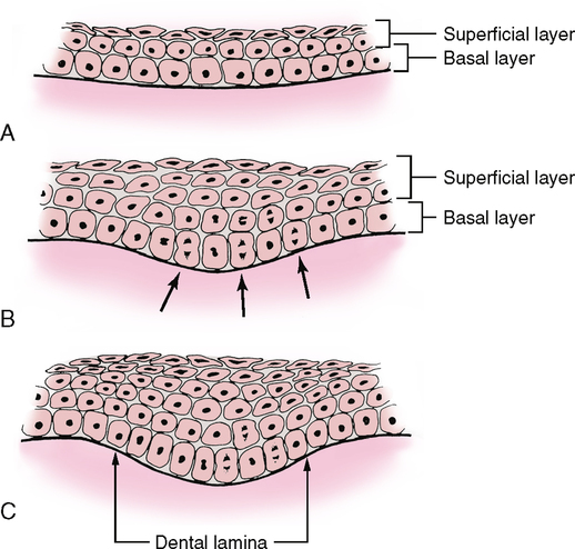
The first signs of tooth development are seen during the sixth embryonic week. At that time the embryonic oral (stratified squamous) epithelium begins thickening. As the epithelium thickens, it grows downward into the underlying connective tissue and does not create a visible ridge in the oral cavity at the time. This thickened oral epithelium is known as the dental lamina. It is a U-shaped thickening of the epithelium of the primitive oral cavity and is found in a position corresponding to the future arch-shaped arrangement of the upper and lower teeth (dental arch). This thickening does not begin all at once throughout the mouth but is first seen in the anterior midline and slowly spreading posteriorly toward the molar region (Fig. 19-1). It takes only several weeks for this thickening to extend to the future position of the primary molars.
ENAMEL ORGAN
Bud Stage
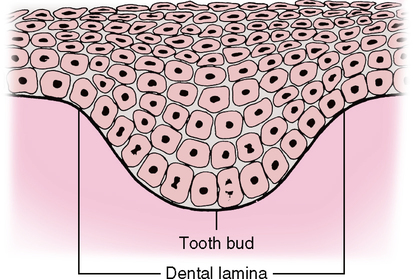
The initial budding from the dental lamina at the 10 thickened areas in each arch is referred to as the bud stage (Fig. 19-2), the first stage in the development of the enamel organ that forms the enamel of the teeth. At first the buds look like blobs of cells from the dental lamina projecting deeper into the underlying connective tissue. The cells in the middle of the buds come from the outer or superficial layers of the oral epithelium, whereas the cells in the periphery of the bud come from the deep or basal layers of the oral epithelium. The buds seem to stretch out from the dental lamina as they grow. As development continues, the deepest parts of the buds become slightly concave. It is at this point that the developing enamel organ goes from the bud stage into the cap stage.
Stay updated, free dental videos. Join our Telegram channel
VIDEdental - Online dental courses


