18: Examination, Diagnosis, and Treatment Planning

Examination, Diagnosis, and Treatment Planning

Diagnosis for Nonorthodontic Problems

The examination of the 3-year-old child often represents a youngster’s first dental experience, although earlier examinations are advocated by most pediatric dentists and the American Academy of Pediatric Dentistry for diagnostic, preventive, and treatment purposes. For a child who has not had a dental examination previously, the new environment, new people, and manipulation of tissues can be difficult or overwhelming.

An initial examination of a child this age can also be stressful for the dentist, who is faced with a potential behavior problem, no clinical baseline, and the challenge of providing both immediate and long-term planning and treatment.

The initial examination is a first experience for both dentist and patient and provides an opportunity to establish a course of dental health for years to come. Of particular interest in the examination of the 3- to 6-year-old are the following factors:

All of the above must be addressed during evaluation of a child this age, especially if this is the child’s initial dental visit.

Patient Records

The nature of health care record keeping in dentistry has evolved from a historical or financial repository to a vital working document. The bare essentials for a pediatric dental record are a health history, examination record, treatment plan, and series of visit notes. Parental or guardian consent should be obtained and recorded at the initial visit. Adjunctive records such as study casts and preventive and dietary forms or analyses also should be kept with the record, if indicated. The history form should permit updating and summary.

The possibilities for a dental examination record for children are endless.

Many practitioners opt for a standard form or use the one they employed in dental school. No clear-cut guidelines exist for choice of a pediatric dental tooth chart, but there are some basic requirements to satisfy medicolegal needs and to provide a complete developmental history. The examination record should do the following:

The tooth chart need not be anatomically correct, and in many cases a diagram of teeth is of more value. Third party reimbursement focuses currently on surfaces and tooth number and the concept of diagramming caries extent on individual teeth is of little value, largely because at this time dentistry does not use a meaningful disease-based coding system, as medicine does. It is critical that the charting system address both primary and permanent teeth so that each record entry provides an up-to-date developmental profile. In addition to a notation of the presence or absence of a tooth, as is done with adults, the mobility of primary teeth and clinically evident eruption of teeth are noted in the pediatric dental chart. Current child safety and forensic concerns strongly suggest making an initial chart of the dentition, including restorations and abnormalities.

Periodontal probing of all teeth is not routine, but the dental chart should provide an area for noting deep pocketing or loss of attachment in some manner, if present. The nature of this notation requires simply adequate baseline data to accomplish treatment and follow-up. A periodontal pocket depth at six points on selected primary teeth is adequate.

Many practitioners develop individual approaches to prevention that can be efficiently addressed on the examination record. A serial chart of oral hygiene performance or gingival scores can be helpful. Other helpful items on the examination record are vital signs, medical alerts particularly important allergies, behavior notes, and unusual findings. Reasons for deferring radiographic examination should be noted in the record. These data provide quick reference for the dentist at chairside.

Recent changes in patient privacy procedures, procedure coding, expected assessments, and dental disease risk assessment suggest that today’s dental examination record (history and physical) should incorporate other measurement scales. These might include any or all of the following:

1. A caries risk assessment based on the Caries Assessment Tool of the American Academy of Pediatric Dentistry (see Chapter 13), or some other instrument that accounts for clinical and historical risk factors. This can be a list of factors or simply a choice of high, medium, or low risk.

2. An initial pain or behavior assessment that provides the dentist with a sense of the child’s cooperation or state of oral health upon initial examination. Some clinicians use a set of diagrammatic “faces” ranging from happy to sad (see Figure 6-3). Others use a version of the Frankl behavior scale, which is a four-point selection ranging from definitely positive to definitely negative. The Frankl scale offers the benefits of simplicity and behavior categories that are relevant to chairside dentistry.

The treatment plan should indicate the sequence of care and permit notation of the date of completion of individual procedures. Each visit’s progress note indicates what was done and any notable occurrences. The advent of the computerized record has made use of dental coding a must. Treatment plans generated in the office ought to include current dental codes to facilitate communication both within and outside the office.

The History

The parent or guardian is the historian for the child. The dentist needs to address both real and perceived problems. Parents may provide erroneous and unverified information simply because the information has not been tested by the health system. Two such examples are reported heart murmurs and allergies. Parents may have been informed of a murmur but are unaware of its seriousness. Parents also may confuse nausea with a true allergic reaction. The dentist may be required to address these concerns directly with a physician to obtain accurate information. In other situations, a long-established or past problem may have been forgotten or dismissed as unimportant.

A general health history form can be used to determine a child’s health background if attention is given to specific elements that relate to children. The dentist should be well versed in conditions that relate specifically to children. Table 18-1 provides a list of health items that are particularly common in the 3- to 6-year-old group. The American Dental Association offers a contemporary history form specifically designed for children that covers most childhood health issues.

A short and noncontributory history was unusual in this age group in the past, but with the improvement in infant health practices and home care, immunization, and early medical intervention, many routine problems and illnesses have been prevented or resolved. On the other hand, a growing number of infants survive who would have perished previously. Although some develop normally, a substantial number are physically or mentally compromised and require alternative and more complex health care approaches. It is not uncommon in this age group to have parents note normal development or simply indicate a vague delay in speech or motor skill. This may occur because a disability has not been clearly diagnosed or a parent is reluctant to accept that the child may have a problem.

The dentist’s review of a checklist with annotations can be used to complete an accurate medical history. Significant findings should be explained in the record. Any health history should be finalized by a summary of the status of the child, especially in the areas of drug allergies, surgical procedures and related problems, cardiac abnormalities, and developmental status. Many pediatric dental records are designed so that this summary appears on the examination form to preclude paging through the record for important information. This summary of positive responses is also a function of some electronic dental records. A dated notation also serves to confirm that the dentist has reviewed the history and made a decision about its impact on treatment at that point in time.

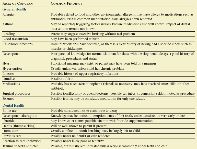
The dental history should be comprehensive. Many parents do not think about recording their child’s dental history other than the eruption of the first tooth. The dental history should cover, at a minimum, past problems and care, fluoride experience, current hygiene habits, and an eruption-developmental profile. Table 18-1 addresses the essential elements of the dental history. The most contemporary approach to the dental history uses a developmental model that permits the parent to address age-specific issues. For example, the health history may ask about bottle use, weaning, access to sweets, and other dietary issues to cover a range of ages on one form. The checklist approach to the health history permits a “not applicable” choice when a child has outgrown a set of questions. This developmental approach provides an age-specific set of findings that can be converted to preventive instruction in the anticipatory guidance counseling by dental staff. Some pediatric dentists tailor the dental history to serve as a screen for caries, orthodontic, gingival, and injury risk factors, asking questions whose answers can later be addressed with take-home preventive advice.

The Examination

The examination encompasses six major sections: behavioral assessment; general appraisal; and head, neck, facial, intraoral, and radiographic examinations.

Behavioral Assessment

A complete behavioral assessment is discussed in Chapter 23. The general appraisal and chairside examination provide two opportunities to observe behavior and initially assess potential cooperation.

General Appraisal

The general appraisal addresses the child’s physical and behavioral status. The classic areas of this appraisal include gait, stature, and presence of gross signs and symptoms of disease. The normal 3- to 6-year-old is ambulatory, well-coordinated in basic tasks, engaging, and physically healthy in appearance. Table 18-2 lists physical and behavioral milestones for the 3- to 6-year-old child. The dentist should incorporate these markers mentally into a profile for evaluation of the child’s status. The general appraisal of the child is best accomplished in the waiting room or a similar nonthreatening environment. This appraisal should be followed by clarification of any abnormal findings and discussion of potential behavior problems with the parent. The role of vital signs in the general appraisal is twofold. The first purpose is to identify abnormalities, and the second is to satisfy the medicolegal role of providing baseline health data for emergency situations. Vital signs may be distorted if the child is upset or anxious. Taking vital signs of blood pressure, pulse, and respiration may be put off until the child has become accustomed to the environment, but these data must be obtained before any drugs are administered. Weight should be obtained and recorded in a conspicuous location on the chart so that the information is available in an emergency. Height should also be recorded and, together with weight, should serve as an index of physical development. Because of concerns about childhood overweight and obesity, the dentist may choose to calculate a body mass index (BMI) for the child or enter height and weight on standardized growth curves and include these if referring the family to a physician for weight concerns.

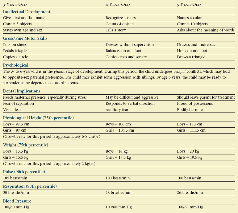
Head and Neck Examination

Examining a 3-year-old requires attention to both clinical findings and the patient’s behavior in the dental setting. Stated differently, the product (dental findings) cannot be separated from the process (patient’s behavior) of the examination. The examination provides a moderately threatening environment for development of behavioral interactions between dentist and child.

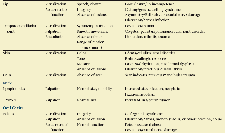
Table 18-3 outlines the elements and expectations for a thorough head and neck examination. The process begins with an orientation about what is to occur. The dentist should describe what will take place at each step in the examination. This tell-show-do technique, which involves explanation, demonstration, and finally completion of a step, is usually the way the diagnostic process is handled. Positive or negative responses from the child should be encouraged. Children also should be warned and supported before the dentist makes positional changes or begins intraoral manipulation.

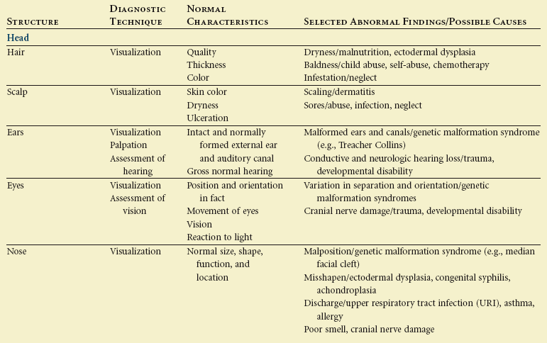
Parental presence is always a matter of controversy. Initial parental involvement may be encouraged to allow a transition from a dentist-parent relationship to a more direct dentist-child relationship. This supported transition is important for children younger than 3 years of age but is less threatening for children near school age. Each child reacts differently to having a parent in the operatory, and the dentist must assess the benefit of that presence on the developing relationship he or she has with that child. It is within the parent’s purview to request to be present during the examination and treatment, but it is within the practitioner’s purview to choose not to treat the child under those circumstances. A growing number of practitioners allow parents in the treatment setting, and most data indicate that parents are neutral factors.7

The examination must involve evaluation of the head and neck. Palpation to identify enlarged and fixed lymph nodes or other swellings is critical. Many children of this age have swollen nodes, but the nodes are usually movable and confined to the lower face and jaws and only indicate minor infection. Swollen nodes in the neck and clavicular region are rarer and may suggest the child has a more serious ailment.

Critical to a thorough examination of the head and neck is evaluation of form and function. The cranial nerves, speech, and mandibular function should be evaluated. However, a complete cranial nerve examination need not be performed because careful observation of sensory and motor function and the child’s responses can indicate nerve status to a significant degree. Normal conversation can be used to identify gross speech pathosis. While palpating the craniofacial structures, the dentist should talk with the child and observe his or her responses. Asking the child to open and demonstrate maximal opening and maximal intercuspation allows the child to perform simple tasks. Mandibular movements should be observed for deviation and restriction of range of movement. The child should also be asked to move the mandible from side to side and to protrude it. Restriction of these movements may identify functional and morphologic problems that result from developmental anomalies or trauma.

Verbal responses also serve as behavioral signals of the child’s adaptation. A child’s cooperation, nonverbal communication, and physiologic responses often suggest stable, improving, or deteriorating behavior. Because the examination setting is only moderately threatening, it provides a good opportunity to develop cooperation.

The manual examination should address any physical variations as well as the strength and mobility of structures. The visual aspect of the process should address color changes, asymmetry, and marked physiologic responses such as sweating or trembling.

Facial Examination

A systematic facial examination is one portion of a complete orthodontic evaluation that describes skeletal and dental relationships in three spatial planes: anteroposterior, vertical, and transverse. The steps include a description of the overall facial pattern, the positions of the maxilla and mandible, and the vertical facial relationships. Next, the position of the lips is determined. Finally, facial symmetry is assessed, and the maxillary dental midline is located relative to the facial midline.

Overall Facial Pattern

First, the facial profile is evaluated in the anteroposterior plane. An assumption is made that the soft tissue profile reflects the underlying skeletal relationship. To begin the examination, the child should be seated upright, looking at a distant point. Three points on the face are identified: the bridge of the nose, the base of the upper lip, and the chin.

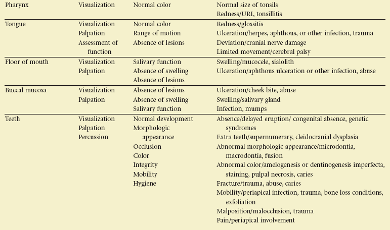
Line segments connecting these points form an angle that describes the profile as convex, straight, or concave (Figure 18-1). A well-balanced profile in this age group is slightly convex. A well-balanced profile in the anteroposterior dimension has an underlying skeletal relationship that is labeled class I (see Figure 18-1, A). This terminology is used because most class I skeletal relationships also have flush terminal plane or mesial step second primary molar relationships and Angle class I or end-to-end permanent first molar dental relationships if those teeth are erupted. Furthermore, the canine relationships usually will be class I, and there will be overjet of 2 to 5 mm. The Angle dental classification is described in a later section.
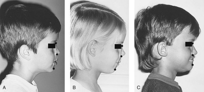
An assessment of the overall profile gives a general feeling for the skeletal relationships, but this evaluation does not diagnose the reason for the relationships. Some children in this age group have extremely convex profiles (see Figure 18-1, B). This is consistent with a class II skeletal relationship, and these patients usually have distal step second primary molar relationships and class II permanent first molar relationships if those teeth are erupted, class II canine relationships, and increased overjet. Other children have straight or concave profiles (see Figure 18-1, C). These are usually found with mesial step second primary molar relationships and class III permanent first molar relationships, class III canine relationships, and negative overjet.

Positions of the Maxilla and Mandible

If the profile is excessively convex or concave, the clinician can try to determine which skeletal component is contributing to the problem. This is a necessary step if orthodontic treatment is being considered. When the problem is known, the appropriate treatment can be performed. It is extremely rare, however, to treat an anteroposterior problem in the primary dentition.

Specifically, in this diagnostic step, the anteroposterior position of the maxilla and mandible are determined. A vertical reference line is extended from the bridge of the nose (the anterior aspect of the cranial base), and the position of other soft tissue points is noted relative to the reference line (Figure 18-2). If the maxilla is properly oriented relative to other skeletal structures, the base of the upper lip will be on or near the vertical line. The soft tissue chin will be slightly behind the reference line if the mandible is of proper size and in the correct position. If the maxilla is positioned significantly in front of the vertical reference line, the patient is said to exhibit maxillary protrusion. If the maxilla is substantially behind the line, the patient exhibits maxillary retrusion. The position of the mandible is described in the same way.
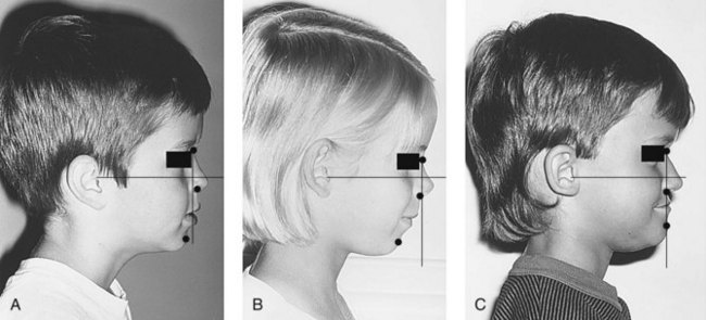
So, if the overall skeletal pattern is significantly convex (class II), the maxilla is positioned in front of the line (maxillary protrusion), the mandible is positioned behind the line (mandibular retrusion), or both. Class II skeletal relationships are sometimes caused by one jaw alone but are often the result of some combination of maxillary protrusion and mandibular retrusion.

Conversely, a facial pattern that is straight or extremely concave (class III) results when the maxilla is positioned behind the line (maxillary retrusion), the mandible is positioned in front of the line (mandibular protrusion), or both. Again, both jaws usually contribute to the skeletal dysplasia.

Some caution must be exercised because soft tissue profile relationships do not always accurately reflect the underlying skeletal relationships. Research has shown that the 3- to 6-year-old age group is especially difficult to classify accurately from a profile analysis.4 In addition, vertical facial relationships influence the anteroposterior relationships. This interaction between the horizontal and vertical planes of space and its effect on the profile is discussed later.

Only gold members can continue reading. Log In or Register to continue

Stay updated, free dental videos. Join our Telegram channel

Jan 14, 2015 | Posted by in Pedodontics | Comments Off on 18: Examination, Diagnosis, and Treatment Planning

VIDEdental - Online dental courses

Get VIDEdental app for watching clinical videos