CHAPTER 15
SALIVARY DUCT CARCINOMA
Salivary glands and breast tissue are of the same embryological origin and are both skin adnexa. Primary salivary duct carcinoma is a high-grade malignancy related morphologically to mammary ductal adenocarcinoma, which is why it is important to exclude the presence of primary mammary carcinoma at the time of diagnosis. This entity represents approximately 3% of all salivary gland tumors. For a long period of time, salivary duct carcinoma was considered under the broader category of “salivary adenocarcinoma not otherwise specified.” However, currently all agree that it represents a specific unique entity. Occasionally, salivary duct carcinoma may develop in pleomorphic adenoma. Moreover, a “low-grade variant” of salivary duct carcinoma has been described. It is also worthwhile to mention that similar tumors have been reported in lacrimal glands.
This carcinoma occurs in the parotid gland with male predominance in the sixth and sevenths decade of life. Because of its high-grade nature, patients present with a recently enlarging, painful, and fixed parotid mass. Cervical lymph node metastases and distant metastases are frequently present at the time of diagnosis.
15.3 CYTOLOGICAL FEATURES AND HISTOLOGICAL CORRELATE
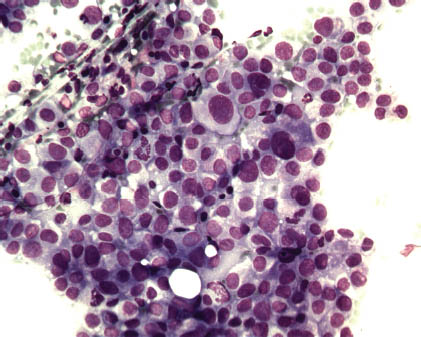
Cytologically, salivary duct carcinoma is similar to high-grade ductal mammary carcinoma. Smears are richly cellular with obviously malignant cells with an abundant background of necrosis. Cells are frequently clustered, but single malignant cells are also observed. The cells are pleomorphic and have well-preserved cytoplasm. Occasionally, tubular, glandular, and papillary formations are observed. Nuclei are large and contain prominent nucleoli. Cellular and nuclear atypia is prominent, and numerous mitotic figures are present. The background contains extensive necrosis, apoptotic debris, macrophages, and numerous leukocytes are observed. Cells around necrotic areas show an oncocytic cytopalsmic change with vacuolization (Figures 15.1–15.4). Table 15.1 summarizes the key diagnostic cytological features of salivary duct carcinoma.
FIGURE 15.1. Marked cellular and nuclear atypia (May–Grünwald–Giemsa [MGG], 200×).

Stay updated, free dental videos. Join our Telegram channel

VIDEdental - Online dental courses


